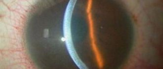
Uveitis that develops with Fuchs syndrome is also called heterochromic Fuchs cyclitis. This chronic non-granulomatous inflammatory disease is characterized by gradual development. At a young age, eye damage is usually detected on only one side.
Quite rarely, it is detected in children or affects both eyeballs. This disease occurs in approximately 4% of patients with uveitis, but it is often misdiagnosed. Heterochromia may be absent or invisible, especially in patients with brown eyes. Therefore, diagnosis should be carried out only in daylight and with a dilated pupil.
Symptoms of the disease
With this disease it is noted:
- Gradual decrease in visual acuity due to the development of secondary cataracts.
- The presence of constant floating opacities.
- Difference in the color shade of the iris in different eyes.
- This condition is usually diagnosed accidentally.
Fuchs syndrome is accompanied by the development and formation of:
1. Corneal precipitates, which are considered pathognomonic signs. They are usually quite small, gray or white, star-shaped or round in shape, and cover the surface of the entire endothelial layer. They may appear or disappear, but are not pigmented and do not merge with each other. Fibrin fibers are usually detected along with precipitates. 2. There is a weak opalescence of the aqueous humor, the number of cells in which is +2. 3. Infiltration of vitreous cells is often a leading sign of the disease.
During gonioscopy, changes are either not detected or are detected:
- Light radial vessels in the anterior chamber of the eye that look like branches. They lead to the formation of hemorrhages on the side opposite to the puncture of the anterior chamber of the eye (Amsler's sign).
- Membrane formations in the corner of the anterior chamber of the eye.
- Small anterior fusions (synechia) of irregular shape.
Causes
You may be interested in: Tired eyes: what to do, how to treat. Methods and tips
The prerequisites leading to the appearance of the syndrome remain unclear today. There is an assumption that there is a connection between the disease and toxoplasmosis, in its ocular form, but there is still no concrete evidence for this hypothesis. Histological studies reveal lymphocytes and plasma cells, which indicates the inflammatory nature of this pathology. Of course, it remains unclear whether Fuchs syndrome can be called an independent disease, or whether it involves only a counter-reaction of the eye structures to certain conditions.
Iris changes
The following changes are detected on the side of the iris:
1. The presence of posterior synechiae, which form after cataract removal. 2. Widespread atrophy of the iris stroma, which includes the absence of iris crypts and a change in color to a faded direction. The dullness of color in the area around the pupil is especially noticeable. Due to the loss of supporting tissue, bulging of the radial vessels of the iris occurs. 3. With atrophy of the posterior pigment layer, a spotty appearance occurs, which is revealed by retroillumination. 4. Nodules form on the iris. 5. Rubeosis of the iris occurs, in which irregularly shaped, delicate neovascularization appears. 6. With atrophy of the pupillary sphincter, mydriasis occurs. 7. The formation of crystalline deposits in the iris is rarely detected. 8. Heterochromia is a very important diagnostic sign.
Forecast
With timely diagnosis and treatment, the prognosis for the disease is favorable. After therapy, visual acuity is restored, the medicine allows the patient to maintain the ability to work. In the absence of therapeutic measures or the patient’s refusal to undergo surgery, the disease continues to progress, developing into complete loss of vision.
It is impossible to prevent the development of pigmentary glaucoma in advance, because specific measures or drugs to reduce the risk of developing the pathology have not yet been developed. When the first symptoms of the disease develop, iridectomy is recommended. Laser correction is carried out as a preventative measure.
People predisposed to developing pigment dispersion syndrome should be checked by an ophthalmologist twice a year. In this case, a person must constantly measure blood pressure to avoid an increase in IOP. In addition to tonometry, ophthalmoscopy, visometry and gonioscopy are performed.
Photos of the iris in Fuchs syndrome
In this case, hypochromia occurs more often and only in 10% of cases does hyperchromia of the iris develop. Quite rarely, hyperchromia is a congenital feature. The heterochromic iris develops as a result of stromal atrophy and impaired pigmentation of the posterior endothelial layer, but it is also determined by the genetically determined color of the iris. With severe stromal atrophy, the posterior pigment located on the endothelium becomes translucent, which leads to the development of hyperchromic coloring. Usually the brown iris becomes lighter, and the blue one, on the contrary, becomes more saturated.
The essence of the disease
The cornea is an important optical structure of the eyeball, thanks to which we are able to see. It is multi-layered, each layer has its own function. The main role of the posterior layer of the cornea - the endothelium - is to ensure the outflow of excess fluid from the corneal tissue. If the endothelium has a normal structure and functions correctly, the risk of swelling and clouding of the cornea is almost eliminated. If disturbances occur in its structure and operation, then fluid accumulates in the tissues, swelling inevitably occurs and the transparency of the cornea is lost. It is precisely these disorders that occur in Fuchs' dystrophy, which is why this disease also has a second name - epithelial-endothelial dystrophy (EED).
Complications
Uveitis developing against the background of Fuchs syndrome is prone to a chronic course. In this case, complications often arise (cataracts, glaucoma), which are associated with the irrational use of local preparations of corticosteroid hormones.
1. Cataracts often complicate the course of the disease and have no distinctive signs from cataracts with anterior uveitis of a different nature. The effectiveness of surgery and implantation of posterior chamber lenses is quite high. However, the development of hyphema is possible. 2. Glaucoma can cause vision loss and usually accompanies a long-term condition.
Fuchs' dystrophy, which affects vision, often affects people over 30 years of age
Author Alexandra Balan-Senchuk
18.09.2019 15:07
Health » Vision
Fuchs' dystrophy is a progressive disease of the cornea that appears as a clear, round dome covering the iris and pupil of the eye. The cornea plays a key role in vision as it helps focus light into the eye, allowing for clear vision.
In patients suffering from Fuchs' dystrophy, and these are often people over 30 years of age, the endothelial cells in the inner layer of the cornea are smaller than normal. These cells are important for processing water in the cellular structure of the cornea.
As a result, people with Fuchs' dystrophy experience inadequate absorption of this fluid and water accumulation may occur. This eventually leads to thickening and inflammation of the corneal tissue, which can affect vision.
Causes of Fuchs' dystrophy
The main cause of Fuchs' dystrophy is heredity caused by a gene mutation. This genetic susceptibility leads to a decrease in the number of corneal endothelial cells, which reduces their ability to process water.
Only one parent must be a carrier of the gene for a child to be at risk of developing the disease. If Fuchs disease affects one parent, each child has a 50% chance of inheriting the gene mutation and disease.
However, some people without a family history of Fuchs' dystrophy may also be affected, suggesting that there are other factors that influence the development of the disease. Scientists have not yet established the exact reason.
Progression of Fuchs syndrome
In the early stages of the disease, patients may not notice certain symptoms. Minor corneal swelling may be present and has some impact on vision, which is worse in the morning but improves during the day.
Symptoms of Fuchs' dystrophy often vary in intensity depending on air humidity. Because symptoms are related to fluid accumulation in the cornea, they tend to improve in dry climates and worsen on rainy days.
Over time, changes in the cells of the cornea can cause vision problems that become chronic over time. This is associated with the development of scar tissue, which can lead to blindness. This progression often occurs gradually over several decades. In some cases, surgery is required to remove scar tissue and improve vision.
Treatment of Fuchs' dystrophy
Treatment for Fuchs' dystrophy depends on the progression of the condition and the severity of symptoms at the time of diagnosis.
For patients diagnosed in the early stages of the disease, annual follow-up examinations are usually sufficient to monitor progression and make appropriate interventions if required. Patients diagnosed in advanced stages of the disease already have some changes in vision. They usually require more frequent examinations and complex treatment regimens.
Because there is no complete cure, treatment for this condition focuses on symptomatic management of visual changes and pain. This may include collecting fluids or using medicinal ointments, drops, or contact lenses applied to the eyes. In severe cases, a corneal transplant may be required.
Photo: novosti-mediciny.ru
Treatment of uveitis in Fuchs syndrome
Treatment of uveitis that develops with Fuchs syndrome includes:
1. Injections of long-acting drugs (triamcinolone acetate) into the posterior sub-Tenon's space help with vitreous opacities. 2. The use of topical steroids is ineffective. 3. Due to the fact that posterior synechiae are formed extremely rarely, mydriatics, as a rule, are not prescribed. 4. Vitrectomy is performed when there is significant opacification of the vitreous body, which is not amenable to treatment with steroid drugs.
Prognosis and prevention
As such, no means or methods for preventing Fuchs syndrome have been found. Patients with this pathology should visit an ophthalmologist every six months to carry out all the necessary diagnostic methods in order to early detect subsequent complications in the form of cataracts and glaucoma. In addition, it is recommended to adjust the diet to include a significant number of vitamins, and organize a sleep and rest schedule.
The prognosis for this disease for the continuation of a full life and the ability to work is often positive. For a long time, the disease proceeds latently, but in the event of the subsequent development of cataracts or glaucoma, complete loss of vision is likely. For a long period of time, the disease has a hidden extent, however, in the case of the addition of a secondary cataract or glaucoma, absolute loss of vision is likely to be assigned to a disability group.
Source


