Episcleritis is an inflammatory reaction that affects the episcleral tissue located between the tunica albuginea and the connective tissue.
The pathology often recurs and is mild.
Episcleritis sometimes goes away on its own, but in most cases it requires therapy. According to studies, it affects women more often than men.
The disease is characterized by the development of redness of the conjunctiva, so it is often confused with conjunctivitis. The true cause of the disease often cannot be determined, since many patients do not seek medical help.
Causes
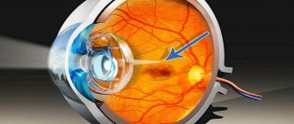
The eyeball is almost completely surrounded by a whitish dermis (sclera), which protects the eye from external influences. Unlike other elements of the eye, the sclera, consisting of a collagen ligament, is relatively vascular. It extends from the entry point of the optic nerve to the cornea.
In this case, the dermis at the junction with the cornea is covered with a small area of conjunctiva. Under the sclera itself there is another layer of connective tissue - the episclera. It consists of thin collagen bundles, loose connective tissue, and separates the dermis of the anterior eye from the conjunctiva.
Episcleritis causes inflammation of this layer. Unlike ordinary scleritis, the corresponding inflammation is present only superficially on the episclera and leaves the adjacent layers of the sclera intact.
The disease is provoked by the following reasons:
- Infectious pathologies. Bacteria and viruses rarely provoke inflammation. The most common are tuberculosis, Lyme disease, syphilis, shingles, and herpes.
- Autoimmune diseases. These include Sjögren's syndrome, rheumatoid arthritis, Liebman-Sachs disease, or SLE.
Metabolic disorders (gout), diseases of the digestive tract (ulcerative colitis), vascular inflammation, irritation of the optic nerve (during stress and eye fatigue) can lead to the development of the disease.
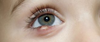
Episcleritis in children is a rare occurrence, especially under 5 years of age. As a rule, inflammation does not last long and does not lead to complications.
In older children, episcleritis is usually associated with rheumatological diseases. Pediatric rheumatologists should regularly evaluate the vision of their patients, as ocular complications of rheumatic diseases can lead to vision loss.
Scleritis
Some people believe that episcleritis and scleritis are the same disease. In fact, these are different diseases that have similar symptoms in the early stages.
Scleritis is less common than episcleritis and is much more severe. Usually combined with an underlying autoimmune or systemic disease. Most often, this pathology affects people aged 30-60 years.
Scleritis can lead to serious consequences such as loss of vision. If treatment is not carried out, inflammation of the anterior choroid in some cases can provoke the appearance of complicated cataracts and secondary glaucoma.
Symptoms of scleritis
Inflammation begins in the upper layer of the sclera (as with episcleritis), then affects deep structures.
The following general symptoms of scleritis can be identified:
- lacrimation and photophobia;
- sudden “shooting” pain radiating to the head;
- a very painful red-violet inflammation in the form of a tubercle on the sclera.
Types of scleritis
Superficial scleritis is characterized by extensive damage to the sclera near the limbus. Often the inflammation affects both eyes at once. Swelling and hyperemia are observed on the surface of the sclera and conjunctiva, as well as pain in the eyeball area. There is almost no lacrimation. The disease is chronic, continuing with periods of remission for several years. Vision does not decrease.
Deep scleritis can be purulent and granulomatous .
With granulomatous scleritis, infiltrates are created in the deep layers of the sclera. Patients experience photophobia, eye pain, and watery eyes. Often becomes chronic. When the iris, ciliary body, cornea and sclera become inflamed, keratosclerouveitis .
Purulent scleritis is characterized by an acute course of the disease. With this pathology, a painful focus is formed at the site of the anterior and posterior ciliary arteries. The source of inflammation is dark red with a yellowish tint, which after a while becomes soft and opens. Then a scar may form in this place, which in some cases resolves on its own.
Classification
There are three forms of the disease:
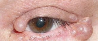
- Nodular. This type of episcleritis is less painful and is rare. It is characterized by the appearance of a nodule in a certain area of the eyeball. The tissues around it become inflamed, and the lesion gradually spreads.
- The migratory form of the disease manifests itself as hyperemic flat lesions near the edge of the cornea. Accompanied by cephalgia and swelling. This form is characterized by a strict frequency of relapses.
- Rosacea. The disease is combined with rosacea around the skin and often recurs. It manifests itself as painful nodules on the organs of the visual analyzer, one by one.
Varieties of episcleritis
Depending on the patient’s sensations and external signs, three types of eye episcleritis are distinguished.
- Nodular episcleritis. It spreads to both eyes at once. In this case, nodular rashes appear in the limbus area (the place where the sclera joins the cornea). When pressing on the eyelid, a person experiences pain. These formations disappear in about a month, leaving dark marks behind them. The disease can recur quite often, with nodules appearing in new places.
- Rosacea-episcleritis. As it progresses, nodules appear on one eye or the other, the whites turn red, and the eyelids swell. The disease is also characterized by acne on the skin of the face.
- Migratory episcleritis. Flat foci of inflammation appear near the limbus, and the eyelids also swell. The disease has a short-term course and goes away on its own after a few days.
Symptoms
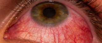
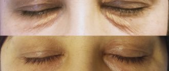
Episcleritis is characterized by bright red spots. The eye becomes covered with a network of inflamed capillaries, and hemorrhages in the form of nodules form.
The nodules move and are sensitive. There is usually no deterioration in visual perception. Episcleritis manifests itself differently. During inflammation, the following symptoms are observed:
- tearfulness;
- swelling;
- increased sensitivity to light;
- eye strain when moving the pupil;
- pain on palpation.
If your eyes become blue, you should immediately go to the hospital. This condition is called scleritis.
Photo
Prevention measures
Prevention of episcleritis
To prevent the development of episcleritis, you need to take a set of preventive measures:
- protect your eyesight from negative factors , especially if you work in the construction industry, where you must always wear personal protective equipment;
- observe the rules of personal hygiene and make sure that all members of your family do this (especially children);
- do not allow small children to play with objects with small parts that can damage their eyes;
- all infectious diseases must be treated promptly;
- in summer, when the sun is very active, wear sunglasses . This will protect your eyes not only from the negative effects of ultraviolet rays, but also from small insects and dust;
- Regularly conduct preventive examinations with a doctor , especially if any problems arise with the functioning of the visual system.
Visit your ophthalmologist regularly
Although episcleritis is mild in most cases, its symptoms cannot be ignored. Any pathological processes affecting the organs of vision in one way or another can lead to serious consequences.
Diagnostics
The diagnosis is made by an ophthalmologist. He examines the affected eye externally for abnormalities such as reddish nodules and excessive tearing.
The patient is asked about other symptoms and existing diseases. A biomicroscopic analysis of the affected eye is then performed. Thus, the doctor determines whether the inflammation has spread to deeper layers of tissue.
If it is difficult to make a diagnosis, use a solution of Phenylephrine at a concentration of 2.5%. It allows you to distinguish episcleritis from deep lesions.
Preventive actions:
- use of protective equipment for the eyes (protection from the ingress of chemical components and foreign bodies);
- strengthening the immune system;
- providing the eyes with decent rest;
- timely therapy for chronic diseases;
- medical examinations;
- taking care of eye hygiene.
The prognosis for the course of the disease is favorable. Periodic relapses are characteristic. The disease lasts 7-15 days , the break is up to 3 months. Complications are rarely identified.
However, it is important to find out the causes of the disease and not self-medicate in order to avoid infection of deeper tissues and a decrease in the quality of vision.
Video on the topic:
Related articles:
- Nystagmus of the eyeball: what is it and how is it treated?
- Colorblind. What colors do these people not distinguish?
- Macular degeneration of the retina: ICD-10 code, treatment, forms
- Uveitis: photos, symptoms, treatment, causes
Treatment
If symptoms of an inflammatory process appear, you should immediately consult an ophthalmologist. However, episcleritis tends to go away on its own; the signs go unnoticed for a long time.
If unpleasant symptoms appear, anti-inflammatory medications are prescribed. The main recommendation is peace. It is important to avoid visual strain so that the condition does not worsen.
If there are complaints, the following treatment regimen is prescribed:
- Hyaluronic acid eye drops are used to hydrate the affected eye. Activities such as long periods of computer work are temporarily suspended and bright light sources are excluded.
- In severe inflammation, steroid eye drops help. For example, Tobradex. The drugs are used for a short period, as they can lead to worsening symptoms and the development of adverse reactions.
- Cooling. For swelling and pain, cool compresses are applied to the affected eye. Cold prevents the spread of inflammation. Any herbal extracts are used for compresses.
If the disease appears due to rheumatoid arthritis or gout, oral administration of salicylates and butadione is prescribed. For infectious lesions, antibacterial agents are prescribed in the form of tablets, drops or ointments.
Episcleritis caused by tuberculosis is treated with immunomodulatory therapy and chemotherapy, which is based on the use of anti-tuberculosis drugs.
For home remedies, it is recommended to use sunflower oil and golden mustache, a decoction of wormwood, chamomile and cornflower flowers. Decoctions are prepared from them for washing the eyes. Use up to 4 times a day for 14 weeks.
Use for the prevention of healthy eyes. Use different towels to wash your eyesight so as not to introduce infection into your healthy eye.
Prices for treatment for episcleritis
The cost of treating episcleritis at MGK is calculated individually and will depend on the volume of therapeutic and diagnostic procedures performed. You can make an appointment or find out the cost of a particular procedure by calling in Moscow or online, using the appropriate form on the website, you can also familiarize yourself with the “Prices” section.
Go to the “Prices” section>>>
Dagaev Adam Huseinovich
How is the diagnosis of episcleritis confirmed?
If the patient complains of redness of the eyes, the ophthalmologist conducts a full diagnosis, including:
- determination of visual acuity;
- biomicroscopy – slit lamp examination;
- tonometry – measurement of intraocular pressure.
If necessary, examination of the fundus, ultrasound of the eyeball and orbit, taking smears from the conjunctiva for flora.
If a systemic cause of redness is suspected, the patient is referred for consultation to related specialists - ENT doctor, dentist, allergist, rheumatologist.
Sometimes it is difficult to determine whether redness of the eye is due to episcleritis, scleritis or conjunctivitis. For an accurate diagnosis, a test with phenylephrine 2.5% is performed. 1 drop of this drug is instilled into a reddened eye, and first the conjunctival vessels turn pale, after 1-2 minutes the episcleral vessels, but the scleral vessels remain unchanged even after 5-10 minutes. Thus, if the redness of the eye is associated with episcleritis, it turns pale 1-2 minutes after the start of the test.
Photo: https://pixabay.com/photos/optometrist-doctor-patient-eye-91751/
What factors provoke the development of episcleritis?
Most often, episcleritis of the eye is an independent disease that occurs spontaneously without good reason.
In rare cases, it can occur with collagenosis, gout, rheumatoid arthritis, systemic lupus erythematosus or other connective tissue diseases, the presence of a herpetic viral infection in the body, Lyme disease or syphilis. Sometimes the disease is associated with other autoimmune inflammations. Episcleritis often recurs, but almost never develops into a more serious pathology – scleritis.
Damage to the episclera occurs when the inflammatory reaction penetrates from the bloodstream into the supporting tissue of the eye. Risk factors may include foreign bodies, thermal and chemical burns of the eyeball, frequent allergic and viral conjunctivitis.
Women are more often susceptible to this pathology than men. Many examined patients with episcleritis showed elevated levels of uric acid in the blood.
Possible complications
Complications with episcleritis occur if the correct treatment is not provided in a timely manner and you do not consult a doctor. The disease can go away on its own, but it is worth considering that in this case it can sometimes become chronic. This threatens that you will have to suffer with this disease constantly and its occurrence will be frequent.
Also, the conjunctiva, cornea, and lens can be involved in the inflammatory process, especially when provoked by infection. When the last two organs are damaged, visual acuity noticeably decreases, and if the choroid of the anterior chamber of the eye is involved, intraocular pressure increases and the lens becomes cloudy.
If the disease has acquired a chronic form, then over time the episclera becomes thinner, which leads to thinning of the sclera; keratoconjunctivitis sicca (dry eye syndrome) and scleral staphyloma may develop.
Episcleritis should be treated in a timely manner, otherwise its chronic form may become complications.
Physiotherapeutic methods in the treatment of disease
Electropheresis
The complex of therapeutic measures to eliminate the symptoms of episcleritis also includes physical methods of treatment:
- Electrophoresis is prescribed in cases of diagnosing a tuberculosis-allergic form of the disease. The medicine is administered through electrodes with a current of up to two microamps for eight minutes. If infections are detected that cause chronic inflammation of the episclera, deep infiltrates, the procedure is carried out using antibiotics, a two percent solution of calcium chloride up to two times a day.
- Phonophoresis is based on the effect of ultrasound energy, which helps the penetration of the drug into the affected tissues. One percent hydrocortisone after its administration relieves swelling, provides pain relief, and reduces hyperemia. Sessions are carried out using baths. The method is used for existing arthritis of various etiologies, causing swelling of the outer layer of the sclera.
- Short-wave irradiation of the affected area with ultraviolet rays will quickly remove foci of inflammation on the episclera and restore tissue. For the procedure, a tube with an oblique cut is used.
- Diadynamic therapy is based on the use of currents of various shapes and frequencies. The positive effect of the method is to activate peripheral blood circulation, weaken the inflammatory process, and relieve tissue swelling. The effect is carried out through closed eyelids for four to six minutes.
Physical methods are an additional means in the treatment of episcleritis.
Learn more about the causes, symptoms and treatment of episcleritis of the eye in the video:
♦ Category: Diseases.
Risk factors
As a consequence of a systemic autoimmune disease, scleritis is more common in middle-aged or elderly women. This usually occurs in the fourth to sixth decades of life. Men are more likely to develop infectious scleritis than women.
Patients with a history of pterygoid surgery with additional mitomytion or beta-administration irradiation are at higher risk of developing infectious scleritis due to defects in the overlying conjunctival tissue with the formation of calcific necrosis of the sclera.
Bilateral scleritis is more often observed in patients with rheumatic disease. Two or more surgical procedures may contribute to the onset of surgically induced scleritis.