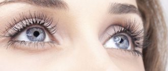
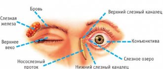
Dacryocystitis is an inflammation of the lacrimal sac, which is not so rare in children of the first year of life. Parents of a sick child, already 1-2 weeks after the birth of the baby, may notice that his eyes are constantly moisturized and tears are flowing from them. After some time, when pressing on the inner corner of the eye, purulent discharge appears, and the child’s eyelashes stick together. Although this disease is not very dangerous, in advanced cases it can result in blindness. Unfortunately, dacryocystitis cannot be prevented. But everything will be fine if you make a diagnosis in time and start treatment.
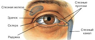
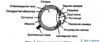
The structure of the lacrimal gland
The lacrimal gland has a lobular structure and is a tubular gland located in the frontal bone. This gland has from 5 to 10 excretory ducts, which pass into the conjunctival sac and secrete tears from the medial canthus to the lacrimal lake. Some of the ducts open into the temporal part of the conjunctiva, and some ducts open near the outer canthus into the conjunctival sac.
If a person's eyes are closed, then tears pass along the tear stream in the back edges of the eyelids. Passing through the lacrimal lake, tears flow into pinholes at the medial edges of the eyelids.
The lacrimal sac is the superior duct, which is located in the bony fossa, near the orbit. From the walls of this sac, bundles of lacrimal ducts begin, which pass through the lacrimal canaliculi.
The tear film has three layers - outer, middle and corneal (next to the cornea). The middle layer is the thickest and is secreted by the lacrimal glands.
The lower part of the lacrimal gland is located under the upper eyelid in the subaponeurotic space. This lower part consists of 25-30 connecting lobes, the ducts of which pass into the main gland.
The palpebral part is separated from the conjunctiva of the lacrimal gland, which is located in the upper eyelid and can be seen through the conjunctiva.
Innervation of blood vessels
Innervation of the glands. Innervation of the lacrimal and salivary glands.
The afferent pathway for the lacrimal gland is n. lacrimalis (branch of n. ophthalmicus from n. trigemini), for the submandibular and sublingual - n. lingualis (branch of n. mandibularis from n. trigemini) and chorda tympani (branch of n. intermedius), for the parotid - n. auriculotemporal and n. glossopharyngeus.
Efferent parasympathetic innervation of the lacrimal gland. The center lies in the upper part of the medulla oblongata and is connected with the nucleus of the intermediate nerve (nucleus salivatorius superior). Preganglionic fibers are part of n. intermedius, then n. petrosus major to ganglion pterygopalatinum. This is where the postganglionic fibers begin, which are part of n. maxillaris and further its branches, n. zygoma ticus, through connections with n. lacrimalis reach the lacrimal gland.
Efferent parasympathetic innervation of the submandibular and sublingual glands. Preganglionic fibers come from the nucleus salivatorius superior as part of n. intermedius, then chorda tympani and n. lingualis to the ganglion submandibulare, from where the spinal glionic fibers that reach the glands begin.
Efferent parasympathetic innervation of the parotid gland. Preganglionic fibers come from the nucleus salivatorius inferior as part of n. glossopharyngeus, then n. tympanicus, n. petrosus minor to ganglion oticum. This is where the postganglionic fibers begin, going to the gland as part of n. auriculotemporalis. Function: increased secretion of the lacrimal and named salivary glands; dilation of gland vessels.
Efferent sympathetic innervation of all these glands. Preganglionic fibers begin in the lateral horns of the upper thoracic segments of the spinal cord and end in the superior cervical ganglion of the sympathetic trunk. Postganglionic fibers begin in the named node and reach the lacrimal gland as part of the plexus caroticus internus, to the parotid gland as part of the plexus caroticus externus, and to the submandibular and sublingual glands through the plexus caroticus externus and then through the plexus facialis. Function: delayed saliva secretion (dry mouth); lacrimation (not a drastic effect).
The degree of innervation of arteries, capillaries and veins is not the same. Arteries, which have more developed muscular elements in the tunica media, receive more abundant innervation, veins - less abundant; v. cava inferior and v. portae occupy an intermediate position.
Larger vessels located inside the body cavities receive innervation from the branches of the sympathetic trunk, the nearest plexuses of the autonomic nervous system and adjacent spinal nerves; the peripheral vessels of the walls of the cavities and the vessels of the extremities receive innervation from the nerves passing nearby. The nerves approaching the vessels run segmentally and form perivascular plexuses, from which fibers arise that penetrate the wall and are distributed in the adventitia (tunica externa) and between the latter and the tunica media. The fibers innervate the muscle formations of the wall, having different ending shapes. The presence of receptors in all blood and lymphatic vessels has now been proven.
The first neuron of the afferent pathway of the vascular system lies in the spinal ganglia or ganglia of the autonomic nerves (nn. splanchnici, n. vagus); further it is part of the conductor of the interoceptive analyzer (see “Interoceptive analyzer”). The vasomotor center lies in the medulla oblongata. The globus pallidus, thalamus, and also the gray tubercle are related to the regulation of blood circulation . The higher centers of blood circulation, as well as all vegetative functions, are located in the cortex of the motor zone of the brain (frontal lobe ), as well as in front and behind it. The cortical end of the vascular function analyzer is located, apparently, in all parts of the cortex. Descending connections of the brain with the stem and spinal centers are apparently carried out by the pyramidal and extrapyramidal tracts.
Closure of the reflex arc can occur at all levels of the central nervous system, as well as in the nodes of the autonomic plexuses (own autonomic reflex arc).
The efferent pathway causes a vasomotor effect - dilation or constriction of blood vessels. Vasoconstrictor fibers pass as part of the sympathetic nerves, vasodilator fibers pass as part of all parasympathetic nerves of the cranial part of the autonomic nervous system (III, VII, IX, X), as part of the anterior roots of the spinal nerves (not recognized by everyone) and parasympathetic nerves of the sacral part (nn. splanchnici pelvini).
Functions of the lacrimal gland
The lacrimal gland performs several main protective and nutritional functions:
- tears contribute to the flow of useful substances into the cornea;
- tears cleanse the eye of foreign objects, dust and various contaminants;
- Tears help eliminate “dry eye syndrome”, which occurs from eye strain, fatigue and severe visual stress;
- The tear fluid contains useful substances - potassium, chlorine, organic acids, proteins and carbohydrates, lipids and lysozyme.
Tears are often a manifestation of positive or negative emotions, but their release always has a positive effect on a person’s overall emotional and mental state.
How to diagnose?
An ophthalmologist will help diagnose the disease.
To understand whether the gland is simply inflamed or more serious disorders are occurring, a number of studies are carried out:
- Palpation. It is carried out to confirm pathologies of the lacrimal sac.
- Tear secretory tests: Shimer's test. The amount of total secretion is determined.
- Jones test. The secretion of secretions from additional glands is assessed.
Abnormalities in the development of the lacrimal gland
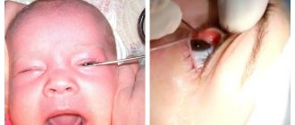
The main cause of abnormalities of the lacrimal drainage system is intrauterine trauma. Often, when examining an infant's eye, an ophthalmologist may find several lacrimal puncta in the lower eyelid that open as a canaliculus and lacrimal sac. Another most common anomaly is displacement of the lacrimal openings and blockage of the lacrimal gland.
Such congenital anomalies require special ophthalmological procedures. If obstruction of the nasolacrimal canal occurs in newborns, it is better not to perform operations, since it spontaneously opens within a few weeks.
There are several types of location of the nasolacrimal canal with anomalies of its development. Location options depend on the type of lacrimal canal, changes in the nasal wall and nasal passage.
Diagnostics
If dacryocystitis is not recognized and treated in time, in the first months of the baby’s life, then chronic eye diseases may occur in the future: inflammation of the conjunctiva, abscesses, external fistulas of the lacrimal sac, lesions of the cornea, which are difficult to treat and require surgical intervention.
What should parents be wary of? The main symptom of dacryocystitis is the presence of tears in the eye when the child is calm. Over time, tears begin to flow from the baby's eyes. If, when pressing on the inner corner of the eye, where the lacrimal sac is located, mucopurulent discharge appears, then the diagnosis of “dacryocystitis” is beyond doubt. Gradually the discharge becomes purulent and profuse. The child begins to worry, sleeps poorly, refuses to eat, and his temperature rises. In this situation, parents should urgently consult a doctor. To make a correct diagnosis, an examination by an ophthalmologist, otolaryngologist and pediatrician is necessary.
It is very important for the doctor to find out from the mother how the pregnancy and childbirth proceeded, and whether there is a hereditary predisposition to dacryocystitis. To exclude diseases or disorders of the structure of the nose, rhinoscopy is performed (examination of the nasal cavity using a special apparatus). At the same time, attention is paid to the condition of the nose, maxillary sinuses, polyps, adenoids, and anatomical features (narrowness of the nasal passages, curvature of the nasal septum). There are tests that help identify pathology of the nasolacrimal duct. They are carried out by otolaryngologists if the attending physician (pediatrician) suspects the child has an obstruction or dacryocystitis.
Tubular test. One or two drops of COLLARGOL solution are instilled into the child's eye and the eyeball is observed. If it turns white, then the tear duct is passing. A test is considered negative if the solution is retained and the eyeball remains stained for more than five minutes, which indicates pathology. The appearance of COLLARGOL in the nose a few minutes after instillation into the eye confirms the positivity of the test.
Mechanism of lacrimation
Tear fluid is produced in the gland of the same name. Then, along the excretory canaliculi, it moves into the conjunctival sac, where it accumulates for some time. Blinking transfers the tear to the cornea, wetting it.
The outflow of fluid occurs through the lacrimal duct (a narrow space between the cornea and the lower eyelid), which flows into the lacrimal lake (the inner corner of the eye). From there, through the canal, the secretion enters the lacrimal sac and is evacuated through the upper nasal passage.
The basis for normal tear production is made up of several factors:
- suction function of lacrimal openings;
- the work of the orbicularis oculi muscle, as well as the Horner muscle, which creates negative pressure in the ducts that drain tears;
- the presence of folds on the mucous membrane that act as valves.
Symptoms
Why should you not trigger inflammation of the lacrimal gland? Photos of patients with this pathology show that it is not so easy to ignore these symptoms. And only a person who is indifferent to his health can allow complications to develop.
At the very beginning, inflammation of the lacrimal gland manifests itself as pain in the inner corner of the eye. Local swelling and redness are clearly visible. The doctor may ask the patient to look at his nose and lift his upper eyelid to see a small section of the gland. In addition to local ones, there are also general signs that characterize inflammation of the lacrimal gland. Symptoms are similar to other infectious diseases: fever, headache, nausea, feeling tired, enlarged lymph nodes of the head and neck.
Patients may complain of double vision, blurred vision, or difficulty opening the upper eyelid. With a severe reaction, the entire half of the face swells, including the affected eye. If the symptoms are left unattended, the situation may eventually worsen into phlegmon or an abscess.
Inflammation of the lacrimal gland in a child occurs in the same way as in an adult. The only difference is that the likelihood of spreading the infection is higher than in adults. Therefore, children are treated in a hospital.