- Laser iridectomy for glaucoma
- Contraindications for surgery
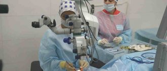
- How is the operation performed?
- Cost of iridectomy
Laser iridectomy is a microsurgical operation, the main purpose of which is to create a through hole in the iris, that is, aimed at restoring the natural circulation of fluid in the eye, by forming an additional path for the flow of intraocular fluid from the posterior chamber to the anterior one.
The main indication for laser iridectomy is narrow-angle or closed-angle glaucoma with pupillary block.
What is laser iridectomy
This is a surgical procedure designed to reduce intraocular pressure. The purpose of the correction is to normalize the circulation of fluid inside the organ of vision. During the operation, the doctor creates several small holes on the surface of the iris. There are three methods of iridectomy:
- One-step correction;
- Staged intervention;
- Layer-by-layer operation.
In the first case, a pulsed laser with a power of 5 to 15 mJ is used. Application is carried out from one to three times. As a result, the surgeon creates a through hole in the iris. The main advantage of the technique is the speed of execution. The operation is performed on patients with any eye color.
Phased intervention is carried out in several stages. The break between each session is two to three weeks. As a result, the process of forming holes in the iris is delayed for a long time. This type of surgery is performed only on patients with light eye shades.
| Step-by-step correction is required in order to eliminate the risk of damage to the thin tissue of the iris and other structures of the visual apparatus. The power of the installation used to carry out the intervention ranges from six hundred to one thousand megawatts. The duration of exposure is up to 0.5 seconds. |
Layer-by-layer surgery is performed in patients with a dark iris color. The power of the laser installation is about one and a half thousand megawatts, the exposure time is up to 0.2 seconds. The hole in the iris is formed gradually, using layer-by-layer destruction of pigment matter. An argon or short-pulse laser is used for correction. Return to contents
Iridectomy technique
Laser basal iridectomy involves creating an artificial hole in the inner corner of the cornea. It allows you to establish the outflow of fluid from the posterior chamber of the eye to the anterior chamber. This procedure is performed under local anesthesia. In addition to the painkiller, it is necessary to instill a 1% solution of Pilocarpine. This is necessary to constrict the pupil during laser intervention.
A lens is applied to the eye to maximize the view of the surgical field. Then select the location where the hole will be formed. It can be located in any part of the iris. It is not recommended to puncture at 12 o'clock, as gas bubbles accumulate in this area. Ophthalmologists prefer to create a hole in the area of a large crypt or in a thinned area of the iris.
The laser beam penetrates the tissue of the eye through the lens. It focuses on a specific place. The laser acts on the selected area for 0.2-0.5 seconds. As a result, a hole of the required depth is formed. The circulation of aqueous fluid is improved, which helps reduce intraocular pressure.
Indications for surgery
To normalize the outflow of intraocular humor, iridectomy is required. The intervention allows you to minimize the risk of developing complications that are dangerous to the health of the organ of vision. The procedure is recommended for anyone diagnosed with fluid stagnation and high intraocular pressure. Such conditions include:
- Angle-closure or mixed glaucoma;
- Increased pigment content;
- Pathologies in the structure of the anterior chamber of the visual apparatus;
- Pupil block.
Laser iridectomy is also recommended as a preventive measure for acute attacks of glaucoma. An increase in pressure inside the eye leads to the formation of “blind spots” and a decrease in visual acuity. If the pathology is ignored for a long time, glaucoma can lead to blindness.
The anomaly is formed due to a violation of the circulation of intraocular moisture, as a result of which the fluid stagnates between the chambers of the eye. The disease can be congenital or acquired. In the first case, glaucoma is a consequence of improper development of the visual apparatus. Acquired high IOP manifests itself when inflammatory diseases recur or there is a failure in the blood supply to the eye.
| Laser iridectomy is most effective in the treatment of angle-closure glaucoma. Excision of the iris improves the outflow of moisture from the anterior chamber and reduces pressure. |
Treatment of an acute attack of angle-closure glaucoma
An acute attack of angle-closure glaucoma is an emergency that requires emergency medical care. If intraocular pressure (IOP), which during the development of an attack can reach values of 40-60 mm Hg. Art. and no longer decrease to normal values during the first 24 hours, the prognosis for visual functions can be very dismal.
The eye is in danger of permanent loss of vision! Therefore, the main goal during the development of an acute attack is to reduce intraocular pressure (IOP). For this purpose, the following is carried out:
- Drug therapy:
- It is necessary to immediately begin instillation of the miotic - 1% pilocarpine solution. The following scheme is used: during the first 2 hours, 1 drop of the drug is instilled every 15 minutes, over the next 2 hours - every 30 minutes, over the next 2 hours - 1 time per hour. Next, the drug is used 3-6 times a day, depending on the degree of reduction in intraocular pressure (IOP). A similar scheme is used if the test result for pilocarpine is positive (constriction of the pupil with one or two instillations of the drug). If there is no pupil reaction due to iris ischemia, continuing treatment with pilocarpine is inappropriate and even dangerous;
- In addition to miotic instillation, a 0.5% solution of timolol is instilled, 1 drop 2 times a day;
- Acetazolamide (diacarb) is prescribed orally at 0.25-0.5 g 2-3 times a day. In addition to systemic carbonic anhydrase inhibitors, you can use a 2% solution of dorzolamide (trusopt) 3 times a day or a 1% suspension of brinzolamide (azopt) 2 times a day;
- Osmotic diuretics are used orally or intravenously (the most commonly used is a 50% glycerin solution at a dose of 1.5-2 g/kg). If the pressure decrease is insufficient, loop diuretics (furosemide 20-40 mg) can be used intramuscularly or intravenously;
- If, despite the therapy, intraocular pressure (IOP) does not decrease, a “lytic mixture” is injected intramuscularly: 1-2 ml of a 2.5% solution of aminazine, 1 ml of a 2% solution of diphenhydramine or 2 ml (50 mg) of promethazine (“pipolfen”) "), 1 ml of 2% promedol solution. After administering the mixture, you should remain in bed for 3-4 hours due to the possibility of developing orthostatic collapse (a sharp drop in blood pressure).
- Distraction therapy:
- Hot foot baths, saline laxatives, cupping, mustard plasters, leeches on the temple area (carried out simultaneously with drug therapy);
Advantages and disadvantages
The procedure has several undeniable advantages:
- Nearby tissues are not affected during the intervention, their integrity is not compromised;
- The correction is carried out on an outpatient basis; hospital stay is not required;
- Low cost of the procedure;
- Short recovery period;
- The laser forms new ducts to remove intraocular fluid;
- The effectiveness of the operation is noticeable immediately after the correction.
Compared to other techniques, the risk of complications with laser iridectomy is several times lower. But still, any operation can lead to unforeseen consequences. The disadvantages of this type of correction include:
- After a while, the formed holes are tightened and it is necessary to carry out repeated intervention;
- In some cases, adhesions form in the area of “holes” created in the iris, which negatively affect the circulation of intraocular fluid;
- During the intervention, there remains a risk of touching blood vessels;
- If the hole is made too large, a “second pupil” effect may occur.
To avoid the development of complications, it is necessary to carefully select a clinic.
Risk groups and contraindications
Contraindications to laser therapy are:
- fungal, bacterial and viral infections affecting eye structures (keratitis, uveitis, conjunctivitis);
- corneal ulcer;
- edema;
- damage to the corneal epithelium;
- paralytic form of mydriasis (pupil dilation);
- slit-like anterior chamber;
- severe narrowing of visual fields;
- damage to the optic nerve head (for trabeculoplasty).
The risk group includes people with retinopathy, vascular pathologies, diabetes mellitus and arterial hypertension.
Maybe
Patient preparation
A few days before the intervention, doctors prescribe miotics, which reduce the size of the pupil. They must be used regularly, strictly adhering to the recommendations of the attending physician. Avoid overdose.
The patient also needs to undergo a series of examinations and tests. Main activities before surgery:
- X-ray of the lungs;
- Electrocardiogram;
- Optical field analysis;
- Measurement of intraocular pressure;
- Ophthalmoscopy;
- Ultrasound examination of the visual apparatus;
- Blood sampling for general and biochemical analysis.
An examination by a dentist and an ENT specialist is mandatory. If necessary, sanitation of the oral cavity is carried out. To assess the condition of the anterior chamber angle and accurately determine the location for iridectomy, gonioscopy is prescribed.
| If an acute infection or inflammation is detected, intervention is postponed until the disease is completely eliminated. |
Return to contents
Examination before iridectomy
The examination differs little from that of other eye surgeries. General and biochemical tests, chest radiography, electrocardiography, and blood testing for antibodies to HIV, syphilis, and hepatitis are prescribed.
A dental examination and, if necessary, dental sanitation, as well as an examination by an ENT doctor are required. Chronic foci of infection can cause complications in the postoperative period.
In the presence of an acute infectious disease or inflammatory processes, the operation will be postponed until recovery.
Special examinations, in addition to tonometry of the eyeball, include gonioscopy, ophthalmoscopy, visual field examination, ultrasound or CT of the eyeball.
During a routine examination, it is impossible to examine the anterior chamber angle; gonioscopy allows you to evaluate it and select the site for iridectomy.
Stages of the operation
Surgery involves several steps:
- Before correction, an anesthetic substance is injected into the organ of vision, which helps the patient avoid pain and discomfort;
- A special optical lens is attached to the eye being treated, which allows the surgeon to examine the surgical area under magnification during the correction process;
- Through the eyepiece, the laser beam is focused on the desired area;
- A microscopic hole is formed, which will further help normalize the outflow of intraocular moisture.
The surgical procedure lasts about twenty minutes. Within an hour after completion of the correction, blurred vision may occur. After sixty minutes, a control measurement of intraocular pressure is carried out. If it has increased, the patient is given drops to normalize it.
results
Laser dissection of secondary cataracts in all patients was carried out without complications with a significant improvement in visual functions the next day after surgery. Further visual acuity did not change during the entire observation period; 8 (18.6%) patients (8 eyes) noted increased floaters within 1 week after laser dissection of the posterior capsule of the lens.
No complications were observed in all patients after laser iridectomy. According to gonioscopy, after the operation the angle of the anterior chamber in all cases of observation opened with visualization of the trabecula - grade II-III according to the Shaffer classification. In the immediate period after laser iridectomy, 2 (3%) eyes had hyphema in the form of blood smears on the iris in the area of post-laser coloboma. In no case was there any deterioration in visual function.
Medication support was not intentionally carried out; it was assumed that medications would be used only in the event of postoperative complications.
1 hour after laser discision, an increase in IOP level was observed (p>0.05), followed by a decrease and normalization by the 3rd day. In all patients, after laser iridectomy, there was a gradual decrease in IOP values within 1 month (p<0.05) and their further stabilization for the entire observation period. In both groups, throughout the study, there was no significant change in the thickness of the peripapillary RNFL and ganglion cell complex (all p>0.05) (Tables 1-4) .
Table 1. Thickness of the retinal ganglion cell layer (in µm) before and after laser dissection of secondary cataracts (n=43)
| Index | Before surgery | 1 hour | 1 day | 3 days | 7 days | 1 month | 6 months |
| Average | 94,33±6,52 | 93,88±7,23 | 94,01±6,75 | 94,04±5,29 | 94,66±6,35 | 93,92±6,46 | 94,19±7,41 |
| Superior | 94,78±6,18 | 94,74±7,27 | 94,63±6,80 | 94,46±5,97 | 94,23±6,83 | 94,28±6,12 | 94,89±7,02 |
| Inferior | 93,74±6,71 | 93,29±7,45 | 93,38±6,18 | 93,62±5,82 | 93,44±6,15 | 93,49±6,61 | 93,38±7,72 |
Note. Here and in the table. 2: average - average thickness of the retinal ganglion cell layer; superior - thickness in the upper hemisphere; inferior - thickness in the lower hemisphere.
Table 2. Thickness of the retinal ganglion cell layer (in µm) before and after laser iridectomy
| Index | Before surgery | 1 hour | 1 day | 3 days | 7 days | 1 month | 6 months |
| Average | 94,14±6,23 | 93,87±5,41 | 94,16±6,03 | 94,03±6,03 | 94,06±6,26 | 94,71±6,03 | 93,52±6,57 |
| Superior | 94,72±6,18 | 94,33±5,94 | 93,92±7,05 | 93,35±5,30 | 94,84±5,39 | 94,78±5,79 | 93,83±6,08 |
| Inferior | 93,56±6,28 | 93,41±4,86 | 94,39±4,95 | 94,72±6,84 | 93,28±7,12 | 94,63±6,27 | 93,22±7,06 |
Table 3. Retinal nerve fiber layer thickness (in µm) before and after laser dissection of secondary cataracts
| Index | Before surgery | 1 hour | 1 day | 3 days | 7 days | 1 month | 6 months |
| Average | 106,49±20,87 | 106,41±18,72 | 106,67±18,80 | 106,09±19,44 | 106,78±17,15 | 106,29±16,14 | 106,38±18,53 |
| Tempo | 89,35±17,11 | 89,13±19,28 | 88,39±18,41 | 89,74±17,32 | 89,10±18,93 | 88,63±18,27 | 89,02±16,56 |
| Superior | 127,82±20,09 | 128,14±18,19 | 127,97±17,88 | 128,21±18,77 | 128,46±16,42 | 128,46±18,58 | 128,82±16,18 |
| Nasal | 71,87±16,27 | 72,09±17,00 | 71,64±16,06 | 71,27±16,42 | 71,79±18,44 | 71,92±16,68 | 71,82±17,81 |
| Inferior | 137,19±16,21 | 137,47±20,64 | 135,63±15,62 | 135,20±16,23 | 135,85±16,93 | 135,71±15,88 | 135,98±16,32 |
| TU1 | 78,26±16,69 | 77,99±14,02 | 79,48±15,29 | 79,75±13,19 | 78,89±15,65 | 78,33±13,82 | 78,47±14,97 |
| TU2 | 107,91±18,64 | 108,18±15,54 | 108,79±14,26 | 108,39±14,49 | 107,55±13,55 | 108,88±16,64 | 108,89±12,63 |
| ST2 | 140,13±22,16 | 140,85±22,97 | 140,72±21,27 | 139,76±21,35 | 142,76±15,68 | 140,88±19,94 | 139,89±20,86 |
| ST1 | 132,93±28,15 | 133,02±26,59 | 130,53±26,96 | 132,91±27,37 | 134,61±26,09 | 131,48±25,67 | 131,14±25,84 |
| SN1 | 122,35±27,99 | 122,73±21,49 | 121,26±25,87 | 121,18±27,21 | 122,63±22,22 | 122,77±19,52 | 122,95±21,62 |
| SN2 | 119,74±23,76 | 119,23±19,37 | 119,96±17,09 | 120,85±22,41 | 119,19±17,09 | 119,33±15,35 | 120,23±14,85 |
| NU2 | 89,40±18,84 | 88,88±16,89 | 89,71±15,86 | 90,21±15,97 | 89,51±15,76 | 89,81±17,69 | 89,12±20,36 |
| NU1 | 61,87±11,79 | 61,92±12,25 | 62,51±18,62 | 62,09±17,05 | 61,94±17,02 | 61,33±13,67 | 61,31±17,28 |
| NL1 | 57,89±11,06 | 58,07±10,64 | 58,21±18,82 | 58,59±16,21 | 58,94±15,98 | 57,21±14,54 | 57,81±17,21 |
| NL2 | 78,67±15,89 | 78,51±14,57 | 78,43±18,66 | 77,71±16,71 | 77,04±17,26 | 78,76±14,43 | 78,14±17,82 |
| IN2 | 112,39±18,13 | 112,9±19,91 | 111,7±12,94 | 112,71±15,71 | 112,84±15,19 | 112,92±14,13 | 112,39±17,04 |
| IN1 | 132,71±23,56 | 132,3±20,32 | 131,4±14,93 | 132,15±18,24 | 131,02±15,29 | 132,93±13,12 | 132,37±18,09 |
| IT1 | 155,65±27,35 | 155,7±21,82 | 155,3±18,75 | 154,07±16,89 | 155,71±16,08 | 155,05±13,24 | 155,41±17,31 |
| IT2 | 148,91±26,09 | 148,6±25,78 | 147,3±20,98 | 147,12±26,72 | 148,08±16,29 | 148,75±15,61 | 148,11±17,52 |
| TL2 | 97,19±23,09 | 96,74±18,59 | 97,26±16,44 | 98,12±15,23 | 97,63±15,47 | 97,47±14,34 | 97,55±17,19 |
| TL1 | 71,62±14,25 | 70,30±10,86 | 70,86±16,02 | 72,44±15,07 | 71,58±14,68 | 71,31±14,69 | 71,08±16,47 |
Note. Average—average thickness of the peripapillary RNFL, tempo—average thickness in the temporal quadrant; superior - average thickness in the upper quadrant; nasal—average thickness in the nasal quadrant; inferior - average thickness in the lower quadrant. TU1, TU2 - upper sectors of the temporal quadrant of the RNFL; ST1, ST2 - temporal sectors of the upper quadrant of the RNFL; SN1, SN2 - nasal sectors of the upper quadrant of the RNFL; NU1, NU2 - upper sectors of the nasal quadrant of the RNFL; NL1, NL2 - lower sectors of the nasal quadrant of the RNFL; IN1, IN2 — nasal sectors of the lower quadrant of the RNFL; IT1, IT2 - temporal sectors of the lower quadrant of the RNFL; TL1, TL2 - lower sectors of the temporal quadrant of the RNFL.
Table 4. Peripapillary RNFL thickness (in µm) before and after laser iridectomy
| Index | Before surgery | 1 hour | 1 day | 3 days | 7 days | 1 month | 6 months | |||||||
| Average | 106,03±20,79 | 105,90±21,22 | 106,09±21,39 | 106,49±21,94 | 104,62±21,72 | 105,92±23,53 | 105,90±23,31 | |||||||
| Tempo | 89,13±16,81 | 89,76±17,07 | 88,73±18,82 | 89,03±18,64 | 89,15±17,09 | 88,82±21,63 | 89,25±20,68 | |||||||
| Superior | 128,19±16,78 | 127,88±17,75 | 127,47±18,61 | 128,37±19,68 | 128,89±19,14 | 128,35±20,54 | 128,63±19,82 | |||||||
| Nasal | 71,22±16,69 | 71,59±16,87 | 71,49±16,38 | 71,76±17,15 | 71,48±16,07 | 71,34±18.39 | 71,19±17,31 | |||||||
| Inferior | 135,21±21,54 | 134,36±21,59 | 135,82±18,69 | 135,24±18,49 | 135,13±20,20 | 135,21±21,79 | 135,48±22,62 | |||||||
| TU1 | 78,43±15,32 | 79,85±15,92 | 79,28±15,41 | 79,34±15,79 | 78,32±14,81 | 78,39±22,73 | 78,96±24,82 | |||||||
| TU2 | 107,62±20,03 | 107,31±19,49 | 108,81±19,63 | 108,78±22,15 | 107,27±20,16 | 108,28±26,41 | Retinal thickness, according to OCT, significantly increased 1 hour after laser dissection of secondary cataracts in the fovea and parafoveal areas. 1 day after laser exposure, retinal thickness returned to its original values, except in the paranasal quadrant, where it was significantly higher than before surgery. On the 3rd day, the thickness of the retina in all areas was normalized and did not fundamentally change throughout the entire observation period (Table 5). Table 5. Retinal thickness (in microns) before and after laser dissection of secondary cataracts Index | Before surgery | 1 hour | 1 day | 3 days | 1 week | 1 month | 6 months |
| Fovea | 242,49±17,64* | 246,93±19,49* | 243,28±16,34 | 242,29±16,66 | 243,11±14,58 | 242,36±15,87 | 241,94±16,38 | |||||||
| Parafovea | 311,46±13,94* | 313,76±16,77* | 312,64±15,25* | 311,18±15,55 | 311,44±14,47 | 311,60±13,54 | 311,70±13,25 | |||||||
| Paratempo | 306,39±14,74* | 309,04±16,72* | 307,57±19,29 | 306,64±18,62 | 305,67±15,44 | 306,62±14,88 | 307,49±14,37 | |||||||
| Parasuperior | 313,63±14,48* | 315,65±15,83* | 313,55±13,29 | 313,02±14,87 | 312,91±15,36 | 313,34±13,92 | 313,71±12,85 | |||||||
| Paranasal | 316,39±12,23* | 319,71±19,48* | 318,72±13,04* | 315,81±14,29 | 316,24±13,47 | 316,28±11,83 | 316,13±11,38 | |||||||
| Parainferior | 309,44±14,29 | 310,61±15,04 | 310,72±15,38 | 309,24±14,41 | 310,94±13,59 | 310,16±13,52 | 309,45±14,41 | |||||||
| Perifovea | 269,83±13,72 | 269,64±14,13 | 269,19±14,42 | 269,36±13,30 | 269,36±13,85 | 268,96±13,73 | 269,65±13,60 | |||||||
| Peritempo | 271,86±13,79 | 272,13±12,37 | 272,03±14,22 | 271,73±13,28 | 270,91±13,39 | 271,39±13,81 | 271,26±13,24 | |||||||
| Perisuperior | 267,58±12,92 | 266,84±14,39 | 266,19±13,04 | 267,85±13,39 | 266,84±14,72 | 266,28±13,34 | 267,43±13,05 | |||||||
| Perinasal | 274,23±11,84 | 274,25±11,77 | 273,98±13.03 | 274,73±11,22 | 273,45±12,81 | 273,58±12,34 | 274,62±12,74 | |||||||
| Periinferior | 265,65±16,33 | 265,34±17,98 | 264,55±17,38 | 265,03±15,29 | 266,22±14,48 | 264,57±15,42 | 265,29±15,38 |
Note. Fovea - average retinal thickness in the foveola; parafovea - retinal thickness at a distance from the foveola 3 mm; perifovea - the thickness of the retina at a distance from the foveola is 6 mm. Quadrants: superior (upper, 46-135°); nasal (nasal, 136-225°); inferior (lower, 226-315°) and tempo (temporal, 316-45°).
Retinal thickness significantly increased 1 hour after laser iridectomy in the fovea and parafoveal areas and continued to increase for 1 day. On the 3rd day, the thickness of the retina in the upper and lower parafoveal quadrants did not differ significantly from the preoperative values, and the thickness of the retina in the fovea, paranasal and paratemporal quadrants decreased compared to that on the 1st day, but continued to remain significantly greater than before surgery .
After 7 days, retinal thickness returned to initial values and did not change during 6 months of observation.
Indicators of retinal thickness in the perifoveal areas did not undergo significant changes after laser intervention (Table 6) .
Table 6. Retinal thickness (in microns) before and after laser iridectomy
| Index | Before surgery | 1 hour | 1 day | 3 days | 1 week | 1 month | 6 months |
| Fovea | 235,54±13,32* | 244,23±17,87* | 249,31±16,09* | 242,16±16,34* | 235,91±13,43 | 235,67±13,18 | 235,18±12,21 |
| Parafovea | 309,60±15,61* | 314,92±19,05* | 317,29±17,36* | 313,13±17,25* | 310,57±13,79 | 309,80±14,83 | 309,54±13,55 |
| Paratempo | 304,14±13,75* | 312,34±17,31* | 316,72±21,23* | 309,34±19,26* | 305,23±13,18 | 304,61±12,72 | 304,17±16,72 |
| Parasuperior | 311,39±18,23* | 315,26±19,52* | 316,37±17,48* | 313,73±15,82* | 312,02±15,91 | 311,31±18,33 | 311,57±12,29 |
| Paranasal | 310,45±12,64* | 316,37±20,62* | 320,61±17,28* | 315,81±17,42* | 311,83±13,27 | 311,16±13,33 | 310,18±12,37 |
| Parainferior | 312,43±17,82* | 315,72±18,73* | 315,44±13,43* | 313,65±16,49* | 313,21±12,78 | 312,12±14,92 | 312,26±12,83 |
| Perifovea | 269,36±14,42 | 269,72±14,31 | 269,14±14,58 | 269,10±14,69 | 269,09±14,09 | 269,36±13,60 | 269,25±14,64 |
| Peritempo | 273,49±12,68 | 274,82±15,69 | 273,96±14,85 | 273,82±16,93 | 272,63±14,57 | 273,64±16,72 | 273,43±13,54 |
| Perisuperior | 265,72±15,85 | 265,25±14,84 | 266,06±14,73 | 265,16±13,37 | 265,45±14,76 | 265,71±12,41 | 265,82±13,77 |
| Perinasal | 273,58±14,39 | 274,11±12,44 | 273,26±13,56 | 273,37±14,05 | 273,82±12,41 | 273,69±11,79 | 273,54±14,32 |
| Periinferior | 264,64±14,74 | 264,69±14,25 | 263,27±15,18 | 264,03±14,39 | 264,46±14,62 | 264,41±13,48 | 264,21±16,92 |
Note. * — p<0.05; Fovea - average retinal thickness in the foveola; parafovea - retinal thickness at a distance from the foveola 3 mm: perifovea - retinal thickness at a distance from the foveola 6 mm. Quadrants: superior (upper, 46-135°); nasal (nasal, 136-225°); inferior (lower, 226-315°) and tempo (temporal, 316-45°).
A slight thickening of the retina in the early postoperative period was not subjectively felt by the patients; they did not complain of decreased vision or metamorphopsia.
There was a high correlation between the thickness of the retina in the fovea in the postoperative period and the total values of energy expended during laser dissection of secondary cataracts and laser iridectomy; the correlation coefficient was 0.87 and 0.94, respectively (p<0.05).
Rehabilitation period
Since the correction is classified as bloodless and the patient does not require stitches, there are no particular difficulties or restrictions on the recovery period. After the operation, the person feels well and does not experience discomfort or pain.
To eliminate the risk of infection, the doctor selects antibacterial medications. They also help speed up the rehabilitation process. In some cases, medications are prescribed to reduce intraocular pressure.
During the recovery period, avoid excessive physical activity and try to avoid overstraining your vision. Also follow simple rules:
- Sleep on your side (opposite the operated side) or back;
- Do not rub the organ of vision;
- Avoid getting dust and water in your eyes;
- Avoid contact with direct sunlight, wear protective glasses;
- Do not visit the bathhouse or solarium;
- Do not communicate with people suffering from infectious pathologies;
- Avoid drinking alcohol and spicy foods.
| During the first month after correction, you must visit your ophthalmologist every week for a follow-up examination. |
Postoperative period
During the rehabilitation period it is recommended:
- use of non-steroidal anti-inflammatory drugs for 1 week or until pain disappears;
- use of Diacarb or other decongestants;
- monitoring intraocular pressure through regular diagnostic testing.
It is strictly forbidden to eat highly salty foods before and after the intervention.
In the postoperative period it is prohibited:
- create increased strain on the eyes (work a lot at the computer, watch TV, etc.);
- injure the eye (including scratching, rubbing, etc.);
- fall asleep on the side of the eye after the intervention;
- visit a bathhouse or swimming pool;
- train intensively;
- wash your hair for the first 2-3 days;
- include in the diet foods that cause fluid stagnation in the body (salted, pickled, dried, etc.).
It is necessary to adhere to the regimen prescribed by the doctor to avoid complications. If the patient used any eye drops before the operation, it is necessary to consult with the treating ophthalmologist before using them.
An effective remedy for combating glaucoma is Xalacom eye drops.
Nonsteroidal anti-inflammatory drugs are indicated for use after iridectomy
A proven medication for lowering intraocular pressure is Xalatan eye drops.
Possible complications of iridectomy
The risk of severe consequences after surgery always remains. With this method it is minimal. Possible complications after laser iridectomy:
- Damage to parts of the structure of the visual apparatus;
- During the correction process or immediately upon its completion, intraocular pressure may rise sharply;
- Bleeding (occurs when the integrity of blood vessels is violated);
- After some time, repeated surgery is required;
- Fusion of the tissues of the cornea, which causes the opposite effect of the procedure;
- A large hole in the iris can cause serious harm to visual function.
In some cases, the correction does not bring the expected result. Most often this is due to the individual structural features of the eye. Before performing an intervention, the doctor is obliged to warn the patient about possible complications. The operation is recommended to be performed in the early stages of glaucoma development, since the risk of scar tissue formation increases in the future. Return to contents
Possible complications
Possible complications may include:
- Short-term blurring of the observed picture (in particular);
- Corneal bulge;
- Bleeding;
- Increased intraocular pressure.
Further complications may include:
Severe glaucoma usually affects both eyes at the same time. If a severe form of glaucoma suddenly develops in one eye and laser surgery is performed on it, the same operation is performed on the second eye in order to prevent the development of the disease.
If severe glaucoma is not treated promptly, it has a 50% chance of leading to vision loss. High pressure inside the eye may also persist after laser iridotomy.
Even after the procedure, a person may need careful care. Some people may require additional treatment in the form of eye drops or a surgical iridotomy to relieve pressure in the eye.
Patient reviews of laser iridectomy
Mostly the feedback is positive. General opinion of people:
- The procedure does not cause pain;
- Discomfort persists only for a few hours after correction, then disappears without a trace;
- The intervention has a high level of effectiveness;
- Complications develop in isolated cases;
- If the hole closes, the procedure can be repeated;
- During the operation, the patient feels only bright flashes of light.