Phosphenes in ophthalmology
Phosphenes appear as spots, formations, or colored figures of varying configurations, sizes, and colors .
It is worth noting! Such visual images can be visualized with different intensities and frequencies and appear in different quantities, differing in brightness and shape.
Such phenomena, even if they change with great frequency and appear constantly, are never repeated in configuration , each time being unique optical phenomena.
Mostly such formations have soft pastel colors and can be yellow, green, orange and blue, while different people may have one or another color as their main color.
This phenomenon does not happen in people who are blind from birth, but if blindness is acquired, by irritating certain parts of the brain, visual images can appear.
In sighted people, this disorder can manifest itself both with closed and open eyes.
In the second case, phosphenes are superimposed on the contours of visible objects and can take on their outline and similar color.
Study
- The researchers studied a visual prosthesis for the blind, which involves a set of electrodes implanted in the skull above the occipital lobe to produce phosphenes. This type of implant has been around for a long time. Risks such as infections and seizures prevented their development.
- The possible use of phosphenes as part of the brain-to-brain communication system has been reported. The system, called BrainNet, produces phosphenes using transcranial magnetic stimulation (TMS). The goal of the study is to connect thoughts to the brain using a system in which signals are detected using electroencephalography (EEG) and delivered using transcranial magnetic stimulation (TMS). The experiment was conducted with 5 different groups of three people each. The subjects were divided into two groups. Two subjects acted as senders and were connected to EEG electrodes, and a third person acted as a receiver who wore a TMS helmet. Each was placed in front of a television screen playing a Tetris-style game. Senders had to determine whether the falling blocks needed to be rotated, but without the ability to rotate them - only the recipient could perform this operation. At the edges of each screen were two icons with two flashing lights at two different frequencies (one at 15 Hz and the other at 17 Hz). The sender focused on one icon or another to indicate that the block should be rotated to the right or left. The EEG receives a unique signal, which was transmitted to the TMS, sends a receiver, which is perceived by phosphenes that differ for the 15 Hz and 17 Hz signal, and rotates the unit in the appropriate direction. The success of the experiment was 81%. These results could eventually lead to the creation of mind-to-mind communication technology.
Reasons for appearance
Keep in mind! The appearance of phosphenes is not an ophthalmological pathology, the causes of which lie in physical diseases of the eyes.
The source of such phenomena is the brain, but in general, such manifestations may have the following pathological causes :
- a sharp increase in intraocular pressure;
- traumatic brain injuries;
- hemorrhages in the eyeballs;
- eye injuries;
- sudden turns of the head;
- intoxication of various origins;
- oxygen starvation (including carbon monoxide poisoning).
Often such visual images are seen by people whose activities involve long periods of work at the computer .
In such cases, a few minutes of rest are enough for the visions to disappear.
Often this disorder appears in people over 40-45 years of age , since the tissues of the organs of vision and brain are subject to dystrophic, atrophic and sclerotic processes, as a result of which such optical disorders often occur.
Newspaper "News of Medicine and Pharmacy" Ophthalmology (363) 2011 (thematic issue)
Introduction
Among the numerous treatment methods in therapeutic ophthalmology, a new method has become widespread in recent years - transcutaneous phospheneelectric stimulation of the visual analyzer. Electric current, being a universal irritant of nerve cells, increases the volume of afferent impulses in nerve conductors, which led to its use as a therapeutic factor in neurology. In ophthalmological practice, direct electrical stimulation (ES) of the visual pathways was first used for the treatment of optic nerve atrophy (ON) of various origins [1]. Currently, the number of indications for its use in the practice of an ophthalmologist has increased significantly and includes myopia, spasm of accommodation, amblyopia, dystrophic and degenerative diseases of the retina, and optic nerve atrophy [1, 2]. On the basis of the Institute of Eye Diseases and Tissue Therapy named after. V.P. Filatov in the functional-diagnostic ophthalmic center, 9 modifications of ophthalmic therapeutic and therapeutic-diagnostic electrical stimulators of the original design “Phosphen” were created and successfully used in clinical practice (Fig. 1), and a method for treating various ophthalmopathologies using these devices was developed. A special feature of the technique is the individual selection of the strength of the therapeutic electric current, which causes the occurrence of a phosphene phenomenon in the patient, which served as the basis for the name of the treatment method itself - phosphene electrostimulation (FES).
In this work, we analyze 20 years of experience in treating patients with varying degrees of myopia, accommodative dysfunction, amblyopia and optic atrophy using the phospheneelectric stimulation method. And also, based on neurophysiological and clinical studies, we present some pathogenetic mechanisms of the effect of this treatment method on the above-mentioned ophthalmopathologies.
Mechanisms of FES
What mechanisms underlie the improvement in resolution and other functions of the visual analyzer (VA) as a result of electrical stimulation?
Under the influence of a low-intensity electric current at the level of the retina, synchronous excitation of neurons and their fibers occurs, as a result of which the functionality of those elements that remained viable but did not transmit visual information (were in parabiosis) is restored [1, 5, 9]. Data on the presence of such fibers are given in a number of works [3, 5]. The improvement in impulse conduction along the visual pathways was reflected in the positive dynamics of the neurogram parameters of ON fibers, electrical sensitivity and lability of the ON [1]. According to available data, with ES there is an increase in mobility, breakdown and renewal of membrane phospholipids (primarily myelin), and the synthesis of DNA and collagen increases. The density of membrane Ca decreases, and the concentration of K in the intercellular space increases, which leads to cell depolarization. The number of glial cells in the optic nerve increases, and the flow of ions in the fibers of the optic pathway increases.
There is an opinion that these processes play a certain role in restoring the conductivity of the optic nerve when it is partially atrophied [5, 8].
The next mechanism of action of ES involves the development at the level of the visual cortex of a persistent focus of increased excitability (long-term post-tetanic potentiation), which leads to the restoration of the functionality of deprived cells of the visual centers of the cortex, improving the conductivity of sensory and autonomic nerve fibers, some of which emerge from the state of parabiosis. Broken connections are restored and the reserve capabilities of brain centers are expanded [4, 6, 8]. There is an opinion in the literature that the obvious effectiveness of electrical stimulation is due to the fact that this treatment method activates the entire visual system, simulating a holistic visual act [1, 9]. ES enhances the plastic and adaptive properties of neural networks at various levels and improves the possibility of their more effective interaction [3, 5]. It is the second mechanism that is most important in amblyopia, which is accompanied by significant changes in the functional activity of neurons in the visual centers of the brain.
Also, under the influence of electrical stimulation, the functioning of the neuromuscular system improves. At the same time, the vasodilating effect inherent in unidirectional current and improvement of blood supply to tissues are manifested, metabolic processes are activated, and redox reactions are enhanced. With improved blood and lymph circulation, better absorption of nutrients and elimination of under-oxidized products is ensured. The ATP content increases, the energy potential of tissues increases, and the trophism of nerve conductors improves. Through training with the help of rhythmic electrical stimulation, nervous and humoral regulation improves [4, 5, 8].
Low-intensity pulsed electric current has a multidirectional action. In addition to the above points, electric current also affects the activity of enzymes and neurotransmitters. In addition to the direct effect of FES on the pathways of the visual analyzer, it can be assumed that electrical stimulation has an indirect effect on other structures through the activation of retinoencephalic and encephaloretinal connections. In this aspect, the most significant, obviously, is the effect of ES on the hypothalamus through retinohypothalamic fibers. Which leads to significant changes in the general immunological reactivity of the body and systemic hemodynamics of the eye and brain [4, 8].
FES for partial optic nerve atrophy
Clinical and physiological studies on the use of electrical stimulation for the treatment of partial optic nerve atrophy (PANA) were first carried out at the Research Institute of Experimental Medicine of the USSR Academy of Medical Sciences and at the Military Medical Academy named after. Kirov. Electrical stimulation was used by the direct method with electrodes being applied to the optic nerves either during neurosurgical operations in the chiasmal-sellar region or transorbitally. An analysis of observations revealed a positive effect of electrical stimulation on the restoration of visual functions in 75–80% of patients with PAI according to visual acuity, threshold values of electrical sensitivity, state of electrical lability and visual fields. The level of restoration of visual functions depended on the degree of their damage and the duration of the disease.
E.B. Kompaneets [1] proposed a method of indirect transcutaneous electrical stimulation of the optic nerve with rectangular pulses in burst mode with a pulse duration of 10 ms and a pulse repetition rate of 30 Hz. The course of treatment included 10 daily procedures of 10–15 minutes each. The positive effect when using this method was, according to various authors, from 30 to 85%.
A.P. Chupriko used zonal-lateral transcerebral electrical stimulation. In the course of experimental studies, the author showed that by means of lateral electrical stimulation with a weak pulse current, an induced focus of excitation is created in one of the hemispheres. The creation of a focus of induced activity helps to suppress the activity of the pathological determinant. The mechanism of action of this therapy, according to the author, is similar to the mechanism of action of electrosleep, which is essentially bilateral transcerebral electrical stimulation. By analogy with electrosleep, low frequencies of zonal-lateral electrical stimulation (1–5 Hz) introduced a relaxing component, and high frequencies (20–30 Hz) gave a stimulating effect.
To treat PAI, we used the FES method through a closed eyelid in both eyes simultaneously in the pupil area with rectangular pulses lasting 10 ms, with a pulse repetition rate in a burst from 5 to 35 Hz. The threshold value of the therapeutic current was determined in each specific case individually. The course of treatment consisted of 7–15 daily (or alternating every 1–2 days) sessions of 10 minutes each [11]. FES was performed in 909 patients (1957 eyes) with various lesions of the inner layers of the retina and optic nerve fibers. FES contributed to the improvement of the following functional indicators of the visual analyzer:
— visual acuity increased from 0.21 ± 0.02 to 0.34 ± 0.03 (p < 0.001);
— the electrical sensitivity of the visual analyzer improved from 178 ± 8 μA to 135 ± 7 μA (p < 0.001);
— the electrical lability of the visual analyzer improved from 35 ± 0.5 Hz to 42 ± 0.4 Hz (p < 0.001);
— cone photosensitivity increased from 1.3 ± 0.04 log.un. up to 1.7 ± 0.04 log.un. (p < 0.01);
— the total boundaries of the visual field along the sum of 8 meridians expanded from 270 ± 10° to 337 ± 10° (p < 0.05);
— in 90% of patients there was a positive change in vegetative status—approximation to a state of normotension (p < 0.01). The severity of dysfunction of the autonomic nervous system according to the Kerdo integrative autonomic index (IAI) decreased from 19.5 ± 0.3 units. up to 12 ± 0.3 units. (p < 0.01). In patients with CHAZN against the background of moderate vegetative dysfunction, VIC approached normal by 34% (p < 0.01). In patients with CHAZN against the background of severe vegetative-vascular dysfunction, VIC improved by 30% (p < 0.01);
— under the influence of FES, patients with PAI had an immunomodulatory effect, which consisted of: a decrease by 60% relative to the initially increased level of the immunoregulatory index Tx/Tc (p < 0.05); an 85% decrease in the initially increased sensitization of the body to neurospecific antigens of the retina (p < 0.01).
Thus, the effect of electrical impulses on the retina, the excitation of neurons of which is transmitted further to the hypothalamus, pituitary gland and autonomic centers of the medulla oblongata, provides a regulatory effect on the vegetative and immune status of patients with various ophthalmopathologies.
The significant effectiveness of FES even in cases of severe visual impairment is due precisely to the fact that the method meets the requirements of systemic vision correction, since the treatment process itself includes activation of the visual system.
Phosphenelectrostimulation in children with amblyopia
Amblyopia occurs as a result of visual deprivation during the development of the visual analyzer (VA) and is associated with a persistent inhibitory process in the visual cortex, which represents the central fovea of the retina.
Deprivation leads to disruption of the development of the brain at all levels - from the eye to the visual centers of the cerebral cortex, as a result of which functional and morphological changes develop in the subcortical and cortical centers of the brain, confirmed experimentally and clinically. With the development of AD, in parallel with resolution, other visual functions also develop - color, light, electrical sensitivity, which makes it possible to assume their involvement in the process of amblyopia. In recent years, these assumptions have been confirmed by several studies. However, when assessing the effectiveness of a particular treatment method, these disorders are rarely taken into account.
To treat amblyopia, among many other methods, the method of transcutaneous electrical stimulation of the visual analyzer is used, which is based on the effect of low-intensity pulsed electric current on the visual pathways. The effectiveness of this method turned out to be quite high, but it was assessed solely by the magnitude of the change in the resolution of the visual analyzer without taking into account its influence on other visual functions. All of the above determined the goal of our work - based on a comprehensive study of visual functions, to determine the therapeutic significance of phosphene electrostimulation in children with amblyopia.
Material and methods
The study included 176 patients (229 eyes) with refractive, anisometropic and dysbinocular amblyopia aged 5 to 13 years. Fixation was central in all patients with refractive and anisometropic amblyopia and in 55% of cases with dysbinocular amblyopia. Binocular vision was present in 60% of patients. Before and after treatment, all patients underwent a comprehensive functional diagnostic study, which included: determination of visual acuity in normal and mesopic lighting conditions (MLC), light sensitivity (LS) and color sensitivity (CS), electrical sensitivity (ES) and lability using standard methods.
Phosphene electrostimulation was carried out using the ophthalmological therapeutic stimulator KNSO "Phosphene", developed at the Institute of Eye Diseases and Tissue Therapy named after. V.P. Filatov, according to the standard method.
results
A functional diagnostic study carried out before treatment revealed that in children with amblyopia, along with a decrease in visual acuity, other visual functions were reduced (compared to the norm): mesopic visual acuity - by 69.1%, photopic light sensitivity - by 15.7 %, EC threshold - by 19.8%, lability - by 36.2%, color sensitivity to red - by 28.9%, green and blue - by 89.7% (Table 1).
After a course of phosphene electrostimulation, an improvement in visual functions was noted in all groups of patients. The degree of improvement was different for different types of amblyopia (Table 1). With amblyopia with hyperopic refraction, visual acuity increased more significantly (in 84.6% of cases with refractive amblyopia (by 0.13) and in 84.4% with anisometropic (by 0.17)) than with amblyopia with myopia ( in 48.1% of cases by 0.08). In dysbinocular amblyopia, treatment results depended on the type of fixation and were naturally higher with central fixation - in 88.0% of cases (by 0.21) versus 73.1% with eccentric fixation (by 0.06). The presence of myopia and eccentric fixation turned out to be unfavorable initial factors for improving the following visual functions: light sensitivity, electrical sensitivity and lability, which were below normal even after treatment, in contrast to the normalized indicators in amblyopes with hypermetropia [3].
The degree of improvement in visual function as a result of treatment also varied. The most improved electrical lability (by 0.47 ± 10.6%), mesopic visual acuity (by 0.47 ± 16.0%), color sensitivity to green (by 21.2 ± 1.9%) and blue color (by 19.2 ± 3.7%).
Thus, the studies have shown that in patients with different types of amblyopia, in addition to resolving power, other visual functions are also impaired - mesopic VA, light, color, electrical sensitivity and lability, the degree of impairment of which depends on the nature of amblyopia. The positive effect that phosphene electrostimulation has on the indicators of these functions is a reflection of the high effectiveness of this method in patients with amblyopia and allows us to recommend it in wide clinical practice.
FES for myopia
FES was performed on 123 patients (246 eyes) with varying degrees of myopia, aged from 8 to 20 years, including 60 people with low myopia, 52 with moderate myopia, and 11 with high myopia.
After a course of FES, there was an improvement in visual acuity without correction in both eyes in 57% of patients by 0.09 (from 0.30 ± 0.01 to 0.39 ± 0.02) (p < 0.01) with mild myopia, which amounted to 30% of the initial level; by 0.05 (from 0.13 ± 0.02 to 0.18 ± 0.02) with moderate myopia (38% of the initial level). With high myopia, the improvement in VA was less pronounced, amounting to only 0.01 (from 0.07 ± 0.005 to 0.08 ± 0.006). Visual acuity in mesopic lighting conditions (MV) in people with mild myopia increased in 35% of patients by 0.24 - from 0.59 ± 0.02 to 0.83 ± 0.02 (p < 0.01) and not changed in 74% of cases, which is associated with normal indicators of mesopic visual acuity in these patients (0.78). With moderate myopia, the initial MVA was below normal, which led to a greater percentage of cases of its improvement after treatment - 56% of patients by 0.2 (from 0.41 ± 0.03 to 0.62 ± 0.03). With high myopia, positive changes in MVR were noted in 35% of cases, from 0.47 ± 0.03 to 0.59 ± 0.03 (p < 0.01). On average, the strength of the corrective glasses decreased by 14% in the group with low myopia, by 6% in those with moderate myopia, and by only 3% in those with high myopia (p < 0.01). At the same time, the accommodation reserves, determined by the Dashevsky method, for mild and moderate myopia increased from 4.15 ± 0.24 to 4.65 ± 0.24 (12% of the initial level) and from 4.36 ± 0 .32 to 5.77 ± 0.38 (by 32%), respectively. The reserve of relative accommodation increased in all three groups by an average of 21–24%.
A parallel study of the effect of FES on cerebral hemodynamics during the treatment of myopia revealed a decrease in the tone of large arterial vessels in the ICA and SMA in 47.9% of patients. A decrease in the tone of arterioles and venules as a result of FES by 36.8% was noted in 52% of cases (15.2% from the initial level). In 32.3% of cases, the tone of large vessels did not change and increased only in 19.9% of patients (p < 0.01). The tone of small vessels increased in 36.4% of patients by 20.1%, reaching normal values. In 60.9% of cases, an increase in blood supply to the cerebral vessels was noted (by 32%). Changes in cerebral hemodynamic parameters depended on their initial level and were of a normalizing nature [7].
The frequent combination of myopia with disorders of the immunological status of the whole organism prompted us to study the possible influence of FES on the immunological reactivity of patients with myopia. The following immunomodulatory effects were revealed: a decrease in the immunoregulatory index, increased before treatment, to a normal level (from 13.0 ± 1.3 relative units to 8.0 ± 0.5 relative units) due to an increase in the functional activity of T-suppressors, suppressed before treatment; a tendency towards normalization of specific reactivity to antigens of the retina and choroid; decrease in the increased sensitivity of immunocompetent cells to norepinephrine in 41.3% of cases (from 16.0 ± 0.5 relative units to 9.4 ± 0.6 relative units).
Application of a new method of phospheneelectropuncture in the treatment of patients with accommodative dysfunction
Treatment of accommodative dysfunction (ADF) has been a problem for more than a decade.
We have proposed a new method of phospheneelectropuncture (PEP), based on total simultaneous electrical stimulation of the visual analyzer (with the appearance of the phosphene phenomenon) and exteroceptors (endings of the vegetative perivascular plexuses) at biologically active paraorbital points (BAP) according to the scheme (Ukrainian patent (11) 39768 (13 ) A).
The FEP method consists of sequential transcutaneous electrical stimulation of BAP zones with rectangular pulses lasting 10 ms, in series of 45 s with 30-second intervals and a current strength equal to triple the threshold of electrical sensitivity, determined in each BAP separately with a pulse repetition rate of 20 Hz. (A higher current amplitude can lead to extreme inhibition of the visual sensory system.) The choice of the BAPs recommended by us, as well as the scheme of influence on them, is based on literature data and our own experience, taking into account the maximum effectiveness of these points for myopia and other disorders of refraction and accommodation (spasm of accommodation ), as well as the compatibility of points depending on their belonging to different meridians, their activity mainly in the daytime and the representativeness of recommended BAPs for spasms of accommodation, confirmed by the lowest electrical resistance of the skin and the high electrical conductivity of these points according to the Ryodoraky method. Already at the level of the skin projection of the BAP, under the influence of electrical stimulation, an impulse is formed, directed along the retinogeniculostriate-cortical pathways, as well as to the autonomic centers in the hypothalamus and in the region of the 1st–3rd thoracic vertebrae. Then - afferently to the peripheral vegetative receptors in the region of the ciliary muscle, dilatatora and sphinctera pupillae, to the retina, thereby correcting the state of the above-mentioned sections towards vegetonormotonia, which plays a major role in eliminating the spasm of accommodation. As a result of treatment with the FEP method in patients with ADF and all types of refraction, the functional state of the visual analyzer improved according to the following indicators: visual acuity increased by 70%, maximum mesopic visual acuity - by 26%, absolute accommodation reserves - by 193%, relative accommodation volume - by 54%, fusion volume by 22%, light sensitivity by 17%, electrical sensitivity by 9.5% and electrical lability by 5% (Fig. 2). The FEP method was effective in 98% of patients with ADP; The duration of the effect obtained in terms of visual acuity and absolute accommodation reserves persisted for 6 months in 95% of patients [6, 10].
Thus, the FEP method is pathogenetically substantiated and highly effective.
Effect of FES (ES) on hemodynamics in rabbits (in vivo experiment)
One of the elements of the mechanism of the therapeutic effect of phosphene electrostimulation (FES) (stimulation with dosed low-intensity electric current of the peripheral part of the visual analyzer) is the functional induction of excess anabolism, which manifests itself in the activation of reparative processes of intracellular and tissue regeneration. At the same time, regional and local blood flow is activated (Fig. 3).
In clinical and experimental studies [4], we found that the improvement in the functional state of the visual analyzer in people with pathology of the visual system under the influence of transcutaneous FES is based on vasoactive and cardiogenic effects. The fact of dilatation of cerebral arteries and changes in heart rate in response to electrical stimulation indicated a nonspecific effect of FES on autonomic subcortical formations, possibly the hypothalamus as a center for integrating responses to sensory stimulation.
The purpose of this study was to study the neurosecretory activity of magnocellular cells of the supraoptic nucleus of the anterior hypothalamus of rabbits under the influence of transcutaneous electrical stimulation.
The experiments were carried out on 20 butterfly rabbits of both sexes. The Phosphen-mini stimulator (Storm Research Institute, Odessa) was used in the experiment. Stimulation parameters: rectangular pulses with a duration of 10 ms, with a burst frequency of 30 Hz. Course - 10 sessions of 15 min stimulation. The first group (8 rabbits) received a course of electrical stimulation with a current of 100 μA, the second (8 rabbits) - with a current of 300 μA. The control group consisted of 4 intact rabbits.
After completing the FES course on days 2–3, the experimental and intact animals were removed from the experiment with an overdose of 15% Nembutal at a dose of 500 mg/kg, which was injected into the ear vein. The isolated brain was fixed in a 10% buffered formalin solution. After 24-hour fixation, frontal sections were prepared at the level of the chiasm, lateral to the cut optic tracts. Embedding in paraffin was carried out according to the generally accepted method of G.A. Merkulova. Microtome sections 3–5 μm thick were stained with hematoxylineosin, according to Nissl; histochemical detection of PAS-positive substances was carried out using the L.A. method. Shabadasha [4].
Cells in different phases of secretion were observed on micropreparations of both intact and experimental animals. Typing of neurons was carried out according to a number of characteristics: cell shape, size of the body, nucleus and nucleoli, number of grains of neurosecretion according to the classification of S. D. Knorre [4, 5].
In the supraoptic nucleus of the hypothalamus of intact rabbits, 5 morphofunctional types of neurons are noted, of which neurons at the stage of neurosecretion synthesis predominate (51%). After a course of exposure to currents of 100 µA and 300 µA, the preservation of all morphological types of neurons was noted, while the number of neurons in the synthesis stage decreased by 25%, but at the same time the content of neurons in the resting phase after removal of the neurosecretion and in the accumulation phase increased by 20 and 7% in relation to the intact group (p ≤ 0.05).
Thus, under the influence of FES, we noted the activation of the accumulation and excretion of neurosecretion by magnocellular cells of the supraoptic nucleus of the anterior hypothalamus of rabbits, which confirms the participation of the neurohumoral mechanism in the implementation of the vasoactive effect of FES. It has been shown that under the influence of phosphene electrostimulation with a current of 100 μA and 300 μA in the magnocellular cells of the supraoptic nuclei of the anterior hypothalamus of rabbits, a dose-independent increase in the number of neurons occurs in the resting phase after the removal of neurosecretion, which indicates activation of the secretory activity of neurocytes. Thus, the neurohumoral mechanism takes part in the implementation of the vasoactive effect of phospheneelectric stimulation.
The influence of FES on immunology
The paper presents the results of a study of basic visual functions and neuroimmune relationships in 165 patients with myopia before and after electrical stimulation of the visual analyzer, based on the phosphene phenomenon.
Carrying out therapeutic phosphene electrostimulation with various frequency modes (10, 15 and 30 Hz) leads to positive effects in terms of optimizing the functioning of the visual sensory system and the autonomic nervous system (the initially reduced visual functions increased, and the initially increased integral vegetative Kerdo index decreased, i.e. the level of sympathotonia was corrected towards normotonia). A direct correlation was noted between an increase in the integral vegetative Kerdo index and the sensitivity of T cells to norepinephrine (r = 0.51, p < 0.01), and a direct correlation of VIC with indicators of cellular immunity, namely with the absolute content of T- lymphocytes (r = 0.23, p < 0.03), in particular with the content of such a subpopulation of T-lymphocytes as T-helpers (r = 0.23, p < 0.01) (Table 2).
The decrease in the sensitivity of immunocompetent cells to adrenaline and norepinephrine revealed after FES indicates that the implementation of the therapeutic effects of FES is mediated by the influence of electrical stimulation on the adrenergic thymus-dependent mechanisms of immune homeostasis.
The most optimal FES mode for using this method for therapeutic purposes is the frequency of electrical stimulation of the visual analyzer of 15 Hz, which is confirmed by a comparative analysis of the degree of increase in visual functions, indicators of the accommodative-convergent system of the eye, a reduction in the time of the sensory-motor reaction, as well as the dynamics of the parameters of the body’s immunological reactivity with various modes of phosphene electrostimulation in patients with myopia.
Differences in the immunomodulatory effect were established when using different FES modes, which indicates the advisability of using the optimal adequate electrical stimulation mode ZA - FES frequency of 15 Hz, and indicates the need for a differentiated approach to the administration of FES for the purpose of immunocorrection, taking into account the individual characteristics of the body's immunological reactivity.
The immunocorrective effects of electrical stimulation of the visual analyzer when using the optimal frequency mode of FES are manifested in an increase in the nonspecific reactivity of the body, a decrease in the autosensitization of the body to antigens of the lens and retina. Thus, the results of our research confirm that the therapeutic effects of FES are complex, since they are mediated by the optimization of neuroimmunomodulation processes in the body.
conclusions
Thus, analysis of the results of treatment of various pathologies of the visual analyzer using a new method - phospheneelectric stimulation - showed its high efficiency, and also revealed the multidirectional effects of low-intensity electric current depending on the type and degree of pathological disorders.
In addition to the direct influence on the afferent component of the visual pathways, FES has a definite regulatory influence, presumably through extensive retinocortical connections on other body systems, in particular, on the system of cerebral hemodynamics and the body’s immune defense. The presented data indicate the prospects for the widespread use of FES in ophthalmological practice and the need for further research into the mechanisms of its effect on various systems of the body and parts of pathological processes with the aim of a differential approach to the treatment of various ophthalmic pathologies.
Types and symptoms of the disorder
Note! The appearance of phosphenes may be accompanied by the following symptoms (or some of them):
- eye fatigue;
- pain in the head , eyebrow area and shoulders ;
- frequent blinking;
- difficulties when trying to focus the gaze on certain objects and doubling of objects;
- feeling of the presence of foreign bodies and “sand” in the eyes;
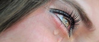
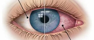
- increased sensitivity to light and uncontrolled tearing;
- redness of the conjunctival membrane.
Symptoms can vary and appear more or less intense - it depends on the reasons for the appearance of the images.
Such optical images can most often appear (lines, dots, circles, ovals, spots).
Classification
How severe the symptoms will be and what they will be depends on the root cause of the appearance of visual images. Visual disturbances may appear as simple geometric shapes such as lines, dots, circles, ovals, or regular spots. As a rule, they have different sizes, configurations and colors.
Each time such visual hallucinations appear with different intensity, the frequency and number of visual images differ. It must be taken into account that even with frequent repetitions, phosphenes always have a different configuration and do not repeat with each other, which makes each new appearance a unique optical phenomenon.
Most often, these color manifestations are dominated by pastel tones of yellow, green, orange or blue.
Artificially induced phosphenes
There is no practical sense in intentionally causing visual images to appear.
But in a healthy person, such phenomena can be easily caused by external stimuli, for example, electromagnetic fields .
Thus, phosphenes are often observed among workers of power plants and nuclear power plants.
The artificial appearance of this optical disorder can be provoked by introducing electrodes into certain areas of the cerebral cortex .
You can independently create such an optical illusion by pressing your fingers on the drooping eyelids of your eyes.
Interesting points
Perhaps the most interesting consequence of this is the fact that people who have lost their sight continue to see such phophens. Which, in principle, is logical - the ganglion cells haven’t gone anywhere. True, with the complete loss of an eye, the ability to see phosphenes is also lost. And in the case of congenital blindness, when ganglion cells, in principle, cannot respond to stimuli, phosphenes are also not observed.
Photo: Draft.blogger
There are other ways to evoke similar images. For example, exposure to magnetic fields or small electrical discharges. And some chemicals can also directly affect ganglion cells, provoking various visual images. And no, we are not talking about hallucinogenic drugs now - with them everything is a little more complicated, since they most often directly affect the visual areas of the brain.
Another effect, visually similar to phosphenes, is called the Ganzfeld effect and can be observed in people living for long periods in conditions resembling sensory deprivation. That is, when you see and hear the same thing day after day, the brain itself begins to complement the picture of what is happening with various “noises”. Both visual and visual. Up to pronounced hallucinations.
We also think you might be interested in hearing about some unusual professional tasters. That is, about people who have brought the ability to distinguish the taste and smell of certain substances to the highest level.
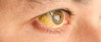
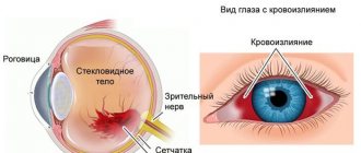
Possible concomitant diseases
Need to know! Such phenomena may be a secondary sign of some ophthalmological diseases associated with disturbances in the structures of the visual organs.
But in such cases, additional symptoms appear in the form of headaches and eye pains, increased blood pressure, dizziness, and loss of balance.
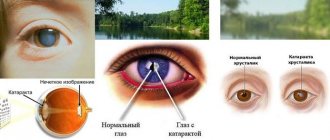
Typically, in such cases, choroiditis, retinitis, retinal rupture or and pathological processes in the vitreous body .
Diseases in other areas include the following pathologies, accompanied by the described phenomenon:
- the appearance of tumors and neoplasms of various origins in the brain area;
- migraine;
- hypertension;
- diabetes;
- pathologies of the vascular system.
Diagnostics
It is not possible for specialists to diagnose the appearance of hallucinations, and a doctor can only learn about the appearance of a disorder from the words of the patient , who describes the intensity and type of formations.
Important! With such symptoms, the ophthalmologist should always prescribe certain diagnostic procedures that will identify concomitant diseases:
- angiography (x-ray examination of blood vessels);
- ophthalmoscopy;
- measurement of intraocular pressure;
- Ultrasound of the organs of vision;
- determination of visual acuity;
- for medical reasons - MRI and CT.
Sometimes it is not possible to determine the exact cause of the images even after these procedures, and in such cases the patient needs to undergo blood tests and undergo a complete examination of the body .
Treatment and prevention
There is no symptomatic treatment for the appearance of phosphenes, since this is only an indirect manifestation of certain pathologies, with the treatment of which the symptom disappears.
Treatment is always selected individually based on the diagnosis , the patient’s age and the manifestation of other symptoms.
In some cases , the presence of an ophthalmologist alone is not enough, and cardiologists, neurologists and angiologists (specialists in vascular diseases) may be involved in the treatment.
If the reason lies in ophthalmological pathologies , are prescribed for instillation into the eyes.
If there is no effect, surgery may be prescribed .
Remember! Throughout the course of treatment, vitamin complexes are used as supportive and strengthening therapy.
Every fifth case of visual hallucinations associated with ophthalmological disorders is a sign of maturing or progressive retinal detachment or disorders in the vitreous body.
In such situations, only surgery will help, which must be performed as early as possible.
Prevention of the appearance of phosphenes has no specific features and consists only of maintaining a healthy lifestyle and, if possible, avoiding junk food, alcohol and tobacco.