The structure of the eye muscles
There are six extraocular muscles:
- Four straight lines - internal, external, upper and lower
- Two obliques - upper and lower.
They are called so because of the peculiarity of the course of the muscle in the orbit and the method of attachment to the eyeball. The work of the eye muscles is controlled by three cranial nerves: abducens, trochlear and oculomotor.
All muscles, except the inferior oblique, begin in a dense connecting ring around the opening in the optic canal. Here, five muscles are twisted into a kind of funnel through which the optic nerve and blood vessels pass. Next, the superior oblique muscle smoothly moves upward and deeper, penetrating the trochlea - the place where the muscle transforms into a tendon, changes its direction to oblique and is attached to the upper part of the eyeball under the superior rectus muscle.
The inferior oblique muscle begins at the inferior internal orbital rim, passes under the inferior rectus muscle and is attached to the lower part of the eyeball.
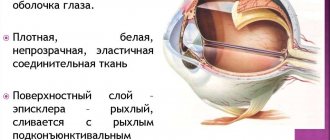
Near the eyeball, the muscles are covered by Tenon's membrane (a dense capsule) and are attached to the sclera (the white coat of the eye).
This complex system of muscle attachment determines how our eyeball moves.
Oculomotor NERVES
VI pair, n. abducens - motor nerve (see Table 11). Core (motor) n. abducentis is located dorsally in the pons at the bottom of the rhomboid fossa, under an elevation known in descriptive anatomy as colliculus facialis, or eminentia teres (see Fig. 50). The elevation is formed due to the presence of fibers of the facial nerve entwining here from above the core of the VI nerve, as mentioned above. The root fibers are directed from the nucleus to the base and emerge as a stem at the border of the pons and medulla oblongata at the level of the pyramids (see Fig. 48). Next, the nerve goes forward and through the fissura orbitalis superior leaves the cranial cavity into the orbit, where it innervates the only muscle - m. rectus externus (lateralis - PNA), turning the eyeball outward.
Nuclear lesions are usually accompanied by peripheral paralysis or paresis of the facial muscles and alternating (on the opposite side) central paralysis of the limbs - the so-called Foville syndrome. In addition, nuclear or perinuclear lesions entail not only paralysis of the m. recti externi (lateralis - PNA), but also gaze paralysis in the direction of the affected muscle and lesion (see explanation below).
When a nerve or root is damaged at its base, isolated paralysis of the m. recti externi (lateralis - PNA), which causes convergent strabismus, the inability to rotate this eyeball outward (Fig. 59), double vision (diplopia), especially when looking towards the affected muscle, sometimes dizziness and forced position of the head.
IV pair, n. trochlearis - motor nerve. The nerve fibers originate from the nucleus located in the bottom of the Sylvian aqueduct at the level of the posterior tuberosities of the quadrigeminal (see Fig. 49, 50). The peculiarities of the exit of this nerve from the brain are that the fibers from the cells
Rice. 59. Paralysis of the left abducens nerve (external rectus muscle). When looking away to the left, the left eyeball does not move outward
the nuclei are directed upward, bypass the aqueduct of Sylvius and emerge not at the base, but dorsally, crossing in the anterior medullary velum (see Fig. 79). Coming out behind the quadrigeminal, the nerve goes around the cerebral peduncle (see Fig. 48) and along the base of the skull passes to the fissura orbitalis superior (see Fig. 53), through which it leaves the skull, innervating the only muscle in the orbit - m. obliquus superior, turning the eyeball outward and downward.
With an extremely rare isolated lesion of n. trochlearis, convergent strabismus and diplopia are noted only when looking down. A very typical complaint is that the patient complains of double vision only when he looks at his feet (for example, when going down the stairs); When looking straight ahead, up and to the sides, diplopia does not occur.
III pair, n. oculomotorius - motor nerve. Core n. oculomotorii is located in the bottom of the Sylvian aqueduct, at the level of the anterior tubercles of the quadrigeminal (see Fig. 49, 50, 80). Fibers from the nuclear cells go mainly to their own (partially to the opposite) side, go down and exit to the base of the brain, at the border of the bridge and legs on the medial side of the latter (see Fig. 48). The nerve leaves the skull along with the IV and VI nerves and n. ophthalmicus n. trigemini through fissura orbitalis superior, innervating 5 external (striated) and 2 internal (smooth) muscles.
Nuclei n. oculomotorii (Fig. 60) consist of five cell groups: two outer large-cell nuclei, two small-cell nuclei (Yakubovich) and one internal, unpaired, small-cell nucleus (Per-lea).
Rice. 60. Diagram of the nuclei of the oculomotor nerve (according to L. O. Darkshevich - simplified).
From the paired external magnocellular nucleus originate fibers for the following external muscles:
1) levator palpebrae superioris - raises the upper eyelid;
2) rectus superior - turns the eyeball upward and somewhat inward;
3) rectus internus (medialis - PNA) - moves the eyeball inward;
4) obliquus inferitor - turns the eyeball upward and somewhat outward;
5) rectus inferior - moves the eyeball downward and somewhat inward.
From the paired small cell (parasympathetic) nucleus of Yakubovich, fibers go to the smooth internal muscle of the eye - m. sphincter pupillae, which constricts the pupil.
Parasympathetic fibers for m. emerge from the unpaired internal parvocellular (accommodative) nucleus. ciliaris (accommodation function).
Rice. 61. Fig. 62.
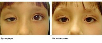
Rice. 61. Palsy of the right oculomotor nerve (ptosis of the upper eyelid).
Rice. 62. Palsy of the right oculomotor nerve.
Fibers from both paired and unpaired small cell nuclei do not directly reach m. sphincteris pupillae and m. ciliaris, and are interrupted in the ganglion ciliare, from where the fibers of the second neuron (non-pulpate) conduct impulses to the named muscles. With complete paralysis n. oculomotorii the following is observed (Fig. 61, 62).
1. Ptosis - the eye is closed by a drooping upper eyelid (see Fig. 61).
2. The eyeball is turned outward and slightly downward (as a result of the action of the preserved m. rectus externus (lateralis - PNA) and m. obliquus superior from the VI and IV nerves); therefore, there is divergent strabismus (see Fig. 62).
3. When the upper eyelid is raised, diplopia is noted.
4. The pupil is dilated (antagonistic effect of m. dilatatoris pupillae from n. sympathicus).
5. There is paralysis of accommodation (m. ciliaris is affected), which is why vision at close distances deteriorates.
6. Convergence, upward and inward movements of the affected eyeball are impossible; Its downward movements are significantly limited (see diagram of eye movements - Fig. 63).
7. The eye is somewhat protruded from the orbit (due to loss of tone of a number of external muscles of the eye) - exophthalmus.
During processes inside the brain stem (in the cerebral peduncle) paralysis of n. oculomoiorii is usually accompanied by central paralysis of the opposite limbs (Weber's alternating syndrome) due to damage simultaneously with the fibers of the third nerve of the pyramidal tracts that cross below. With lesions located at the same level, but more dorsally, with the involvement of the red nucleus in the process, alternating Benedict syndrome is observed (third nerve palsy and cerebellar ataxia in opposite limbs).
If complete paralysis of the third nerve is accompanied by a lesion of n. abducentis and n. trochlearis, then there is no movement of the eyeball at all ophthalmoplegia totalis, or completa.
Fig. 63. Scheme of movements of the eyeballs during contraction of the external muscles. R. ext. - m. rectus externus (lateralis, VI n.); 0. sup. - m. obliquus superior (IV n.), R. inf. - m. rectus inferior (III n.); R. int. - m. rectus inlernus (medialis, III n.); R. sup. - m. rectus superior, (III n.); 0.inf. - m. obliquus Inferior (III n.).
With isolated damage to only the small cell nuclei, the so-called ophthalmoplegia inierna is observed (loss of functions of only the internal muscles), and with damage to only the external large cell nuclei - ophthalmoplegia externa. If n itself is damaged. oculomotorii, too, the picture of its complete paralysis is not always observed; a more limited loss of function is possible due to disruption of the conductivity of only part of the nerve fibers.
Palsies of individual oculomotor nerves (III, IV and VI nerves) are always peripheral. Only with bilateral and, moreover, extensive supranuclear processes that turn off the central neurons going from both hemispheres to the nuclei, can bilateral ophthalmoplegia of the central type occur, since, by analogy with most motor nuclei of the cranial nerves, the nuclei of the III, IV and VI nerves have bilateral cortical innervation.
When examining the nerves of the eye muscles, some data can be obtained from a simple external examination. So, with damage to m. levatoris palpebrae superioris (III nerve) ptosis is visible - drooping of the upper eyelid1: divergent strabismus indicates insufficiency of m. recti medialis (III nerve), converging on insufficiency of m. recti lateralis (VI).
It is important for the patient to indicate that he has double vision (diplopia) - the sign is sometimes more subtle than the objectively determined deficiency of one or another external muscle of the eye.
When complaining of diplopia, it is necessary to find out which muscle (or nerve) is affected by this disorder. Let us remember that diplopia. occurs or intensifies when looking towards the affected muscle. Deficiency of the external and internal rectus muscles will cause diplopia in the horizontal plane, and in other muscles in the vertical or oblique planes. Diplopia is revealed even more clearly if one of the patient’s eyes is covered with colored glass. A sometimes immediately noticeable difference in the size of the pupils (anisocoria), as well as their deformation, should alert the examiner’s attention, but they do not always prove the presence of a lesion n. oculomotorii (possible congenital features, consequences of an eye injury or an inflammatory process, asymmetry of sympathetic innervation, etc.).
Subsequently, the subject is asked, without moving his head, to follow with his gaze a finger or hammer moving up, down, right and left, and paralysis or paresis of any external muscle or eye or gaze may be detected. When the eyeballs are abducted to the sides, nystagmus is usually examined. Convergence and the accompanying constriction of the pupils (the reaction of the pupils to accommodation with convergence) are studied by moving the gaze from a long distance to an object placed close to the eyes (the examiner’s finger, a hammer).
The study of pupillary reaction to light is extremely important. Both direct and conjugate reactions of each pupil are checked separately. The patient's face is turned towards the light source, the eyes are open; the examiner, first tightly covering both eyes of the subject with his palms, quickly removes one of his hands, thus observing the direct reaction of a given pupil to light; The other eye is also examined. To detect a friendly reaction, one eye of the subject is closed with the palm of his hand; in the one that remains open, pupil dilation is observed; when the hand is removed from the closed eye, a simultaneous sympathetic constriction of the pupils occurs in both. The same is done for the other eye. An electric lamp with a switch or a pocket electric flashlight are very convenient for studying light reactions (especially in bedridden patients). Complete loss of pupillary reactions to both light and acco-
1 Partial ptosis and some narrowing of the palpebral fissure can also be observed with damage to the cervical sympathetic nerve, cervical ganglion or centrum ciliospinale (see Chapter IV): in these cases, ptosis is never complete and is accompanied by constriction of the pupil and enophthalmos, i.e. retraction of the eyeball - Horner-Claude Bernard syndrome (see Fig. 23). modulation with convergence is called complete immobility of the pupil. Loss of response to light with preserved possibility of narrowing during convergence with accommodation 1 is called Areail Robertson's sign and is observed almost exclusively with tabes dorsalis and progressive paralysis. Much less often the opposite is observed: preservation of the pupil's reaction to light in the absence of a reaction to accommodation with convergence.
Symptoms of eye muscle diseases
The muscular system of the eye is a complex mechanism, and the acuity of our vision depends on its proper functioning. With pathological changes in the muscles of the eye, various diseases can occur, which significantly reduce the quality of vision and cause physical discomfort.
The main symptoms of eye muscle disorders:
Nystagmus is involuntary eye movements with high frequency fluctuations (up to several hundred per minute). This is a sign of impaired ability to fixate gaze.
Miosis is a strong constriction of the pupil (up to 2.5 mm or less). This feature may appear when the muscle that constricts the pupil contracts or when the muscle that dilates it is paralyzed.
Blepharospasm - intense blinking - is a symptom of a type of facial spasm. It develops due to uncontrolled contraction of the orbicularis oculi muscle.
Diagnosis and treatment
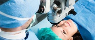
At the appointment, the doctor examines the eyeball, measures pressure, and determines visual acuity. Then, using special equipment, it evaluates the thickness of the cornea, lens, and assesses the condition of other membranes.
After making a diagnosis, the ophthalmologist selects the most effective treatment method. Of course, therapy depends on the disease. This can be either eye drops, ointments or injections, or laser intervention. In addition, physiotherapeutic procedures are used. For example, electrophoresis and magnetophoresis are effective.
It is important to treat eye diseases on time, because otherwise you can live the rest of your days in complete darkness. Make an appointment with an ophthalmologist as soon as the first signs appear.
Express Your Reaction
Similar articles:
Age-related changes in vision: how can you help?
The human eye weighs approximately 8 g. Of course, this figure may vary slightly in one direction or another...
Retinal angiopathy - causes, symptoms, types and treatment
Retinal angiopathy is a change in the blood vessels of the fundus. They expand, contract, or their distortion arises...
Contact lenses
Today, the most affordable and convenient way to correct vision is contact lenses. H...
Horner's syndrome - when the eyelid does not obey
Horner's syndrome In the lives of each of us there are good friends from whom we want to follow an example. Eat …
Demodex mite - how to get rid of it?
Most often, when it comes to demodicosis, we mean a skin disease that causes condition…
Age-related macular degeneration: how to avoid?
No one needs to explain the value of good vision to a person. This is not only an opportunity to enjoy...