Almost 90% of headache complaints during pregnancy are not the result of a serious illness.
Article outline
- Causes of headaches during pregnancy
- Symptoms and treatment of different types of headaches during pregnancy Tension pain
- Migraine
- Vascular headache
Pregnancy and the retina
During pregnancy, the main threat to the visual system is the condition of the retina. The retina is a thin layer of nerve tissue located on the inside of the back of the eyeball that absorbs light. It is a complex formation, the main one of which is a thin layer of light-sensitive cells - photoreceptors. The retina of the eye is responsible for perceiving the image that is projected onto it using the cornea and lens, and converting it into nerve impulses, which are then transmitted to the brain. The main problems with the retina are: retinal dystrophy, retinal tear, retinal detachment.
To prevent possible eye complications during pregnancy and childbirth, it is necessary to determine in advance the state of the visual system of the expectant mother and be sure to check the retina. Ophthalmologists strongly recommend, regardless of how you see and whether you have vision complaints, at 10-14 weeks of pregnancy
.
In addition to a general examination of the visual system, diagnostics of the fundus with a dilated pupil is mandatory. If the diagnostic results do not reveal any abnormalities, then a repeat examination
vision specialists advise to undergo it closer to the end of pregnancy -
at 32-36 weeks
.
However, if you have myopia, then ophthalmologists recommend that you be monitored monthly. During pregnancy, a woman’s entire body undergoes changes, including her vision. Therefore, the visual system requires special attention from the expectant mother.
What will the birth be like?
Will I be able to give birth on my own or will a caesarean section be required? Any woman who has certain vision problems worries about this. It is very difficult to answer this question unambiguously. After all, in many ways the decision about how childbirth will take place is based on a number of factors. Such as: the condition of the fundus and retina, general condition, age, etc. Caesarean section is a surgical operation in which the fetus is removed through an incision in the anterior abdominal wall and uterus. The risk to a woman's life and health during a caesarean section is 12 times higher than during spontaneous childbirth. Therefore, like any other surgical operation, a caesarean section is performed strictly according to indications.
A caesarean section is performed in cases where spontaneous birth is impossible or is dangerous to the life of the mother or fetus. Unfortunately, one of the most common reasons for recommendations for cesarean section is dystrophic changes in the retina. The risk of retinal detachment in women with myopia and changes in the fundus increases during natural childbirth due to pressure changes in the mother.
, preventive laser coagulation is used to prevent the spread of dystrophic changes in the retina and, accordingly, reduce the risk of retinal detachment.
.
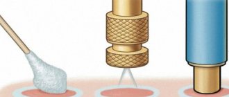
This procedure involves what is called “welding” of the retina
in weak spots and around breaks. Scarring occurs at retinal coagulation points. As a result, a strong connection between the retina and the choroid occurs. The coagulation technique involves applying several rows of coagulates along the periphery of the retina.
When can peripheral prophylactic laser coagulation be performed?
- Any time before pregnancy.
- During pregnancy up to 35 weeks.
The condition of the retina is not always related to the degree of myopia. Often, with a high degree of myopia, the retina remains consistently satisfactory, there are no pre-tears on it, and there are no progressive degenerative changes. It also happens the other way around, when with mild myopia, not exceeding 1-3 diopters, dystrophic foci are observed in the fundus.
If you are planning a pregnancy or are already pregnant, you must undergo an examination by an ophthalmologist with a fundus examination. Remember that a simple procedure to strengthen the retina done in time may well save you from the need for a cesarean section.
A woman’s eyes, like all organs and systems, experience some changes during pregnancy and childbirth. Therefore, expectant mothers should be examined by an ophthalmologist twice while expecting a child. If a woman had certain vision pathologies even before pregnancy, she is under constant supervision not only by a gynecologist, but also by an ophthalmologist. Let's look at what happens to the eyes during pregnancy and what vision problems may arise.
Diagnostics
During pregnancy, all women are recommended to undergo an examination by an ophthalmologist. During the examination, the doctor not only determines visual acuity, but also measures intraocular pressure with a special device. An increase in IOP indicates the development of glaucoma and requires additional examination.
Other methods are also used to diagnose glaucoma:
- ophthalmoscopy (examination of the retina and optic nerve through a dilated pupil);
- perimetry (determination of visual fields);
- gonioscopy (examination of the angle between the cornea and the iris).
Eyes during pregnancy
When carrying a child, the level of the hormone estrogen in a woman’s body increases. It affects the connective tissue, which can lead to a slight elongation of the eyeball and changes in the vitreous body. As a result, intraocular pressure may fluctuate and the cornea may become dry. All this contributes to the appearance of “floaters” in front of the eyes, deterioration of vision, and difficulties in wearing contact lenses.
The most sensitive structure of the eye to change is the retina - a thin layer of nervous tissue that is located on the inner side of the back of the eyeball and absorbs light. Changes that occur in the eyeball and blood vessels sometimes lead to the occurrence of areas of dissection, thinning, areas of dystrophy (malnutrition), and hemorrhages in the retina. In most cases, all these changes are not felt by the woman and can only be determined during an examination by an ophthalmologist. This is why it is so important not to ignore eye exams during pregnancy.
Causes
Glaucoma develops at any age, and pregnant women are not immune to the occurrence of this disease. For the development of a disease, a whole complex of reasons is necessary, which gradually accumulate and at a certain point lead to the formation of a dangerous pathology. The exact mechanisms of glaucoma development have not yet been studied.
Factors that provoke the occurrence of glaucoma:
- disruption of the outflow of intraocular fluid from the eyeball;
- increased IOP;
- impaired blood circulation in the tissues of the eyeball;
- impaired blood supply to the optic nerve;
- compression of the optic nerve or its complete atrophy.
As a result of the development of glaucoma, some of the fibers of the optic nerve atrophy, and some are in a state of a kind of suspended animation. This gives patients suffering from glaucoma a chance to partially restore their vision after an attack.
Eye problems that occur during pregnancy
Sometimes the expectant mother notices frightening manifestations associated with her vision or eye condition. Most of them are not dangerous and are just a “side” effect of pregnancy.
Swelling of the eyelids is quite common in pregnant women, especially often in the morning. To prevent them, a woman must first reconsider her diet and drinking regimen. You should reduce the amount of foods high in salt in your diet, and also drink enough liquid. If such measures do not help, you should consult a doctor.
The flashing of “dots” and “floaters” before the eyes during pregnancy is often explained by vascular disorders, in particular spasms of the fundus vessels. Therefore, in this case, you should not delay your visit to the ophthalmologist.
Changes in the hormonal balance in the body of a pregnant woman provoke a decrease in the production of tear fluid. This contributes to the appearance of dry eyes, photophobia, and the sensation of a foreign body in the eyes. Such symptoms usually go away after childbirth.
Increased sensitivity of the cornea during pregnancy often leads to significant discomfort when wearing contact lenses. Unpleasant sensations are especially annoying in the last three months of pregnancy. In this case, experts recommend temporarily abandoning contact lenses and using glasses.
Another common eye disease during pregnancy is spasm of the accommodative muscle, which is located inside the eyes. Signs of this pathology are eye fatigue, decreased distance visual acuity, and blurred vision. This condition may disappear after childbirth, but in some women it progresses to myopia. Therefore, if these signs appear, it is better to consult a specialist.
Pregnancy sometimes worsens the state of visual functions due to pathologies of the optic nerve and fundus, as well as myopia. This is explained by heavy loads on the body, changes in hormonal levels and the circulatory system. Only regular observation of pregnant women with these pathologies by an ophthalmologist can prevent possible vision complications.
Features of childbirth with eye diseases
If eye diseases are discovered during pregnancy, or a woman had vision problems even before conception, the doctor faces the task of choosing the right way to manage childbirth. In other words, the doctor decides whether it is advisable to perform the birth process using a cesarean section.
Indications for caesarean section are the following conditions:
- High degree of myopia in the only functioning eye;
- Complicated rapidly progressive high myopia;
- A high degree of myopia is combined with obstetric pathology or extragenital pathology (diseases of the cardiovascular, respiratory or digestive systems);
- The appearance of pathological changes in the fundus during pregnancy, such as retinal detachment, swelling of the optic nerve, hemorrhage in the retina.
Symptoms
There are two main forms of glaucoma:
Open angle glaucoma
The disease is inherited. The likelihood of developing pathology increases in women suffering from myopia, diabetes mellitus, hypertension and cervical osteochondrosis. These diseases impair blood supply to the brain and thereby disrupt the outflow of fluid from the eyeball.
Open-angle glaucoma usually affects both eyes. The disease develops gradually, without pronounced symptoms. Periodic blurred vision and the appearance of pain in the area of the superciliary arches are noted by no more than 15% of women. Decreased vision is observed in the later stages of the disease with significant damage to the optic nerve.
Angle-closure glaucoma
The disease occurs with periodic attacks of increased IOP. An acute attack of angle-closure glaucoma is accompanied by the following symptoms:
- sharp pain in the eye;
- blurred vision;
- the appearance of bright circles when looking at a light source.
During an attack of glaucoma, the pupil dilates and does not respond to light. At the height of the attack, a significant decrease in vision is possible. If help is not provided on time, complete blindness develops.
Eye drops during pregnancy
Many women experience eye diseases during pregnancy that require the use of medications. During the period of waiting for the child, it is important not to self-medicate and use only medications prescribed by the doctor. In addition, before starting to use any medications, you should read the instructions for them. You can use those products whose instructions indicate their safety during pregnancy.
In the first trimester of pregnancy, it is not recommended to use eye drops containing beta blockers and carbonic anhydrase inhibitors. These substances have been proven to negatively affect the fetus. 4.9 out of 5 (23 votes)
Blood pressure during pregnancy is an important symptom that characterizes the course of pregnancy. This indicator may vary throughout pregnancy, and this is due to hormonal changes in the pregnant woman’s body. Normal blood pressure in pregnant women is in the range of 90/60-120/80 mmHg.
Blood pressure in early pregnancy
In the early stages of pregnancy, blood pressure often decreases due to hormonal changes. Often the first signs of pregnancy can be: general weakness, loss of consciousness, dizziness, nausea, ringing in the ears, increased drowsiness, etc. These complaints are typical in the morning. Therefore, low blood pressure during pregnancy may be its first sign. Manifestations of toxicosis such as nausea, vomiting, and loss of appetite can help lower blood pressure during pregnancy.
Blood pressure in the last month of pregnancy
In the second half of pregnancy, pressure may increase as the volume of circulating blood increases and a third circle of blood circulation appears. A change in pressure during pregnancy in the later stages in the direction of its increase indicates the onset of preeclampsia, which disrupts the course of pregnancy and childbirth. With the development of preeclampsia, an increase in blood pressure is usually combined with edema and the appearance of protein in the urine. A serious complication of preeclampsia is eclampsia, which is essentially a manifestation of cerebral edema and occurs with loss of consciousness and the development of convulsive seizures. Therefore, in late pregnancy, daily monitoring of blood pressure and pulse is especially important, as well as monitoring proteinuria (protein in the urine) every two weeks. Permissible blood pressure during pregnancy, starting from week 20, should not be lower than 100/60 mmHg. and not higher than 140/90 mmHg.
How does blood pressure affect pregnancy?
Both a decrease and an increase in blood pressure negatively affects the body of the expectant mother and the course of pregnancy. Thus, a decrease in pressure leads to deterioration of blood circulation in the placenta and insufficient oxygen supply to the fetus, leading to hypoxia and intrauterine growth retardation.
Increased blood pressure in the second and third trimester of pregnancy above 140/90 mmHg. is a reason for hospitalization in a specialized hospital. High blood pressure interferes with placental blood flow due to placental edema. Thus, the fetus suffers from a lack of oxygen and nutrients. A rise in pressure above the level of 170/110 mmHg. threatens the development of acute cerebrovascular accident. Alarming symptoms of the growing clinical picture of preeclampsia are difficulty in nasal breathing, flashing spots before the eyes, headache and impaired level of consciousness.
Pressure surges during pregnancy can be a symptom of increased intracranial pressure. Increased intracranial pressure during pregnancy is caused by increased production of cerebrospinal fluid in the blood plexuses of the lateral ventricles. Most likely, the woman suffered from intracranial hypertension before pregnancy, and during pregnancy this pathology worsened. In this case, you need to contact a neurologist and check the intraocular pressure.
Pregnancy is a difficult test for the female body. At this time, the hormonal system undergoes major changes. One of the very fragile organs in our body is the eyes... and at this time all problems with them, even hidden ones, can worsen. What should you do to avoid losing your vision while expecting a baby?
You probably already know that when registering with a gynecologist, a woman has to go through more than one doctor. Among them is an ophthalmologist, whom the majority of the population simply calls an ophthalmologist. Under no circumstances should you ignore this specialist, even if it seems to you that you have no problems with your vision.
Pregnancy is a serious test of the body's strength, and it is not for nothing that the eyes are considered a very vulnerable part of our body. Even minor deterioration that occurs during pregnancy can transform into a catastrophic problem and a severe deterioration in visual acuity.
The first visit to an ophthalmologist is recommended when registering with a gynecologist, namely at the end of the first trimester of pregnancy. During the examination, the ophthalmologist will take a good look at your fundus, evaluate the quality of the fiber, as well as the degree of refraction - this is what pathological changes in the condition of the eye are called. The specialist must determine whether there are any possible tears in the retina and whether there are any changes in the fundus of the eye. In addition, the ophthalmologist will measure eye pressure.
Note!
While expecting a child, almost all women become especially suspicious. Sometimes you may feel like you can't see well enough, but this can be explained quite simply. During pregnancy, connective tissues in the body become more elastic, this also applies to the structures of the eye. However, the organs of vision cannot recover, and therefore vision deterioration during pregnancy by a couple of diopters is quite common.
An effect such as flies flashing before the eyes is not considered dangerous. It can occur in all people and is considered a normal physiological process. The danger of pathological changes is that most often they develop unnoticed. Sometimes a person can see an effect similar to lightning - a bright second flash that periodically appears. If you encounter this phenomenon, it is better to consult a doctor.
Statistics show that vision problems are now a fairly common problem. This is how almost every second person is diagnosed. If you had low vision before pregnancy, you will need to visit an ophthalmologist regularly - once a month. During pregnancy, it is better to stop wearing contact lenses and give preference to glasses. During this period, changes in the structures of the eye occur, so the usual lenses often become uncomfortable.
Obstetrics
All women with vision problems, while expecting a child, ask a completely natural question: how myopia will affect the development of labor, whether it will be natural, or whether a caesarean section will have to be performed. Only the ophthalmologist decides whether a pregnant woman can give birth on her own. In this case, many factors play an important role: age, condition of the whole organism, degree of myopia, characteristics of the retina, etc.
The main prohibition on natural childbirth is the presence of retinal dystrophy in the mother. During labor and labor, strong changes in intraocular pressure occur, which can lead to retinal detachment, changes in the fundus of the eye, and sudden loss of vision.
Statistics show that only in 10% of all cases, vision problems necessitate a cesarean section. But if you are in this percentage, do not be upset, because not only your health is at stake, but also the health of your baby. Remember, as a mom, you need to stay strong and healthy.
Laser correction
In the period up to the thirty-fifth week of pregnancy, laser vision coagulation is allowed. This procedure will take just a couple of minutes; it involves welding the retina located in the weakened layers. With the help of coagulation, scarring of connective tissues occurs and a strong relationship between the cells of the retina and the choroid occurs. Carrying out such a simple procedure can prevent further deterioration of vision and avoid a caesarean section.
Prevention
If you have impaired vision, you should be especially careful about your health when carrying a child. You should not lift heavy objects; in addition, it is recommended to refrain from situations that cause a feeling of a rush of blood to the head, as this leads to an increase in intraocular pressure, which in turn has a negative effect on the condition of the retina.
In order to prevent the progression of visual impairment, do special eye exercises, sit less in front of a computer monitor, and walk more in the fresh air. Don’t forget to attend special courses on preparing for childbirth; they will teach you how to control yourself during labor, which will significantly reduce the load on your visual organs.
Poor vision while expecting a baby requires close attention from doctors, as well as a competent and responsible approach from the woman herself.
Rose cookies the most correct recipe Homemade rose cookies recipe
28.02.2021
Making pesto sauce according to a classic recipe at home
28.02.2021
Instant pickled zucchini
28.02.2021
Glaucoma treatment
Glaucoma is a chronic disease, and it is almost impossible to get rid of it completely. All existing treatment is aimed only at reducing the frequency of attacks and preventing optic nerve atrophy. Ultimately, glaucoma leads to significant vision loss and complete blindness.
Drug therapy
Drugs used to treat glaucoma:
- beta blockers;
- alpha-2 adrenergic agonists;
- prostaglandins;
- carbonic anhydrase inhibitors.
A woman suffering from glaucoma should visit an ophthalmologist as early as possible in her pregnancy. Many medications used to treat this pathology are prohibited for use in expectant mothers. After the examination, the doctor will adjust the treatment regimen or suggest replacing the drugs with relatively safe analogues.
In the first trimester, beta blockers and carbonic anhydrase inhibitors are prescribed with caution. These drugs have a teratogenic effect and can lead to fetal developmental abnormalities. If possible, the expectant mother should avoid using products from this group until 12 weeks of pregnancy.
The drug brimonidine from the group of alpha adrenergic agonists is considered relatively safe. The medicine is available in the form of eye drops. This is what is usually prescribed to pregnant women suffering from glaucoma.
In the second and third trimesters, drugs from the group of beta blockers are added. Fetal heart rate should be monitored regularly when using these medications. Regular use of beta blockers leads to the development of bradycardia (decrease in heart rate) and arrhythmia (irregular heart rhythm).
At the end of pregnancy you should be very careful with prostaglandins. Drugs from this group can trigger premature labor. The use of prostaglandins is possible only under strict indications and under constant medical supervision.
In case of an acute attack of glaucoma, it is necessary to urgently call an ambulance. Treatment is carried out in a specialized ophthalmology department.
Surgery
Surgery for glaucoma is possible in the second and third trimesters. In this case, the effect of anesthetics on the fetus should be taken into account. In the later stages, prolonged supine position during surgery can lead to compression of the inferior vena cava and fetal hypoxia. Laser correction of glaucoma is performed at any stage of pregnancy.
All women planning pregnancy with glaucoma should visit an ophthalmologist before conceiving a child. It is recommended to undergo a full examination by a specialist. After the examination, the doctor will be able to change the treatment regimen and select drugs that are safe for the child. Surgical treatment of glaucoma is also best performed before pregnancy.
Glaucoma in any form can be an indication for cesarean section. The final decision is made after a complete examination of the patient using special ophthalmological equipment.
Eye pressure during pregnancy is quite rare. This is mainly caused by glaucoma, inflammatory processes in the membranes of the eye, and surges in blood pressure. This pathology does not pose a big threat to babies. For pregnant women themselves, this can significantly reduce vision. To prevent a pathological condition, it is necessary to adhere to proper nutrition, avoid stress, and reduce the time you watch TV by 2 times. It is recommended to walk more in the fresh air.