The general term “retinopathy” means a pathological condition of the functional tissue of the retina, which developed under the influence of certain reasons. Almost all forms and types of retinopathy are secondary, i.e. are caused by a lack of blood supply to the retina, and therefore, nutrients and oxygen. In turn, the cause of the development of retinal ischemia is most often damage to its choroid due to a more general disease (atherosclerosis, diabetes mellitus, etc.). Such a primary etiopathogenetic factor serves as the basis for a generally accepted classification: for example, diabetic retinopathy, hypertensive, traumatic, etc. are distinguished.
A special form of retinopathy is considered separately, which is not caused by wear and tear or diseases of the adult body, but, on the contrary, by underdevelopment of the vascular system of the eye. We are talking about retinopathy of prematurity (ROP).
In modern ophthalmology, this pathology is considered severe and represents an acute and painful problem: all the mechanisms and patterns of RP are far from being sufficiently studied, there is no single effective approach to therapy (much still depends more on the natural course of events than on the efforts of doctors), but, at the same time, ROP remains one of the main causes of early childhood blindness.
Development mechanisms
It is known that the formation of the choroid that nourishes the retina begins at the 4th month of gestation and continues until childbirth. Thus, the birth of a very premature baby automatically means an unformed blood supply system to the retina. However, if previously, for example, a 7-month-old newborn with severe underweight was practically doomed, then since the mid-twentieth century the development of neonatology has sharply increased the survival rate. At the same time, ophthalmologists talk about well-groomed children (the first description of the pathology appeared in the 40s).
It was found that the main reason was the mutually exclusive needs of the unformed organism: on the one hand, for the normal development of the vascular (blood supply) system of the retina, it must be nourished through glycolysis, that is, the oxygen-free breakdown of sugars. But on the other hand, the strategy for life support and nursing of premature babies clearly requires intensive oxygenation in the incubator, which suppresses the processes of glycolysis. As a result, at 3-6 weeks of life (but no later than the 10th), severe, in some cases irreversible, organic changes may begin in the retina, fraught with profound insufficiency or loss of visual functions.
Risk factors
As can be seen from the above, the main risk factor for ROP is prematurity itself (delivery at 26-28 weeks). In addition, the likelihood of developing retinopathy increases:
- body weight less than 1.4 kg at birth;
- life-saving artificial ventilation for three or more days after birth;
- active oxygen therapy lasting more than a month;
- instability of general condition, incl. respiratory, vascular, neurological disorders;
- intrauterine infections.
Possible complications and consequences in children
Even if the surgery was successful and the baby managed to preserve his vision, the risk of health problems remains. As the baby grows and develops, parents may encounter the following complications:
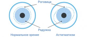
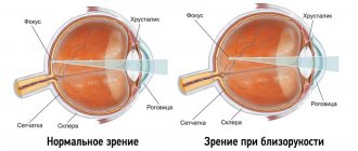
- myopia;
- astigmatism;
- strabismus;
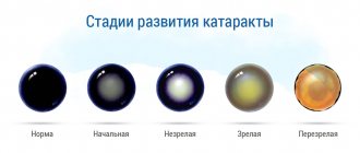
- cataract;
- glaucoma;
- retinal detachment;
- amblyopia.
| It is worth recognizing the fact that the retina of a premature baby will never be the same as that of a baby born at term. Therefore, any negative factors can provoke unpredictable consequences. |
Symptoms and diagnosis
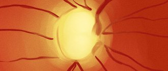
After premature birth, an ophthalmological examination of the child is strictly mandatory and should be carried out no later than 1-2 weeks of life. In the future, inspections are carried out weekly; if there is reason to suspect the onset of ROP, the fundus of the eye is examined carefully using mydriatics (drops that dilate the pupil). For diagnostic purposes, ophthalmoscopy, electroretinography, ultrasound and other methods are used as necessary. The diagnostic criteria are the location, nature, stage, dynamics of the lesion, the presence/absence of neovascularization (i.e., the growth of the newly formed vascular system with its penetration into the surrounding ocular tissues). Thus, there are 3 zones of distribution of retinopathy around the optic nerve head, and 5 stages of damage:
- Stage 1 - characterized by a thin light gray line between the unformed periphery of the retina and the blood-supplied central part;
- Stage 2 – isolated foci of neovascularization, a tendency to shunting (fusion) of retinal arterioles and veins;
- Stage 3 – growth of a new vascular network with penetration into the vitreous body, a tendency to replace functional cells with connective tissue, branching of blood vessels, hemorrhages in the retina and/or vitreous body;
- Stage 4 – a tendency to detach the retina from the periphery to the center (usually at 10 weeks of life);
- Stage 5 – total retinal detachment.
The dynamics of neonatal retinopathy are difficult to predict: up to 80% of cases end in spontaneous restoration of fundus structures (sometimes without residual consequences). With an unfavorable development option, called “plus disease,” the pathology progresses and is manifested by increasing rigidity (insensitivity to light, immobility) of the pupil, opacification of the vitreous, severe neovascularization, extensive hemorrhages and scarring, and the rate of worsening varies significantly: for example, between 1 and 4 stages may take several weeks or just a few days.
Retinopathy of prematurity - immaturity of the retinal blood vessels
Children born prematurely are special babies. They come into this world very small and defenseless. But at the same time, they are the real fighters for life. Still would! After all, they learn to live independently much earlier than ordinary children. However, their early birth is fraught with many health problems. One of the most serious diseases affecting premature babies is retinopathy of prematurity. We asked ophthalmologist, head of the emergency room of the Scientific Center of Pediatrics and Children's Surgery in Almaty Elena Vladimirovna TYAN to tell our readers about what this eye disease is, why it develops, whether all premature babies are susceptible to it, and also give recommendations to parents whose children were born prematurely.
– Elena Vladimirovna, please tell us what retinopathy of prematurity is?
– Retinopathy of prematurity is a very serious disease characterized by immaturity of the retinal blood vessels in premature infants. That is, in children born at gestational age - up to 35 weeks. In a full-term newborn baby, the retina of the eye is fully formed. In a premature baby, the retinal blood vessels do not completely complete growth and form an (ischemic or avascular) zone. The longer the period of prematurity, the higher the degree of vascular underdevelopment. The disease usually affects both eyes, but there are cases of one eye being affected.
Retinopathy of prematurity develops in five stages
During the first stage, a demarcation (dividing) line begins to form between the vascular and ischemic parts of the retina. It interferes with the normal development of blood vessels. Most often, the first stage regresses on its own and does not require surgical correction. Or drug therapy is used to eliminate it.
During the second stage, the demarcation line becomes thicker and denser, forming a so-called ridge. As a result, the boundary between the vascular and avascular zones becomes more obvious.
The second stage in most cases (depending on the location) does not require surgery. It is quite amenable to conservative treatment by instillation, i.e. drip administration of drugs. Today we have already moved away from many of the primitive methods of treatment that were used before. Old, ineffective drugs have been replaced by new, more effective retinoprotectors. They improve the growth of retinal blood vessels.
In the third stage, new vessels begin to form. But due to the demarcation shaft, they do not have the opportunity to grow to the periphery of the retina. Here we are already resorting to surgical laser treatment in order to allow the vessels to develop normally.
If laser surgery is not performed on time or the disease process is so aggressive that the operation did not help, then retinopathy will begin to progress and enter the fourth stage, at which partial retinal detachment begins. At this stage, vitreoretinal surgery is already used.
At the fifth stage, partial retinal detachment becomes total, i.e. complete. The child no longer focuses his gaze; his pupil glows gray. At this stage, even successful surgical treatment becomes ineffective.
There is also a posterior aggressive form of retinopathy of prematurity; it is not included in the general classification. It is believed that this is the most severe form, which practically does not respond to surgical treatment. However, the world has already found a solution to this problem. A drug has been developed for the conservative treatment of the posterior aggressive form of retinopathy by intraocular administration, which has already been introduced into treatment protocols in the USA. Today this drug is being introduced into the CIS countries. It was recently tested in Russia. And I hope that in the near future it will appear here in Kazakhstan.
Treatment
Retinopathy of prematurity in stages 1-2 usually does not require intervention: the chances of spontaneous regression of the pathology are statistically high. Stage 3 is considered threshold and creates indications for intensive measures, conservative and/or ophthalmic surgical. Depending on the clinical picture and dynamics, drugs with angioprotective and antioxidant effects, vitamin complexes (for example, Emoxipin drops), and corticosteroid hormones are prescribed. To eliminate the neovascular vascular network, as well as to prevent retinal detachment, photo- and laser coagulation, cryoretinopexy are used (however, in relation to detachment, these methods often provide only a temporary effect; the percentage of therapeutic success is 80-85%).
Prevention
Preventive measures include:
- Prevention of premature birth (obstetrician-gynecologist observation, timely treatment of pregnancy pathologies, preserving therapy).
- Proper nursing of a baby born prematurely.
- Regular examination of the eyes of a premature baby.
- Competent treatment tactics that prevent progression and relapse after surgery.
All premature babies are subject to careful monitoring and a full range of examinations. Since the organs and systems of a premature baby have not yet had time to form, it is necessary to create nursing conditions that are as close as possible to the intrauterine period.
Share the article on social networks. Tell us in the comments what you know about retinopathy of prematurity? Maybe you have encountered such a disease? Health to you and your baby. All the best.
Surgeries for retinopathy
The most common ophthalmic surgical choice in severe cases is some version of vitreoretinal surgery (on the vitreous body and retina), but in some situations, for example, with traction detachment, when the retina is literally torn off by newly formed ties and adhesions, even such radical intervention is ineffective . In general, stages 4-5 of neonatal retinopathy usually result in irreversible blindness.
Intense research into the problem of RN, given its severity and relevance, continues all over the world. On the one hand, data are published on the tendency towards an increase in cases of ROP and the severity of its forms. On the other hand, reports periodically appear about “revolutionary breakthroughs” in this area - for example, Australian ophthalmologists propose using infrared light to stop the development of ROP, emphasizing the effectiveness and comparative cheapness (which is important) of such a technique. All this information, however, needs additional verification and large-scale clinical trials.
The main conclusion for parents today is that any premature birth poses a threat to the child’s visual system (possible complications and consequences of ROP include not only retinal detachment, but also myopia, amblyopia and other severe anomalies), and the more “ahead of time ", the higher the risk of the most unfavorable development of the situation - up to loss of vision. Considering that sometimes the count is literally days, due to the huge number of other problems of prematurity, the need for a thorough ophthalmological examination and monitoring should in no case be neglected. At the slightest sign of the development of retinopathy, decisions and measures should be taken rationally, reasoned, balanced, but without unnecessary hesitation and delay.
Symptoms of the disease
It is quite difficult to independently detect the symptoms of retinopathy in newborns. The first three stages can only be detected through an ophthalmological examination. Obvious symptoms appear in the later stages.
There are several indirect behavioral signs that allow one to suspect pathology:
- A newborn examines objects with one eye.
- Fixation of gaze on objects is impaired.
- The child does not see people and objects that are at a short distance.
- The baby brings toys very close to the organ of vision.
- The child allows you to close one eye, but when closing the second, he prevents you from closing it.
At the scarring stage, external manifestations appear: strabismus, the pupil becomes gray and reacts poorly to light.