According to the World Health Organization and observations of practicing ophthalmologists, the percentage of children with vision problems is steadily increasing. This is especially true for schoolchildren and students who experience maximum stress due to the need to read, write and use a computer a lot. Half of the students have some degree of myopia. By the end of school, almost 80% of children acquire functional or organic pathology of the organs of vision.
This disappointing statistics forces us to look for new, more effective and safe methods of prevention and treatment in ophthalmology. One of the methods recommended for restoring vision in children is hardware exposure and training using ophthalmological devices. When treating and correcting vision in children, it is advisable to combine conservative and physiotherapeutic measures. Surgery is performed only as a last resort in cases where other types of care are ineffective and there is a likelihood of further progression of the disease. Hardware treatment in many children shows stable positive results, especially if the onset of the disease is associated with excessive or prolonged visual strain and increased eye fatigue.
The duration of the course and intensity of exercise is determined by the doctor based on diagnostic data. One cycle can include from 10 to 15 workouts. Classes are held daily, each lasts about an hour and covers exercises on several (4-5) different devices.
Diagnostic combine Technolas ZDW3 (Germany)
The latest diagnostic optical device for a detailed examination of the anterior segment of the eye.
Most accurate in its class. Consists of an Orbscan 3 keratotopograph and a Zywave wave aberrometer. In just 3 seconds, the device takes measurements of the cornea at 9000 points and builds a detailed three-dimensional topogram of the cornea and simultaneously measures refractive power and thickness. This data is transferred to the Teneo excimer laser computer for laser vision correction.
Read more about the Technolas ZDW3 diagnostic combine.
Alcon Verion Diagnostic Analytical System
The Verion diagnostic analytical system allows for a complete preoperative diagnosis of the eye in patients with cataracts. During this digital study, the device automatically takes several dozen eye parameters and produces the most accurate calculation of the parameters of the artificial lens. This allows us to take into account all the individual anatomical features of the patient’s eye and the specifics of his visual functions. In addition, Verion receives a photographic image of the patient's eye with maximum resolution. This allows the surgical equipment to recognize the patient's eye, create a digital map and, using digital markings, display the optimal course of the operation in the eyepieces of the Luxor microscope. Never before has cataract surgery been so precise and effective.
Learn more about the Verion diagnostic station.
What diseases are treated with hardware treatment?
Some diseases that can be treated with hardware treatment:
- Impaired binocular vision;
- Strabismus;
- Amblyopia;
- Asthenopia;
- Spasm of accommodation;
- Postoperative period;
- Partial atrophy of the optic nerve;
- Dystrophy of the fundus of the eye;
- Farsightedness;
- Myopia;
- Astigmatism;
- Computer syndrome.
Computerized perimeter
Designed to diagnose the state of the visual field. Often a person does not notice the appearance of defects (losses) in the field of vision due to the ability given by nature to look at the world with two eyes (the better eye compensates for the “problems” of the worse). The built-in computer program allows you to analyze the dynamics of the state of the visual field and eliminates the possibility of accidents as a result of this examination.
The latest full-field computer perimeter Centerfield II from Oculus (Germany) performs static, kinetic and color perimetry across the full field of view, as well as retinal threshold sensitivity testing. In addition, the device has the ability to change program parameters and create individual programs.
Flowmetry – measurement of individual norm of intraocular pressure
Specialists from our center and the Federal State Budgetary Institution Research Institute of Eye Diseases have developed an original method for determining the individual norm of intraocular pressure. Many years of scientific research have shown that the same level of IOP can be detrimental and lead to glaucoma in some patients and be safe in others. You can determine your individual tolerant norm of eye pressure only in our Center.
Read more about the method for measuring individual IOP norms.
Hardware vision treatment for children
In our ophthalmological clinic you can make an appointment with a pediatric ophthalmologist (ophthalmologist) in Sevastopol and Evpatoria. Our highly qualified pediatric ophthalmologist (ophthalmologist), after conducting a diagnostic examination, selects an individual course for each small patient, depending on the state of vision and disease. Thanks to regular exercises on the devices, children's vision is significantly improved and maintained without any surgical intervention or drug therapy. The earlier classes start, the faster a more lasting result is achieved, and your child’s vision will be preserved for many years.
Tips for parents
- Regularly bring your child for preventive ophthalmological examinations in order to detect pathology in time, and if detected, control it.
- Complete the course of treatment prescribed by the attending physician in a timely manner and in full. The course may include not only hardware treatment, but also classical drug treatment, eye exercises, and diet.
- As a rule, the child is prescribed glasses for the duration of treatment. You need to constantly monitor the child, convince him of the need to wear them constantly and not take them off.
- Be patient, because the courses of treatment are long, and the positive effect may not occur immediately.
Therapeutic treatment using devices is completely painless and safe for children.
Heidelberg retinal tomograph HRT3
The Heidelberg retinal tomograph HRT3 was developed by the German company Heidelberg Engineering (Germany). This modern, ultra-precise diagnostic device provides rapid topographic measurements of the optic disc and retina.
The high resolution of the tomograph allows for dynamic monitoring of patients with glaucoma and other diseases of the optic nerve, and to choose treatment tactics based on changes in the stereometric data of the optic nerve.
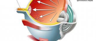
The operation of HRT3 is based on the principle of confocal laser ophthalmoscopy (CSLO). This is a modern technology for obtaining high-quality three-dimensional images of the fundus of the eye using scanning with a specially focused laser beam. The main clinical use of HRT3 is to visualize elements of optic neuropathy caused by glaucoma, as well as disorders of the optic nerve head that occur in other diseases.
The most popular and effective devices
Vizulon is a modern color pulse therapy device with several programs, which allows it to be used not only for the prevention and complex treatment of vision diseases, but also for pathologies of the nervous system (migraines, insomnia, etc.). Available in several colors. Go to the device page >>>
Sidorenko glasses (AMBO-01) are the most advanced device for self-use by patients for various eye diseases. Combines color pulse therapy and vacuum massage. Can be used both in children (from 3 years old) and in elderly patients. Go to the device page >>>
Pankov glasses (Rainbow of Insight) are the most famous and popular eye device based on color pulse therapy methods. It has been produced for about 10 years and is well known to both patients and doctors. It is characterized by low price and ease of use. Go to the device page >>>
We have presented a complete list of ophthalmological devices with their descriptions, characteristics and patient reviews. Which one is right for you will be advised by your attending ophthalmologist, especially since regardless of the method of exposure used, all instructions contain a mandatory requirement - “before use, you must consult with a specialist.”
Retinal Thickness Analyzer (RTA)
The Retinal Thickness Analyzer (RTA), from TALIA TECHNOLOGY, is a unique system that combines the capabilities of a digital fundus camera, a computer scanning slit lamp and a retinal thickness analyzer. The laser beam safely scans the retina in the current mode according to the program specified by the doctor. The results obtained are analyzed by a computer program and presented on the display in the form of a report. The device records retinal thickness and optic disc topography in the living human eye, providing objective and reliable diagnosis and monitoring over time. The most important advantage of this unique device is the possibility of “pre-diagnosis” - i.e. early diagnosis of eye pathology, when the disease has not yet “manifested itself” clinically.
RTA is used in a number of cases: for accurate and early diagnosis of glaucoma (even before the appearance of traditional visual field defects), retinal pathologies (such as macular holes, cysts and membranes), diagnosis of early and ongoing changes in the eyes of patients with diabetes (diabetic retinopathy), assessing the condition of the retinal blood vessels, etc.
Indications for hardware vision treatment:
- Spasm of accommodation
- Myopia
- Computer vision syndrome
- Chronic visual fatigue
- Asthenopic syndrome after refractive surgery on the cornea
- Amblyopia
- Presbyopia
- Glaucoma
- Cataract
- Retinal dystrophy.
For preventive purposes, hardware treatment can be used in the form of courses of training exercises:
Computer users who spend more than 1-2 hours a day on a screen (at risk for developing computer vision syndrome)
For workers engaged in high-precision work - to prevent visual asthenopia and myopia
For schoolchildren - to prevent myopia
Athletes of shooting and team sports - in order to improve athletic performance
For patients over 40 years of age to prevent presbyopia.
Indications for use of Visotronic M3 as part of complex treatment:
- Primary cataract
- Amblyopia
- Retinal dystrophy
- Partial optic atrophy
- Compensated glaucoma.
Contraindications to hardware vision treatment:
- Acute and chronic inflammation of the eyeball and eyelids
- Exotropia
- Marked impairment of the transparency of the optical media of the eye
- Malignant formations of the eye and its appendages
- Glaucoma with uncompensated IOP (intraocular pressure).
Hardware treatment of vision in children and adults with amblyopia and strabismus
For the complex treatment of amblyopia, as well as the development and consolidation of binocular vision in case of strabismus, the computer programs CONTOUR, PLEOPTICS 3, and eYe are used. The programs are used on an outpatient basis as an independent method of hardware vision treatment and in combination with other programs.
Keratotopograph with wavefront distortion analyzer Keratron Scout
The device simultaneously measures the topography and refractive power of the cornea at each point, and also allows for selective aberroscopy of the cornea (i.e., individual defects in the optics of the cornea). The aberrations of each eye are individual. If it were possible to reduce the level of these aberrations, then the human eye would have an optical system closer to ideal, and human vision would be much closer to eagle vision.
The study lasts several seconds, during which the entire surface of the cornea is scanned. The information obtained by this device is necessary when performing excimer laser correction of myopia, farsightedness and astigmatism (after all, the effect occurs directly on the cornea). A powerful computer displays the received information in the form of digital characteristics and allows you to predict visual acuity as a result of surgical correction. The topographer reveals the first signs of keratoconus (a disease in which the shape of the cornea changes) and a number of other diseases of the cornea.
Operating room equipment for vision correction
This is a photo tour
; then there will be 70 photographs from two operating units for laser vision correction. Different options for eyelid dilators:
Packaging with a disposable blade, which is tucked into a microkeratome to form a valve in the cornea during mechanical (non-laser) operations:
And here is the head of the microkeratome, where the blade is inserted:
Here is a set of “accessories” for the working part - it is reusable:
Motor for driving the head with the blade:
And this is the console with an indicator of operating parameters:
Now let’s compare it with the tools for SMILE. This is what a disposable package with a cone looks like, and there is also a vacuum tube for gas removal:
Separation spatula to scale:
The very first spatula was made by Dr. Bloom, who, together with Walter Secundo, invented the technology. Today they don’t use it because it was sharp, because back then they cut at other frequencies, there were no modern lasers. Now the cut is much cleaner, and there are already about 10 different modifications of the tools. The most popular is according to Chansu (this is a Thai surgeon who performs a wild number of operations, 8 or 10 thousand per year, he has a “conveyor” type clinic in Bangkok). There are interesting Russian ones, in particular, the Kostin spatula from Yekaterinburg. There is no huge difference between them. Walter uses the one in the photo, this is his author’s model exactly for his hand. Let me remind you, this is what it looks like (in the photo below, the Chansu spatula is used during the operation):
Microsponges made of hydrophilic material, which scared someone in the second post (these are not blades):
What the patient takes home after correction:
This is what a disposable scalpel for the cornea looks like, which is practically not needed (its functions are performed by a laser):
For the patient, everything begins after the corridor with a bed like this.
Here the patient lies down for a while before the correction (to calm motor skills) and rests for a couple of hours after.
There are two buttons on the wall: call sister and SOS. SOS triggers an alarm in all rooms except other operating rooms.
There are pictures on the wall to look at:
There is a video camera monitoring the patient's condition in the ward. The image is broadcast to the nurse's station so that we always know what is happening with the patient. It is especially needed for those long operations after which the patient wakes up after anesthesia in a not entirely normal state of consciousness.
First operating room with excimer laser:
Couch (more precisely, an operating table, also known as a smart bed):
Excimer laser:
He is “facing” the patient (this part is located under the microscope):
This is how the laser is seen by the surgeon:
Observation is carried out through this microscope. The data is also displayed for the assistant on a screen on the wall of the operating room.
The doctor has the laser stop button at hand:
This is the key to start and the pre-calculated profile for the operation:
Vibration protection:
Laser control pedal (during the operation it is located under the doctor’s foot):
Vacuum suction to remove excess moisture and fatty secretions of the glands:
Excimer laser control knob:
Dioptremeter for assessing laser calibration (the laser burns special test calibration lenses of various dioptres).
Here are the cards, on which the laser burns through lenses of different diopters:
Statutory warnings:
Card with a license (on a femtosecond laser the license is already completely digital):
Guess who's underneath:
The keyboard is almost ordinary, but with a trackpad - needed to work with the interface. Before the laser itself operates, it is a Windows program, then it is the laser firmware.
Testing:
Here you can see the markings for aiming:
Activated by pressing the key at the top left of the joystick. It is positioned on the patient’s cornea during surgery - it is also a “flashing red dot” (its intensity can be changed) during excimer laser correction surgery:
There are three buttons in the left row. The upper left includes a red line, which is projected onto the patient's face for placement in a strictly longitudinal position without lateral displacement. There is a mark on the side of the head end for positioning at eye level. This ensures precise positioning of the head along the XY axis. The vacuum pillow quite firmly fixes the head in the chosen position. The position changes from left to right eye by pressing a button on the bedside by an assistant. Next, the surgeon begins to center the eye under a microscope.
The lower left control button for iris image capture is the iris recorder control button (controls the XY position). The middle left is a button for controlling cyclotorsion (rotation of the eyeball around its axis when the patient changes position from vertical to horizontal).
On the right are buttons for regulating the illumination of the surgical field - direct and side lighting. Often, the surgeon almost completely turns off the illumination of the surgical field, since the tracking camera for the individual iris map operates in the infrared range and the “pattern” of the iris and the pupil in the dark are determined by the device faster.
Here is the basic position of the table during surgery:
It is adjustable in height and other parameters:
The laser screen is completely ordinary:
Excimer lasers are extremely sensitive to changes in temperature and humidity in the operating room. Also, there should be no ether or alcohol solutions in the excimer laser operating room. Constant monitoring with thermometers and hygrometers is required.
License card:
Gas cylinders for laser:
Rausch anesthesia tool Service tool kit. Everything in the laser is attached to the place of use, for example, there is a screwdriver on the holder below, which is needed for the bolts on the bottom panel:
Assistant screen:
That's it, we're done with the excimer for now. We move on to the next operating room, where the VisuMax femtolaser is located.
It is used to form a flap during femtoLASIK (then the patient goes to the operating room described above under an excimer), for laser correction FLEX and SMILE (then you don’t need to move anywhere - everything is done under one laser), as well as for any corneal incisions, for example, for intracorneal rings for keratoconus). In the case of a common manufacturer of femtolaser and excimer laser installations, in some cases one bed is used for two devices, then it is moved during the execution of different stages.
So, here is VisuMax:
The laser was created to impress with its appearance - Zeiss ordered the ergonomics from the Mercedes design bureau (this laser has both design and cost - like Maybach, they not only made it very convenient to operate, but also worked on the appearance). This was motivated very simply: if the patient feels like he is getting into a Maybach, then he is somehow calmer than when getting into something from a movie about Alien.
The ergonomics are really wonderful, everything is at hand (or rather, with your hands):
There is almost no need for a keyboard here, the screens are touch-sensitive:
The pedal is a little different. From the moment it is clicked, the license for a specific operation is considered used:
The landing is like this:
Notice how everything is at hand.
As for the human factor, there are a couple of important points for successful surgery:
- The surgeon must have stereoscopic, i.e. three-dimensional vision, since it is important to see three-dimensionally - the cut is curvilinear and has depth.
- You also need to focus on the sound and light indication - each laser operating mode and each situation has its own indication.
- This is the type of surgery when at the time of the operation the surgeon is left alone with the patient - at this moment only he controls his behavior
Next - about the software. VisuMax's interfaces are slightly newer than those on the excimer, but still on “ATM” Windows.
During setup, an animation of how the cuts will be placed in the cornea and their geometry is displayed on the left of the screen.
The laser insures against errors: if something in the set of parameters falls outside the best practice, warnings are issued. And yes, of course, the last 100 operations are recorded on a “black box” with all telemetry and video.
And here is the cone that comes with the license (this is a replaceable part adjacent to the eye):
This is how it stands up to the eye during the operation (by the way, this is a soft toy that we give to the patient’s hand to distract motor skills):
This is where the cone is located on the laser:
That's all for now. In the first post you can read about how the femtosecond laser ReLEx SMILE works at the physical level and how a cut is made inside the cornea, in the second - look at the telemetry of a real operation, in the third - what types of operations are there, in the fourth - various questions about vibration protection, what will happen if you shake your head and so on, in the fifth there is a specific FAQ about ReLEx SMILE in Russia, and in the sixth about side effects and diagnostics. This is the seventh, I hope now you have a holistic idea of what it all looks like. Next we will talk about other correction methods (in particular, intraocular lenses), a comparison of contact lenses and correction, and a little later I will talk about other eye diseases.
Wave aberrometer
Allows you to measure the optical system of the eye and determine its complete aberrations, and not just those associated with the surface of the cornea. A built-in highly sensitive camera detects the wavefront reflected from the retina, a computer program analyzes its distortions and the result is displayed on the display in the form of a detailed report on aberrations. Each human eye produces a unique distribution of such aberrations. Using this data to control the laser beam during the excimer laser procedure, a significant reduction in the overall level of aberrations can be achieved.
Retinal camera TRC-50EX IMAGEnet 2000
Used for fluorescein angiography in patients with retinal pathology (diabetic retinopathy, pathology of the macular zone of various origins, etc.). Fluorescein angiography is performed both for diagnostic purposes and to determine the indications, tactics and timing of laser treatment, as well as to evaluate the results of the treatment.
Recently, the name IMAGEnet has become a symbol of the latest ophthalmic technology of the 21st century using digital image processing.
IMAGEnet is a computer system for acquiring and analyzing images, created by Japanese specialists based on advanced computer tools and as a result of many years of research in the field of high-quality image reproduction technology.
The IMAGEnet system consists of a computer, a digital video camera, and special software and hardware that are connected to the ophthalmic device and work together in a single system.
A color or fluorescent image of the fundus, taken by the operator, is automatically sent to the computer and instantly appears on the monitor screen. The ophthalmologist can immediately make a diagnosis and refer the patient for surgery on the same day. The image of the retinal structure is significantly improved by using sharpening and contrast enhancement functions. For an accurate analysis of the effectiveness of treatment, a function can be used to measure the linear dimensions of blood vessels and the area of various areas with an accuracy of 0.001 mm. On a fluorescent image, you can highlight an area that is not visible on a color image, and then combine these images. To study a specific area in detail, you can increase the image size. You can display multiple images on the screen at once. These images can then be printed on a video printer.
Images and patient information are stored on the computer and can be either written to a floppy disk (for example, for transmission to the patient) in a compact JPEG format, or recorded on CD-R discs to create your own compact video library. The advantages of digital imaging over conventional photographic film are obvious.
Additional text and graphic information is entered into the image using the Quick Draw function. For fluorescein angiography, there are special functions for efficient processing and analysis. The result of the study is documentary material that meets the best international standards for this type of examination.
IMAGEnet is a modern diagnostic tool not only today, but also tomorrow, since the technical capabilities of the base system allow IMAGEnet to be easily expanded and improved. The IMAGEnet system has no analogues in its wide functionality; it is the best system.
Histology.RU
Material taken from the site www.hystology.ru
The auxiliary apparatus of the eye includes the eyelids, lacrimal apparatus and eye muscles.
The eyelids are movable skin-muscular folds that perform the function of protecting the eye (Fig. 190).
The eyelid has an outer surface covered with stratified squamous keratinizing epithelium, and an inner surface with conjunctiva covered with stratified epithelium with goblet cells. At some distance from the border with the skin surface, the conjunctival epithelium is pigmented (horse). The conjunctiva of the eyelid continues into the conjunctiva of the eye. There is hair on the skin surface of the eyelid. In the connective tissue under the epithelium near the hair roots there are sebaceous and a small number of sweat glands, well developed in pigs. At the edge of the eyelid, near the border with the conjunctiva, there are eyelashes - highly developed and deep-seated hairs, into the hair bag of which developed special ciliary sweat glands open. In carnivores and pigs on the lower eyelid
Rice. 190. Sagittal incision through the upper eyelid of the foal (according to Ellenberger and Trautman):
1 - skin; 2 - oblique cuts at the roots of the eyelashes; 3 - hair root sections; 4 - terminal sections of the skin glands; 5 - bundles of circular muscle; 6 - tarsal muscle; 7 - tarsal plate; 8 - tarsal gland; 9 - epithelium of the conjunctiva.
no eyelashes. Domestic animals have long and elastic tactile hair.
In the thickness of the eyelids, closer to the outer surface, there are bundles of striated muscle fibers of the circular muscle, well developed in ruminants. The part of the muscle that runs along the edge of the eyelid, behind the eyelashes, is called the ciliary muscle.
Along the conjunctival surface inside the eyelids there is a tarsal plate consisting of dense fibrous connective tissue. Inside it are branched sebaceous glands - tarsal glands. Their excretory ducts open in one row on the inner surface along the edge of the eyelids. The glands produce the eye lubricant that coats the eyelashes. In the eyelids of birds there is no tarsal plate or glands. The third eyelid - the nictitating membrane is a fold of the conjunctiva, covered with stratified squamous epithelium. It is based on the cartilage of the third eyelid (in horses, pigs and cats - from elastic cartilaginous tissue, in animals of other species - from hyaline). In birds, the third eyelid is well developed and is able to completely cover the cornea.
Lacrimal apparatus . Consists of lacrimal glands, canaliculi, sac and nasolacrimal duct. The lacrimal glands are located in the upper outer part of the eyelid and belong to the complex tubular-alveolar glands. The terminal sections of the glands are predominantly serous. In small ruminants and pigs, along with serous ones, there are mucous terminal sections. Secretory epithelial cells are surrounded by myoepithelial cells. The tear secretion consists mainly of water, it contains a small amount of sodium chloride and albumin. Due to the presence of the enzyme lysozyme, tear fluid has a bactericidal effect. The excretory ducts of the glands, lined with double-layer epithelium, open in the conjunctiva of the eyelid. When the eyelids move, the tear fluid moisturizes and cleanses the conjunctiva.
On the free edge of the eyelids at the medial corner of the eye there is a lacrimal tubercle, at the top of which there is an opening of the lacrimal canaliculus, which flows into the dilated lacrimal sac. From the lacrimal sac, fluid enters the nasolacrimal duct, which opens into the nasal cavity. The lacrimal canaliculi are lined with stratified squamous non-keratinizing epithelium, and the lacrimal sac and nasolacrimal duct are lined with double-layered prismatic epithelium. At the end of the duct the epithelium contains goblet cells.
Reviews (0)
Add a review
Optical coherence tomograph Stratus OST 3000
Our Center uses a new ultra-modern diagnostic device - optical coherence tomograph Stratus OCT 3000 from Zeiss.
It is used for early diagnosis and monitoring of glaucoma and other diseases of the optic nerve, various pathologies of the central zone of the retina.
The Stratus OST 3000 tomograph is a computerized precision optical device that forms images of sections (tomograms) of the retina with an axial resolution of less than 10 microns. The tomograph operates on the basis of the optical measurement method.
Stratus OST 3000 – allows you to analyze the posterior segment of the eyeball, including the parameters of the optic nerve cut, the central zone of the retina, and the retinal layer of nerve fibers.
The device is based on a new diagnostic technology that allows one to obtain a high-resolution two-dimensional image of a section of the retina and optic nerve and measure the thickness of their longitudinal section by analyzing the light signal reflected from the boundaries of biological layers. The device makes it possible to conduct examinations even in cloudy media with minimal strain on the patient’s eye.
Benefits of use
The main advantage of the Svetodar ophthalmic simulator is the possibility of using it (including for children) at home and the low price of the device. This eliminates the need to visit a clinic or hardware treatment room for courses of light/laser stimulation for parents with children, especially in remote regions.
For people whose work involves visual stress, this is an excellent way to relieve stress after a hard day. For the older generation, the Svetodar mask device is suitable as a means of preventing presbyopia (age-related farsightedness), cataracts, glaucoma and age-related macular degeneration - the most common eye diseases that lead to blindness. Price of the Svetodar device
The cost of an ophthalmic simulator in retail or online stores may vary. On the website of the official partner of the manufacturer ZDOROVIE-GLAZA.RU, where you can purchase the Svetodar mask apparatus, it costs 35,000 rubles. Delivery by courier within Moscow is free, delivery by express mail within the Russian Federation is 500 rubles.