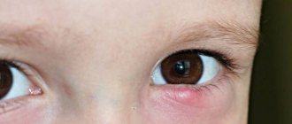
Milia
Milia are not an infectious pathology, and they are not contagious to others. As a rule, millet appears in women.
Milia do not cause discomfort or inconvenience; their main location is the upper and lower eyelids. Compared to papules, millet grass is a little more difficult to eliminate. To do this, you will need to understand the specifics of such formations.
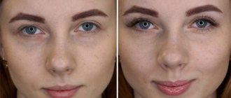
The shape of white spots on the eyelids is similar to small inclusions located on the skin near the eyes, eyelids (lower, upper), and in some cases on the lips. In rare cases, milia can be localized inside the eyelid. The structure of the formations is dense.
Millet, despite the fact that it looks harmless in appearance, is not so safe for life and health. Milia are formed as a result of a violation of the excretory function of the sebaceous glands located in the skin, as well as due to blockage of the pore mouth with keratinized epidermal cells. If the sebaceous glands function normally, excess fat is removed. If there is a violation, the follicle becomes enveloped in sebaceous fat. As a result, whiteheads appear.
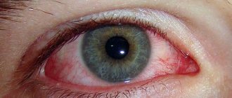
A photo of white spots on the eyelids is presented.
Find the right eyebrow pencil color
It would seem that choosing the color of an eyebrow pencil is quite simple. If you are a brunette, then just take any brown shade. In fact, you can't do this, otherwise you risk making your eyebrows unnaturally dark, which will ruin your entire makeup. With age, eyebrows tend to fade. The hairs become gray and gradually lighten, so an ordinary brown pencil will not work for you. Be sure to choose a shade that will naturally highlight your brows. You can replace it with gel or fondant. They are easier to apply, but at the same time they look more natural and last longer on the eyebrows.
Women's jeans: before you buy them, you need to pay attention to one detail
Why French children behave well: eight ways to raise them
A student at the Vietnam Police Academy shared how she takes care of her facial skin.
Why does pathology occur?
There are a number of different reasons for the occurrence of millet, but most often they are as follows:
- Insufficient hygiene, use of poorly selected care products.
- Hereditary factor.
- Vitamin deficiency, a deficiency of certain microelements in the body.
- Eating junk food, unhealthy diet.
- Diseases of the gastrointestinal tract.
- Reaction of small glands and skin to hormonal changes.
- Pathologies and disorders of the nervous system.
- Hormonal disorders.
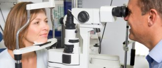
Treatment for milia will only be effective if the root cause is correctly identified. To identify the provoking factor, it is recommended to consult a dermatologist or cosmetologist.
After an accurate diagnosis, which involves conducting a series of studies, therapy is prescribed, which is individual for each patient and depends on the initial causes of the appearance of white spots on the eyelids. If you realize that you have white wen in front of your eyes, we advise you to read what to do with them in this article: https://sammedic.ru/427497a-na-vekah-glaz-belyie-jiroviki-prichinyi-poyavleniya-i-effektivnyie -methodology-treatment.
Varieties of milia
Formations that occur on the eyelids can be classified into two main types:
- Primary. They appear unexpectedly, and there are no compelling reasons for their occurrence.
- Secondary. They occur against the background of trauma, inflammation, and skin damage.
The key feature of such points is that they appear on the eyelids and eyelash area not individually, but in groups. They not only cause aesthetic discomfort, but also need to be removed.
Avoid glitter
Glitter is beautiful and stylish, but cosmetologists believe that they are more suitable for young girls. They tend to clog wrinkles and skin unevenness. Sequins attract unnecessary attention to them, highlight them, which visually makes you look older. If you are going to a restaurant or on a holiday, you can add a little glitter shadow to the center of the eyelids and the inner corner of the eye. But remember that their line should be thin and not too bright.
Millet therapy methods
As a rule, white spots on the eyelids disappear after normalizing the diet and changing cosmetics and care products. In addition, it is important to replenish the lack of minerals and vitamins in the body, which can be done by taking appropriate vitamin complexes. This simple treatment leads to the disappearance of formations within two weeks.
In more severe cases, hardware therapy is indicated. With this approach, white spots on the eyelids are eliminated mechanically. It is important to note that the need for hardware removal should be determined by a doctor after careful diagnostic measures.
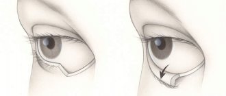
Removal methods
Currently, there are several methods for hardware removal, the choice of one of them is carried out taking into account the situation:
- Electrocoagulation. In this case, excision of the tumor occurs by exposing it to high-frequency current. This procedure is absolutely painless and takes only a few minutes. After removing the milia, a small scar remains on the skin, disappearing within a few days. Thus, electrocoagulation is a modern and effective method for completely removing a growth on the eyelid.
- Laser removal. This procedure is very similar to electrocoagulation, but in this case the cleansing of the dermis is more intense, and the procedure itself is less traumatic and painful. There are no scars or marks left on the skin after laser removal of milia.
- Curettage. This is a more radical method of removing dots. The procedure involves carefully opening the milia and removing the contents with a curette. This procedure is the most traumatic; it leaves red spots and scars on the skin. Curettage is rarely used to remove growths on the eyelids.
You can remove a stain on the eyelid yourself at home, but this will require caution and some skill.
Algorithm of actions
Cosmetologists recommend adhering to the following algorithm of actions:
- It is necessary to clean the skin with an alcohol solution.
- Steam the dermis to open the pores.
- Squeeze out the contents of the milia with your fingers. In such cases, cosmetologists use a special needle for puncture, but doing this yourself is prohibited.
- After removing the formation, it is important to disinfect the resulting wound.
If the milia has reached the size of a wen, self-removal may be ineffective or even dangerous. You should contact a cosmetologist.
Secondary pathologies do not require removal; they can be eliminated by treating the affected skin with salicylic-zinc paste, salicylic ointment, and badyaga.
However, there may be other causes of spots on the eyelids.
Remember that makeup should look natural
Bright makeup is beautiful, but not always appropriate. Remember that moderation is your best friend. You can wear a lot of makeup if you are going to a special event, but you don’t need to do it every day. Makeup should look natural. It is enough to even out your face tone with foundation, cover up skin imperfections with concealer, and then highlight your eyebrows, eyes and lips. It is not necessary to use bright blush, eye shadow or highlighter every day. It's better to save them for a special occasion.
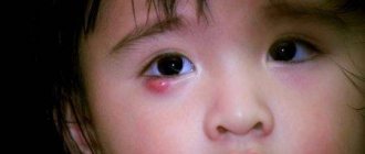
Xanthelasma
In some cases, the formation may not be a simple milia, but a xanthelasma. In this situation, a spot on the eyelid may indicate the presence of some pathological changes in the body.
Xanthelasma of the eyelids is a disease in which yellow, white, and sometimes orange spots appear on the skin of the eyelids. Along with pathology, liver dysfunction, hypertension, and diabetes mellitus are often diagnosed.
Why does a white spot form on the lower eyelid of the eye? Let's look further.
Many diseases can be diagnosed by the eyes.
An attentive observer not only sees in the eyes the mind, the level of a person’s vitality and the emotions that he is engulfed in, but also receives rich information about the state of a person’s health at a given moment. So, for example, the eyes can indicate the following: Cloudy eyes - Suspicion of an infectious disease Reddish eyes - Colds, conjunctivitis Yellow coloration of the sclera - impaired liver function, hepatitis Shiny eyes - hyperfunction of the thyroid gland Red veins in the eyes - Venous stagnation Sunken eyes - exhaustion The look seems dull - weakness, heart disease. A shiny look - fever, excitement. look - extreme weakness A blank look - intestinal diseases, a mortal danger to the body. Pearlescent coloring of the eyes - tuberculosis or anemia Watery eyes - painful emotions. Trembling eyes - in most cases, an indicator of multiple sclerosis. Inability to cry - lack of vitamin A Restless eye movements - fear, neurasthenia Frequent blinking - Vegetative - vascular dystonia; with a strong manifestation of the symptom - hyperthyroidism Rare blinking - hypothyroidism Upper eyelids: Sunken eyelids - high consumption of nervous energy, strong need for sleep Double fold on the upper eyelid - weakness of the connective tissue, diaphragmatic hernia Skin hanging over the eyelid - Remgeld syndrome (syn.: gastro-cardiac syndrome) Swelling of the upper eyelid - disturbance of cardiac activity Drooping eyelid - Heavy load on the heart Drooping eyelid - mineral imbalance, exhaustion, anemia, hypotension Rounded convex areas of yellow -brown color on the upper eyelid - imbalance of hormonal balance and cholesterol balance in the body Lower eyelids: Sunken lower eyelids - nervous exhaustion Swelling of the lower eyelid - impaired kidney function, stagnation of urine The swelling has a pinkish -blue color - impaired bladder function The swelling has a gray-green color - excess uric acid The swelling has a waxy color - heart failure The sunken area of the eyelid has a bluish color - lack of iron in the body The sunken area of the eyelid has darkened - neurasthenia The lower eyelid has a brown color - anemia Plaque on the eyelids - metabolic disorders, hypofunction of the pancreas, hypofunction of the thyroid gland Pigmentation of the lower eyelid - hemorrhoids: possibly internal Drooping of the outer corner of the eyelid - depression, adynamia Absence or loss of eyelashes - insufficient function of the gonads, toxicosis. The condition of the skin directly under the eyelid can determine the condition of the bladder. An area of skin 1 cm below the eyelid indicates the condition of the kidneys. “Bags” under the eyes (in this area) indicate congestion in the kidneys. If the “bags” are located directly on the lower eyelids, this most likely indicates diseases of the adrenal glands and endocrine glands. Large bags that drop even lower indicate the presence of intestinal diseases. The area around the eyes has a yellowish color - diseases of the liver and gall bladder. The area around the eyes has “failed” and has a brownish-black color - neurasthenia, weakness of the nervous system, insomnia. Circulatory disorders The area around the eyes has a bluish color - internal bleeding; in children - infection with worms The area around the eyes has a brownish color - Liver diseases, constipation The area around the eyes has a pale pink color - diseases of the bladder, prostate gland Circles under the eyes - exhaustion of the body Wrinkles in the corners of the eyes or under the eyes - in young people: dysfunction connective tissue
Source: https://www.burmitskiy.com/
Causes of xanthelasma
The nature of the appearance of white or yellow spots on the skin of the eyelids has not yet been clarified by experts. It is generally accepted that the main reasons for the appearance of stains are as follows:
- Disturbances in lipid metabolism.
- Metabolic disorders.
- Obesity.
- Diabetes.
- Pathological changes in the liver.
- Disorders of the pancreas.
It is worth noting that formations are sometimes single in nature, and sometimes appear in groups. Most often they are localized in the corner of the upper or lower eyelid.
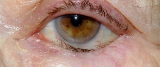
There is a white spot inside the eyelid.
Externally, they may have the form of a white or yellow plaque. The contour of the formation and its surface may be uneven or smooth. When palpated, the spot is found to be quite soft.
It is important to note that xanthelasma is a benign formation that never becomes malignant. The spot itself does not cause any discomfort or pain. Some experts tend to associate the appearance of white or straw-colored spots with disorders in lipid metabolism, but in some cases clinical studies do not confirm this reason. Xanthelasma occurs, as a rule, in older people, most often in females. In fact, the pathology is more of a cosmetic defect than a threat to health.
The size of white or yellow spots on the eyelids can vary from the size of a pea to the diameter of a bean.
Use a pearlescent, not a frosty highlighter
The frosty shade of the highlighter will definitely highlight all your fine lines and wrinkles, so it is better to avoid using it. In addition, such a product itself looks unnatural because it does not blend with the skin. To sculpt the face of women over 40 years old, cosmetologists advise using cream and pearl shades of highlighter. They will highlight your cheekbones and make your skin more radiant, without making you look older.
Diagnosis of yellow spots
Diagnosis of xanthelasma on the skin of the eyelids is carried out by external examination using a glass slide. A glass slide is pressed onto the plaque, which allows you to clearly examine the structure, shape, and color of the formation. White spots are characterized by certain signs, to identify which a laboratory test of a blood sample is prescribed. Based on the results obtained, one can judge the level of cholesterol, which reflects the state of lipid metabolism.
Diagnosis of the disease is carried out by an endocrinologist and a dermatologist, their task is to identify:
- The root causes of white spots.
- Definitions of xanthelasma or tumor formation.
- Determining the absence or presence of atherosclerosis or diabetes mellitus.