Retinopathy is an eye pathology characterized by damage to the retinal vessels. Such lesions cause disruption of the blood supply to the organ of vision, its dystrophy, which leads to atrophy of the optic nerve and blindness. The course of retinopathy is absolutely painless, with the appearance of opaque spots (scotomas) and a veil in the field of vision. In addition, there is a progressive deterioration of vision. If retinopathy is detected, consultation with specialists (ophthalmologist, neurologist, endocrinologist, cardiologist) is required. Diagnosis requires the following ophthalmological studies: checking visual acuity and its fields, performing biomicroscopy, ophthalmoscopy, fluorescein angiography, electrophysiological studies, and ultrasound of the eye. Treatment of retinopathy is carried out simultaneously with compensation for the disease that caused it. Vasodilators, anticoagulants, and vitamins are prescribed. Laser coagulation of the retina is performed, and hyperbaric oxygenation is recommended.
What is retinal dystrophy?
The basis of peripheral retinal dystrophy
eyes (
PCRD
) is a violation of the blood supply caused by various reasons:
- Progressive myopia of varying degrees
- Injuries and contusions of the eyeball
- Head injuries
- Hereditary predisposition.
The risk group for peripheral retinal dystrophy is quite wide and has no age restrictions.
The factors for the development of PCRD can be different, but the result of the disease is the same - decreased blood circulation, impaired delivery of oxygen and nutrients to the retina of the eye, which leads to a change in its density. Thinned areas of the retina can literally “tear” under the influence of any physical activity, sports, or any vigorous activity that a person engages in, not knowing about his asymptomatic disease. Many Patients who were diagnosed with peripheral retinal dystrophy in our clinic did not even suspect that they had such an insidious disease!
Ignorance can continue until retinal detachment occurs and a sharp decrease in visual acuity, up to its complete loss in advanced cases. An additional difficulty in detecting this “silent enemy” is the inaccessibility of the peripheral zone of the retina, which is located deep in the eye and is not visible during a standard ophthalmological examination.
Symptoms
A typical clinical picture of retinal pigment abiotrophy includes the following symptoms:
- hemeralopia (“night” or “night” blindness). Since, as shown above, during abiotrophy, mainly those receptors that are responsible for vision in low light are affected, the early appearance of the symptom of hemeralopia is quite natural. Often its true meaning becomes clear only after many years of development of the process, when changes in the retina are clearly visualized during ophthalmoscopy and a final diagnosis is established;
- gradual narrowing of the field of view from the periphery to the center (in accordance with the typical direction of the process). If at the beginning of the disease this disorder can be established only by a special study of the visual fields (perimetry), then in the later stages visual functions are limited to only a small irregular island in the center of vision, which, like a keyhole, allows a person to see only straight ahead (the so-called tunnel vision) with complete absence of lateral vision;
- decreased visual acuity and color perception ability (in later stages, when the degenerative process spreads to the central region of the retina and destroys the corresponding cone receptors);
- finally, with a non-stop progressing process, at one speed or another, the outcome of the disease is total blindness.
Types of retinal dystrophy
The disease can have benign (peripheral chorioretinal retinal dystrophies) and low-quality (peripheral vitreochorioretinal retinal dystrophies) variants of the course.
If the disease is benign, laser treatment is not required because the risk is negligible. In order to prevent the disease from passing into a low-quality form in peripheral chorioretinal retinal dystrophies, patients are prescribed outpatient monitoring.
Peripheral vitreochorioretinal dystrophy of the retina is dangerous due to ruptures of thin areas and retinal detachments, so in this case laser coagulation of the affected areas is performed - a quick and effective laser operation to relieve the disease.
Diagnostics
The first and mandatory components of the examination for suspected taperetinal abiotrophy are visometry (measurement of visual acuity) and perimetry (examination of visual fields). A visual examination of the fundus structures is performed using a special lens. With a significant duration and intensity of the process, its diagnostic signs are the presence of the so-called. bone bodies (foci of dystrophic cell death), a decrease in the caliber of the retinal veins, blanching of the optic nerve disc extending into the retina.
An informative and diagnostically important method for objective assessment of the functional status of the retina is an electrophysiological study. The preservation of vision and the ability to navigate in darkness are studied. Since the hereditary nature of pigmentary abiotrophy is known and proven, consultation with a medical geneticist will be required; It is also advisable and desirable to perform an ophthalmological examination of the patient’s blood relatives, especially younger ones, to identify the process at the earliest stages.
Treatment of peripheral retinal dystrophy
Laser coagulation is a modern high-precision operation without incisions or punctures in the eyeball. It is performed in a specially equipped room for laser operations under local drip anesthesia. The patient does not experience any discomfort or pain. The entire course of the operation is controlled by a laser surgeon and computer equipment.
The laser coagulation method for the treatment of peripheral retinal dystrophy has long proven itself in Russia and throughout the world. In our clinic, these operations are performed plannedly by laser surgeons.
Laser coagulation technique in the treatment of peripheral vitreochorioretinal retinal dystrophy at the Eye Clinic of Dr. Belikova:
- The Patient's pupil is dilated with medication using drops
- After dilation of the pupil, anesthetic drops are instilled
- The laser device is brought to the Patient's eye
- Coagulation lasts from 5 to 30 minutes depending on the volume of treatment
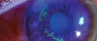
- The patient does not feel pain during the procedure, but sees green flashes - these are laser pulses that coagulate (connect) the retina.
After coagulation there may be lacrimation, the patient may feel a feeling of a foreign body - these phenomena soon pass. For 7 days after laser coagulation, physical activity, sports, and air travel are not recommended. The goal of prophylactic laser coagulation is to reduce the risk of retinal detachment, not to improve vision.
In recent years, indications for preventive laser coagulation have expanded in connection with excimer laser correction using the LASIK method. During this procedure, intraocular pressure can briefly increase to 45–60 mmHg, which requires reliable fixation of the retina and the absence of factors predisposing to its detachment.
For local retinal breaks, the laser beam acts on the edges of the breaks, fusing them with the deep layers located under the retina. To prevent ruptures, laser exposure is aimed at the edges of thinned retinal tissue.
Causes
Inheritance of pathology occurs regardless of the gender of children and parents. It is predominantly transmitted in an autosomal recessive manner, that is, it affects the offspring of parents who are carriers of the disease, but not in 100% of cases. Recent research by geneticists has revealed that gene pathology can also be transmitted through a dominant pathway.
In the case of a dominant transmission route, the disease is milder and rarely leads to disability. Most of the receptor cells in the macula (not the apex of the macula) remain functional, so the pathology has a minimum of manifestations. People retain their ability to work; visual acuity even allows them to drive vehicles.
The main cause of macular cell degeneration is energy deficiency. Due to a gene defect, a defective protein is synthesized, which is involved in the transport of ATP molecules through the membrane of the macula cells - the center of the retina, where the color, graphic image is focused. The macula does not have blood vessels. ATP transport proteins from the adjacent choroid (choroid) take part in the nutrition of cone cells. It is these proteins that transport ATP molecules across the membrane into photoreceptor cells.
Photoreceptor rhodopsin under normal conditions absorbs photons of light, transforming into opsin and trans-retinal. Then, under the influence of ATP energy brought by carrier proteins, trans-retinal is converted into retinal and combines with opsin. This is the process of rhodopsin restoration. A hereditary gene mutation causes the synthesis of an inferior carrier protein. As a result, the restoration of rhodopsin is disrupted, and trans-retinal accumulates. It is transformed into lipofuscin, which has a toxic effect directly on cone cells.
Primary retinopathy
The cause of primary retinopathy is still under study, so in most cases they are considered idiopathic.
Central serous retinopathy . Also called central serous retinitis and idiopathic macular detachment, it typically affects young men aged 20-40 years without serious systemic diseases. Typically, patients note recent emotional turmoil and frequent attacks of migraine headaches. Retinal damage in this pathology is unilateral.
Among the signs of the disease: micropsia (visible objects seem smaller than their actual size), scotomas, decreased visual acuity, narrowing of its fields. An important factor in differential diagnosis is an increase in visual acuity when corrected with glasses with small positive lenses.
Pathomorphological signs of central serous retinopathy: serous detachment in the area of the macula of the pigment epithelium, determined by ophthalmoscopic examination as a limited round or oval protrusion, darker in color than the rest of the retinal tissue. In addition, there is no foveal reflex (there is no light strip around the central fovea on the retina), and grayish or yellowish precipitates appear.
Treatment of central serous retinopathy requires laser coagulation of the retina. Drugs are prescribed to strengthen the walls of blood vessels, reduce retinal edema, and improve microcirculation; oxygen barotherapy is performed. Timely treatment of the disease in 80% of cases gives positive results, the pathological process can be stopped - preventing retinal detachment, improving vision to its original state.
Multifocal acute posterior pigment epitheliopathy . This type of retinopathy can be unilateral or bilateral. It is characterized by the formation of multiple flat subretinal lesions in the retinal tissue of a grayish-white color. Their reverse development leads to the appearance of areas of depigmentation. Examination of the fundus in this case reveals perivascular edema of the vessels in the periphery of the retina, as well as swelling of the optic disc, dilatation and tortuosity of the veins.
The majority of patients with this disease have vitreous opacities, iridocyclitis and episcleritis. An early impairment of central vision occurs due to the appearance of central or paracentral scotomas.
To treat the disease, conservative therapy is sufficient. It includes the prescription of vitamin and vasodilator drugs (Vinpocetine, Pentoxifylline, etc.). Angioprotectors (solcoseryl) are also prescribed for instillation into the eye, and retrobulbar steroid injections are performed. Hyperbaric oxygen therapy is recommended. Active, timely treatment leads to positive results – the pathology reverses.
External exudative retinopathy . Also called Coats disease and external exudative retinitis, the disease occurs primarily in young men, usually affecting one eye. It is characterized by the accumulation of exudate under the retinal vessels, cholesterol crystals, and hemorrhages. Pathological changes are often detected in the periphery; the macular zone is rarely involved in the process. Retinal angiography often reveals arteriovenous shunts and multiple microaneurysms.
This form of retinopathy is characterized by a slowly progressive course. For its treatment, laser coagulation of the retina and a hyperbaric oxygenation procedure are prescribed. If the process has gone far enough, retinal detachment is possible, which requires urgent surgical intervention. In addition, the prognosis may be aggravated by the occurrence of glaucoma and the development of iridocyclitis.
Secondary retinopathy
Diabetic retinopathy . The disease occurs as an ophthalmological complication of type 1 or type 2 diabetes mellitus. The main reasons for its occurrence are: duration of diabetes, lack of compensation for the underlying disease, high blood sugar, nephropathy, hypertension, obesity, anemia, hyperlipidemia.
Diabetic retinopathy is the most common and serious complication of diabetes, the main cause of low vision in patients and their blindness. The process of development of the disease is divided into stages - diabetic angiopathy, diabetic retinopathy, proliferating diabetic retinopathy. The first stages proceed similarly to the stages of hypertensive retinopathy and atherosclerotic retinopathy. In the early period of the stage of proliferating diabetic retinopathy, neovascularization of the retinal tissue begins with the formation of pathological vessels. At a later stage, they grow and become the cause of recurrent hemorrhages into the vitreous body, as well as the spread of glial tissue, which provokes the occurrence of traction. The tension of the vitreous fibers and its deformation cause retinal detachment, which causes incurable blindness.
Symptoms of the initial stages of diabetic retinopathy boil down to a steady decrease in visual acuity, the appearance of floating spots and veils before the eyes, which disappear and reappear. This makes it difficult to read and do work at close range or with small parts. At a later stage, vision disappears completely.
To diagnose diabetic retinopathy, ophthalmoscopy is prescribed. The study involves dilating the pupil, which allows you to examine the fundus in detail and identify characteristic changes. To determine the functional state of the retinal periphery, the perimetry method is used. Ultrasound of the eyes helps to identify areas of hemorrhage, thickening of the tissue, and scars. Electroretinography is also prescribed to assess the viability of various areas of the retina. Detailed study data can be obtained by angiography of retinal vessels or laser scanning tomography. Mandatory studies for this form of retinopathy also include the following methods: visometry, biomicroscopy, diaphanoscopy, CCFM, etc.
To treat diabetic retinopathy, in addition to an ophthalmologist, a diabetes specialist (endocrinologist) is also involved. It carries out the necessary control of blood glucose, ensures timely intake of sugar-lowering drugs, vitamins, angioprotectors, antiplatelet agents, antioxidants, as well as medications that activate microcirculation. For retinal tears, laser photocoagulation is used. In case of large-scale detachment or changes in the vitreous body, surgical intervention is indicated - vitrectomy.
Diabetic retinopathy is often complicated by hemophthalmos; cataracts may develop, opacities of the vitreous body and its cicatricial changes may occur. Retinal detachment and blindness are also possible.
Hypertensive retinopathy . This form is pathogenetically associated with arterial hypertension, renal failure, and toxicosis of pregnant women. It is caused by spasm of the retinal arterioles, which entails elastofibrosis or hyalinosis of the vascular walls. The severity of hypertensive retinopathy directly depends on the duration of hypertension and its clinical degree.
The process of hypertensive retinopathy is usually divided into 4 stages:
- Hypertensive angiopathy of the retina. It occurs with reversible changes that affect the functionality of arterioles and venules of the retina.
- Hypertensive angiosclerosis. It is characterized by organic damage to the retinal vessels, which is caused by sclerotic thickening of the vessel walls and a decrease in their transparency.
- Hypertensive retinopathy. It is characterized by the presence of changes in the retinal tissue of a focal type (hemorrhages, lipid deposition, plasmorrhagia, areas of protein exudate, areas of ischemic infarction), and partial hemophthalmos is possible. At this stage, patients note a decrease in visual acuity, and scotomas appear in the field of vision. All signs regress with antihypertensive therapy.
- Hypertensive neuroretinopathy. At this stage, angiopathy, angiosclerosis, and retinopathy are complicated by swelling of the optic disc, exudations and areas of retinal detachment occur. This condition occurs much more often with malignant hypertension, as well as a persistent increase in blood pressure against the background of kidney disease. The outcome of the stage of hypertensive neuroretinopathy is often atrophy of the optic nerve, which ends in irreversible blindness.
Diagnosis of hypertensive retinopathy helps to consult two specialists - an ophthalmologist and a cardiologist. Methods of ophthalmological examination for this disease are: ophthalmoscopy, fluorescein angiography. The clinical picture demonstrates a change in the width of the retinal vessels, their total or partial obliteration, subretinal exudation, the presence of the Salus-Hun symptom (the vein “goes” into the deep layers of the retina due to the fact that a tense and compacted artery presses on it in the projection of their decussation) and etc.
Therapy of hypertensive retinopathy requires compensation for the underlying disease—arterial hypertension. For this, anticoagulants and vitamins are prescribed, oxygen barotherapy is performed, and laser coagulation of the retina is performed.
Among the complications of hypertensive retinopathy are recurrent hemophthalmos and retinal vein thrombosis. The outcome of hypertensive retinopathy in the absence of timely treatment can be a serious decrease in vision, and in some cases even blindness. Retinopathy complicates the course of the underlying disease and pregnancy, which can be terminated for medical reasons.
Atherosclerotic retinopathy . The main reason for its occurrence is systemic atherosclerosis. Changes in the retina that occur at the stages of angiopathy and angiosclerosis repeat those with hypertensive retinopathy; At the stage of neuroretinopathy, small capillary hemorrhages appear, deposition of crystalline exudate is observed along the veins, and the optic disc becomes pale.
Ophthalmological diagnosis of the disease includes the following methods: direct and indirect ophthalmoscopy, fluorescein angiography. There is no specific treatment for the disease. Treatment and compensation for the underlying disease are of primary importance. For this purpose, antiplatelet agents, antisclerotic drugs, vasodilators, angioprotectors, and diuretics are prescribed. In the stage of neuroretinopathy, courses of electrophoresis, which are performed with proteolytic enzymes, are effective.
Among the complications of atherosclerotic retinopathy, the most dangerous are: occlusion of the retinal arteries, optic nerve atrophy, which can cause irreversible blindness.
Retinopathy in blood diseases . Many pathologies of the blood system can cause retinopathy, these include: anemia, leukemia, polycythemia, Waldenström's macroglobulinemia, myeloma, etc.
Retinopathy caused by each disease differs in its specific ophthalmoscopic picture. For example, the form caused by polycythemia reveals during examination the dark red color of the retinal veins, with a cyanotic tint of the fundus. Thrombosis of the retinal veins and swelling of the optic disc often occur.
With anemia, on the contrary, the fundus of the eye is pale against the background of dilated retinal vessels, with the same color of the arteries and the size of the vascular bed. This form may be accompanied by extraretinal and subretinal hemorrhages, exudative retinal detachment.
In the case of leukemia, tortuosity of the veins in the fundus is observed, diffuse retinal edema, accumulation of foci of exudate, and hemorrhages are detected.
With Waldenström's macroglobulinemia, as with multiple myeloma, due to paraproteinemia, dysproteinemia, and blood thickening, the retinal vessels dilate, thrombotic occlusions occur, microaneurysms and hemorrhages in the retina develop.
To treat this kind of retinopathy, treatment of the underlying disease and laser photocoagulation of the retina are necessary. The prognosis for the outcome of the disease is always cautious.
Traumatic retinopathy . It develops against the background of sudden and sharp compression of the chest, which causes a spasm of the arterioles, provoking the onset of retinal hypoxia and the release of transudate into its area. In the immediate post-traumatic period, hemorrhages in the retina develop and organic changes occur. This form of retinopathy often causes optic nerve atrophy.
With an eye contusion, a complication called Berlin's opacities may occur. It is associated with subchoroidal hemorrhage, accompanied by swelling of the deep layers of the retina and penetration of exudate into the space under the retina.
Treatment of such retinopathy consists of prescribing vitamin preparations, eliminating tissue hypoxia, and recommending hyperbaric oxygenation and other procedures.
Retinopathy of prematurity . This form of the disease is special because it occurs exclusively in premature infants. The reason for this is underdevelopment of the retina in children born before their due time. For the complete formation of the retina and the maturation of the remaining ocular structures, small patients need absolute visual rest, as well as provision of oxygen-free local tissue respiration (glycolysis). However, for full nursing of premature infants, additional oxygenation is required, which helps stimulate metabolic processes in vital organs. Its provision causes inhibition of glycolysis in the retina and choroid, provoking changes in the fundus.
Infants born at or before 31 weeks of gestation are especially at risk for retinopathy of prematurity. Also, their body weight plays an important role; if it is below 1500 grams, the risk of the disease is higher, especially if the condition is aggravated by a general unstable condition and prolonged use of oxygen therapy.
Newborns at risk for this form of retinopathy must be examined by an ophthalmologist 3-4 weeks after birth. Then, examinations by an ophthalmologist must be carried out every 2 weeks until the retina is fully formed. Delayed complications of retinopathy of prematurity include: myopia, strabismus, amblyopia, low vision, glaucoma, retinal detachment.
Often this form of retinopathy ends in complete regression of the pathological process, so treatment is started only if the condition clearly worsens. Basically, the tactics of dynamic observation are chosen. Treatment of the disease consists of laser coagulation of the retina or cryoretinopexy. If the measures taken are not enough, scleroplasty and vitrectomy are prescribed.
Causes of the disease
The exact cause of the pathology has not been established, but experts are inclined to two versions. On the one hand, pigmentary degeneration of the retina is caused by sclerosis of the capillary network. The second reason is endocrinological disorders of the body and insufficient synthesis of retinol (vitamin A). Insufficient nutrition of the retina leads to the destruction of receptor cells - rods and cones, which provide color perception and visual acuity in the dark.