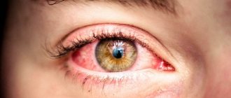
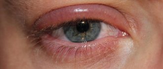
Often a person discovers a symptom of red eyes, the causes of which cannot always be determined independently. The visual organ can become red both with banal fatigue and with eye diseases and pathologies of internal organs.
Description of the symptom of hyperemia of the eyeballs
Eyes turn red for several reasons. If a person does not have the characteristic symptoms of hyperemia, the vessels located in the white of the eye are not visible. Hyperemia is often accompanied by rupture of blood vessels - the proteins partially or completely become red. There is a clear answer to the question why the white of the eye turns red - this happens under the influence of a number of physiological or external factors.
Why do my eyes turn red?
The color of the white of the eye may change due to the expansion of small and large vessels (veins and capillaries) - the blood flowing through them into the organ saturates the eyes with oxygen. As blood flow accelerates, the vascular lumen increases, the walls become thinner and the ophthalmic vein becomes less elastic, which increases the risk of rupture.
The eyes may not be completely red - the location of the spot often depends on a number of reasons. In most cases, red eye syndrome occurs when there is a sudden change in the speed of blood flow.
Environmental factors
Physiological factors that cause the eyes to turn red include direct contact with toxic chemicals and mechanical damage. Harmful substances released by chemicals into the air penetrate the membrane of the eye, accelerating blood flow. This reaction cannot be corrected; the body tries to remove toxins from the surface of the organ as quickly as possible.
Foreign bodies that fall on the surface of the eye white, when in contact with the eyelid, can leave microscopic scratches that provoke irritation. Most often, when the external irritant is eliminated, the redness of the protein goes away on its own and does not require treatment.
Your eyes may get tired when working at a computer for a long time. Hyperemia may be associated with the work activity of a certain category of citizens - most often the pathology is diagnosed in electric welders after ultraviolet radiation. After severe mechanical trauma, the integrity of the cornea and conjunctiva is compromised, the organ swells, and the risk of harmful bacteria and other microorganisms penetrating into the damaged tissue of the organ increases.
Physiological reasons
The whites of the eyes become red for a number of physiological reasons. The body may react inadequately to the ingestion of food allergens, to surges in blood pressure and various deviations from the usual indicators of blood clotting. With prolonged muscle strain, the blood vessels cannot withstand blood pressure and burst. Depending on the physiological causes, hyperemia may not require specific treatment and goes away on its own after the irritating factor is eliminated.
Physiological factors include fatigue, psycho-emotional stress and irritation from contact lenses. Redness of the eye sclera can be observed with hypertension, musculoskeletal disorders, diabetes mellitus and Sjögren's syndrome. An overdose of any medications is accompanied by eye hyperemia.
Common Causes
Most often, redness is caused by excessive fatigue of the body and eye muscles. In the absence of regular sleep, the eyes practically do not rest and begin to stick together, which leads to inflammation. It is worth paying attention to the symptoms of the disease. When a red eye hurts, there may be chronic inflammation of the eyelids or blepharitis (staph infection). A hypertensive crisis can also cause redness due to vascular damage from increased venous pressure.
Diseases such as conjunctivitis are accompanied by redness not only in the morning, but also on an ongoing basis, depending on the type of inflammation of the mucous membrane. Women often experience redness of the white body due to exposure to makeup particles that remain in the eye after evening care.
| Bad habits can have a negative impact on the body as a whole: drinking alcohol leads to increased intracranial pressure, and cigarette smoke irritates the mucous membrane of the eye. |
What reasons can cause this symptom?
An adult's eyes turn red for a number of reasons. An ophthalmologist can determine the exact one after a detailed interview with the patient, collecting an anamnesis and conducting a number of additional studies. If redness of the eyes occurs, the causes and predisposing factors may be as follows:
- pathologies of an infectious nature;
- allergic manifestations;
- direct contact with chemicals;
- consequences of mechanical trauma;
- eye diseases accompanied by inflammation;
- sudden changes in temperature;
- neglect of the rules of personal hygiene;
- dry eye syndrome;
- alcohol abuse;
- rupture of a large or small vessel;
- blood pressure surges;
- diabetes.
Chronic fatigue and systematic sleep disturbances are considered the causes of hyperemia.
Allergic reaction
The concept of “red eye” applies to allergies. Organs are sensitive and easily susceptible to irritating factors. Allergens that can provoke hyperemia are often found in decorative cosmetics, household chemicals, medications, pollen, pet hair and dust. The latter contains a large number of foreign bodies and spores of moldy fungi, which, when they come into contact with the membrane of the eye, cause an allergic reaction. In rare cases, a person may develop an allergy to contact lenses or cleaners.
Xerophthalmia – dry eye syndrome
Redness of the eyes due to xerostomia or dry eye syndrome is often accompanied by dryness of the eye membrane, which appears against the background of impaired secretion. The conjunctiva and cornea partially or completely dry out, so the natural protection of the eyes from pathogenic microorganisms and other harmful factors is reduced. The composition of tears may change; they are no longer evenly distributed over the surface of the eyes. This leads to blurred vision and the development of pathology. The absence of a tear film makes the cornea more vulnerable to atmospheric oxygen.
Alcohol
The whites of the eyes turn pink with systematic alcohol abuse. This occurs against the background of dilation and rupture of blood vessels not only in the eyes, but also in the skin and most internal organs. A critical dose of alcohol contributes to the release of norepinephrine and renin into the blood, which increase blood pressure. In chronic alcoholics, a capillary network appears on the whites of the eyes, and the risk of developing hemorrhage in the eyeball increases.
Eye strain. Computer Eye Syndrome
A person complains of visual fatigue, the white of the eye changes color from pink to bright red, and visual acuity decreases. The patient may be haunted by the sensation of a foreign body in the eye. If your eyes turn red when exposed to a computer, regardless of lighting or time period, this may indicate the development of a number of pathologies, such as myopia. In patients suffering from vegetative-vascular dystonia, the appearance of visual computer syndrome increases. Red squirrels may be observed with this disorder.
Tired eyes
The medical term “asthenopia” means fatigue. Red pupils appear in the late afternoon. The condition is accompanied by a symptom complex, which, in addition to hyperemia, includes dryness, impaired visual acuity, pain in the eyes and temples, dark circles under the eyes. Asthenopia develops for a number of reasons, which include poor lighting, prolonged reading or working at the computer, incorrectly selected glasses or contact lenses, vegetative-vascular dystonia, intraocular or intracranial pressure, osteochondrosis.
Conjunctivitis
Constantly red eyes indicate the development of a form of conjunctivitis. The nature of its occurrence may be different:
- allergic;
- herpetic;
- bacterial;
- viral.
In addition to redness of the white, conjunctivitis is accompanied by irritation, burning sensation, increased lacrimation, photophobia, eye fatigue and swelling of the eyelids. One of the common causes of the disease is failure to comply with personal hygiene rules when using other people's towels, scarves and rare hand washing. The pathology is transmitted not only by contact, but also by airborne droplets (viral type).
The vessel burst
A common cause of redness of the whites of the eyes is the rupture of a small (capillary) or large (vein) vessel. Against the background of predisposing factors (jumps in blood pressure, psycho-emotional stress, intraocular and intracranial pressure), the vascular wall becomes thinner, which loses its elasticity and becomes more vulnerable. With a physiological acceleration of blood flow, the vessel bursts and blood enters the protein. Hemorrhage may be minor or extensive. In the second type, the eye is completely filled with blood.
Irritation of mucous membranes. External stimuli
Red proteins appear with the systematic use of low-quality decorative cosmetics and frequent interaction with chemicals. If foreign bodies get into the eye (sand, smoke, dust), redness may occur. Even minor eye injuries lead to damage to the surface of the eye and rupture of blood vessels. External irritants include plant pollen and feather particles.
Glaucoma
Glaucoma is a common cause of red eyes. It appears when there is increased pressure inside the organ. Late diagnosis and treatment of pathology increases the risk of complete loss of vision. An acute attack of the disease, in addition to redness of the protein, is accompanied by uncontrolled vomiting, photophobia, decreased heart rate and attacks of nausea. Hyperemia is considered the main sign of glaucoma; the disease can develop slowly, and auxiliary symptoms do not appear immediately.
Inflammation of the cornea
Red pupils in humans occur due to inflammation of the cornea. The pathology is accompanied by profuse tearing, decreased transparency of the cornea and severe swelling. The inflammatory process in the cornea can be caused by viruses or colds, pathogenic fungi or mechanical injuries to the visual organ. Self-detection of pathology is impossible. In order to differentiate the disease, you need to make an appointment with an ophthalmologist and undergo a number of additional procedures that allow you to determine the disease with 100% accuracy.
Hypertension
Red proteins appear in people suffering from hypertension (or hypertension). This is a common disease that can affect the condition of the retina. Hypertension may be accompanied by retinal angiopathy - in this case, the tortuosity and branching of the veins is increased, small capillaries are clearly visible. The pathology is characterized by pinpoint hemorrhages. Most often, angiopathy manifests itself at the initial stage of hypertension and is easily treatable.
Angiopathy of the 2nd degree with arterial hypotension develops when the vascular lumen narrows and thickens. This can cause blockage of capillaries or veins. When the disease occurs, the patient experiences peculiar spots before the eyes, pain and a burning sensation. The severe stage of hypertension is accompanied by eye bleeding.
Injury. Chemical and mechanical injuries
Many people are interested in the question of why their eyes hurt and are red after a blow. The cause of hyperemia is mechanical trauma, which could provoke rupture of a blood vessel.
Red eyes after a shower are observed when water enters the organ. The liquid causes a burning sensation and redness.
Chemical injuries include any burns caused by contact with a toxic substance on the surface of the eye.
Diabetes
Red proteins in people with diabetes appear due to regular fluctuations in blood glucose levels. Diabetics experience pathologies of the hematopoietic systems, in which the elasticity of the walls decreases. Angiopathy that develops in diabetes mellitus is accompanied by destruction of the retina and its clouding. The sclera turns red, and the risk of developing hemorrhages (point or extensive) increases.
Experts recommend that people with diabetes regularly monitor their cholesterol and blood sugar levels.
How to remove signs of eye redness?
You can relieve the symptoms of red eyes in different ways:
- natural (rest, functional rest of the visual apparatus);
- folk treatment;
- medicines.
Sometimes it’s enough to just relax and limit the time you spend watching TV and computer. In mild cases, traditional medicine (anti-inflammatory compresses, lotions) also helps.
Quickly removing red eyes is treating the symptom, not the cause of the disease. Therefore, drops for red eyes are only an aid. Primary therapy should be aimed at the cause.
Drug treatment for red eye syndrome consists of medications that will help get rid of the causes of red eye:
- Fatigue of the visual apparatus. For redness caused by fatigue, you need to drip vasoconstrictor moisturizing drops “Vizin”, “Okumetil”, “Systane” into the eyes.
- The most effective moisturizing drops against dryness and redness of the eyes: - Cationorm - a unique cationic emulsion designed to treat intense manifestations of dry eye syndrome. These drops are suitable for you if you experience discomfort in your eyes not only during the day, but also in the morning. Cationorm restores all three layers of the tear film, deeply moisturizes and relieves redness of the eyes, and prevents the subsequent development of the syndrome. Without preservatives, approved for use on contact lenses. - Ocutiarz - eye drops with ultra-high molecular weight hyaluronic acid without preservatives, suitable for people with occasional dry eyes. Most often, such dryness, and as a result, redness of the eyes, occurs after prolonged visual stress (when working at a computer, while driving, also in people who often fly on an airplane). Ocutiarz can also be dropped onto lenses without removing them. They are often prescribed to eliminate discomfort in the eyes after LASIK, PRK, or cataract extraction. — Oftagel is an eye gel with carbomer in maximum concentration, which moisturizes for a long time, eliminates lacrimation and redness of the eyes. Oftagel can be used once at night, if it is not possible to instill moisturizing drops during the day.
- Bacterial infections are treated with antibiotics: Albucid, Floxal drops, Tetracycline, Tobrex ointments.
- Viral inflammations are well relieved with Actipol and Ophthalmoferon drops.
- Instilling antihistamine drops “Allergodil” and “Opatanol” will help relieve allergic redness. In severe cases, glucocorticosteroids will be required: Hydrocortisone ointment, Prednisolone tablets or injections.
Additionally, we invite you to read the article about ointments against inflammation and redness of the eyes.
See 10 tips on how to relieve red eyes:
What diseases can be identified with this symptom?
Many people are interested in the question of what signs of diseases include redness of the protein of the visual organs. Hyperemia accompanies a number of pathologies, which include:
- keratin;
- blepharitis;
- iridocyclitis;
- dacryocystitis;
- episcleritis;
- scleritis;
- glaucoma.
It is extremely difficult to diagnose the disease on your own, so experts recommend not to self-medicate. It is important to remember that only an ophthalmologist can make a diagnosis after a detailed interview and examination of the patient.
Keratitis
Inflammation of the cornea, or keratitis, has several causes. These include physical and external factors, mechanical damage to the eyes and head, and the penetration of harmful bacteria into the layer of the cornea. The disease can also be provoked by vitamin deficiency or an allergic reaction, food, medicinal and other irritants.
There are several main forms of keratitis:
- general;
- superficial;
- bacterial;
- fungal;
- herpetic (viral).
Each form is accompanied by characteristic symptoms, which include:
- ulcers on the cornea;
- purulent discharge from the conjunctival sac;
- hyperemia of the eye;
- rough or superficial clouding of the cornea;
- partial or complete loss of vision.
Corneal syndrome is a sign of the development of inflammatory processes in the cornea. It is expressed in the form of spasmodic compression of the eyelids, lacrimation and severe pain.
Blepharitis
With blepharitis, the edges of the eyes become inflamed. The pathology is accompanied by severe pain - the affected area is constantly itching. The disease is of infectious origin; it develops when pathogenic microorganisms enter the visual organ. Blepharitis is characterized by purulent discharge that sticks together the eyelashes and prevents a person from seeing normally.
There are several main forms of blepharitis - acute or chronic. According to the location, blepharitis can be located:
- In the anterior edge of the eye - the mildest form; the eyelids are affected along the edge of the eyelashes.
- In the posterior edge, the inflammatory process affects the meibomian glands.
- In the corner of the eye - the inflammatory process is localized in the corners of the organ.
Predisposing factors that provoke the development of blepharitis include barley, vitamin deficiency, iron deficiency in the blood, allergies, the use of low-quality cosmetics, and dust getting into the eyes. Myopia, demodicosis, pathologies of the digestive system, caries and other lesions caused by inflammation are considered predisposing factors.
Iridocyclitis
With cyclitis, the ciliary body and the iris become inflamed at the same time. Iritis and cyclitis separately are extremely rare due to the fact that there is general blood circulation in the anterior part of the choroid. The pathology is characterized by inflammation of the posterior part of the vessel lining the inside of the eyeball.
Iridocyclitis can develop against the background of other pathologies accompanied by autoimmune, endocrine or metabolic disorders.
The causes of the disease include:
- bacterial, protozoal or viral infections that the patient has a history of;
- the presence of chronic infectious foci - otitis media, sinusitis, sinusitis, tonsillitis.
- pathologies of the musculoskeletal system - arthritis, ankylosing spondylitis, rheumatism.
The diseases are also associated with disruption of endocrine processes in the body (diabetes mellitus, gout). At risk are people with weakened immune systems and those patients whose work activities involve heavy physical activity.
Dacryocystitis – inflammation of the lacrimal sac
Dacryocystitis can be acquired or congenital. The first form develops against the background of infection in the body or damage to organs. This type has several forms, which include:
- parasitic;
- bacterial;
- traumatic;
- viral.
The disease can be caused by chlamydia. Regardless of the type, it occurs in chronic and acute forms. The congenital type is diagnosed in a child in the first days of life. The disease manifests itself in the form of hyperemia, damage to the semilunar fold. Characteristic bruises appear around the organ of vision, purulent secretion is released from the eyes at night, and the palpebral fissure partially or completely closes.
Scleritis and episcleritis
Scleritis and episcleritis can be bilateral. At the initial stage, pathologies develop slowly and often asymptomatically. The anterior part of the sclera swells and the covering becomes bluish. When palpating the organ of vision, the patient feels acute pain. With annular scleritis, inflammation spreads to the pericorneal region. Episcleritis and scleritis are diagnosed in middle-aged and elderly patients.
Glaucoma attack
An acute attack of glaucoma most often appears at night. A person wakes up with characteristic symptoms of the disorder - the eye swells, pain appears on palpation. An acute attack of glaucoma occurs during the chronic course of the pathology. Predisposing factors include:
- psycho-emotional overexcitation;
- stressful situations;
- overheating or hypothermia;
- long stay in a room with insufficient lighting;
- drinking large amounts of liquid.
When intentionally dilating the pupil with mydriatics, the risk of developing acute attacks of glaucoma increases.
Red eye syndrome
- Differential diagnosis
- Patient complaints
- Anamnesis data
- Inspection data
- Surveys
- Evaluation of the information received
- Differential diagnosis
- Key aspects of patient information
- Treatment and monitoring of the disease
- Red eye without visual impairment
As such, “red eye” syndrome does not exist; it is an artificially created concept that includes a wide range of ophthalmological diseases of various natures, united by the presence of just one symptom - redness of the eye, caused by dilation of superficial blood vessels.
In the vast majority of cases, the diagnosis of redness in one or both eyes is based on anamnesis and objective examination. It is extremely important that the general practitioner (GP) can distinguish between harmless conditions accompanied by redness of the eye and serious diseases (especially keratitis, iridocyclitis and glaucoma) that require immediate specialist attention.
Diseases included in the “red eye syndrome”
- Blepharitis (simple, scaly, ulcerative): scaly, swollen edges of the eyelids with the presence of hyperemia of the vault (in the presence of bubbles and vesicles, assume herpetic origin).
- Stye/chalazion
- Subconjunctival hemorrhages (eg, overdose of drugs that reduce blood clotting)
- Keratitis/keratoconjunctivitis
- Scleritis / episcleritis
- Conjunctivitis (often a bilateral process), superficial redness in the vault, lacrimation, sensation of itching, burning, foreign body like “sand in the eyes”, gluing of the eyelids with exudate, runny nose. Conjunctivitis is predominantly viral in nature (adenovirus), except in cases where there is purulent discharge (bacterial conjunctivitis) or itching, sneezing, runny nose in combination with allergic rhinitis or asthma (allergic, atopic conjunctivitis). In case of itching, differentiate with contact allergies (cosmetics, eye drops, contact lens care products). Dry conjunctivitis (conjunctivitis sicca): feeling of sand in the eyes or burning in elderly patients.
- Iridocyclitis
- Acute attack of glaucoma (sharp and very painful increase in intraocular pressure, which leads to visual disturbances)
- Dacryoadenitis/adenitis
- Edematous endophthalmos
Other reasons:
- redness of the eyes is possible due to eye fatigue;
- with physical impact - particles of dust, sand, smoke, cosmetics, etc. getting into the eyes, improper use of contact lenses
- due to chemical exposure (chlorinated water, cosmetics, soap, chemical reagents, etc.
- when exposed to allergens;
- for dry eye syndrome.
- Cluster headaches (the eye is often bloodshot, the pupil becomes temporarily constricted, vision may be blurry, and the eyelid on the affected side of the head droops and may appear swollen)
- Sjögren's syndrome (Due to impaired secretion of tear fluid, patients experience chronic inflammation of the outer membranes of the eye - the conjunctiva and cornea. Patients are concerned about the red color of the whites of the eyes, pain and burning, sensation of a foreign body in the eye, itching, photophobia. When crying, tears are not released)
Differential diagnosis
Patient complaints
- Pain. One of the characteristic complaints with red eye syndrome is pain, accompanied by photophobia, lacrimation, and blepharospasm. This is one of those complaints that serves as an indicator of the severity of the process. It is this complaint that is key for such emergency conditions as part of the “red eye” syndrome such as: acute attack of glaucoma, keratitis, adenoviral conjunctivitis. Analysis of the nature of pain in combination with data from anamnesis, clinical examination and data from additional research methods allows us to gradually exclude a number of pathologies, settling on the only correct option.
- According to the scheme, one of the first and most urgent pathologies in the red eye syndrome is an acute attack of angle-closure glaucoma . The leading signs of an ongoing attack are, first of all, severe pain radiating to the corresponding half of the head and shoulder, as well as diffuse congestive injection of conjunctival vessels, cloudy edematous cornea, and rigid pupil. The absence of discharge and the rather rare occurrence of this pathology serve as additional confirmation in favor of an acute attack. Anamnesis data on the presence of glaucoma in close relatives are a risk factor for the manifestation of this pathology. However, the most significant differential sign that objectively confirms an acute attack of glaucoma is the presence of high intraocular pressure (IOP).
- With keratitis, the intensity of pain can vary from moderate to severe. A characteristic symptom in this case is a violation of the integrity of the corneal epithelium, accompanied by pronounced pericorneal injection, purulent or mucopurulent discharge, and sometimes infiltration of the cornea. Secondary evidence may include a history of recent stress or trauma. The differential sign in this case will be the presence of a defect in the corneal epithelium when stained with a fluorescein solution and the identification of microorganisms during bacteriological examination.
- One of the most common pathologies with a fairly acute onset is adenoviral conjunctivitis (AVC). The intensity of the pain is less pronounced (from moderate to weak) than with the pathologies described above, but in combination with intense lacrimation and photophobia it brings significant inconvenience to patients. As with conjunctivitis of other etiologies, AVK is characterized by the presence of serous discharge and diffuse injection. But the key signs of this process (although not always apparent) are characteristic films on the conjunctiva of the eyelids and infiltrates on the cornea. A recent history of acute respiratory viral infection or contact with a patient with VKA may provide additional evidence in favor of this process. A differential sign can be the characteristic dynamics of the clinical course of the disease: a bright onset, gradual involvement of the second eye in the process, regional lymphadenitis, somatic malaise.
- This complaint is key at an early stage in the differential diagnosis of conjunctivitis. The combination of itching sensation with signs: diffuse small folliculosis of the conjunctiva and severe swelling on biomicroscopy and with anamnesis data: characteristic seasonality (spring or autumn) is specific for allergic conjunctivitis. Of course, various variations in the course of the disease are possible depending on the severity and severity of the process. Moreover, if the acute allergic process rarely raises doubts, then chronic allergies (especially due to irrational pharmacotherapy) require careful analysis. In this case, the differential sign can be considered rapid (from several minutes to several days) relief of clinical signs with antihistamines.
- However, itching can accompany the course of AVC . Differential diagnosis is difficult at the initial stage of development of AVC before the appearance of characteristic clinical signs (formation of films on the conjunctiva and the appearance of pinpoint infiltrates on the cornea). In contrast to the allergic process, with VKA the main symptom is more pronounced hyperemia of the conjunctiva, sometimes with small petechial hemorrhages. In combination with medical history (recently suffered from acute respiratory viral infection or contact with a virus carrier), the results of biomicroscopy suggest the viral nature of the inflammatory process. The differential feature that ultimately determines the nature of the process is the further characteristic stage of the disease with a specific clinical picture and the involvement of the second eye in the process.
- Complaints of itching are also common when dry eye syndrome is combined with blepharoconjunctivitis . Signs characteristic of this process are the absence of a “tear meniscus” visible during biomicroscopy (a sign of “dry eye”) and the presence of muffs and scales around the roots of the eyelashes (a sign of blepharoconjunctivitis). Anamnesis data indicate a significant duration of the process, sometimes lasting for years. The differential sign will be the patient's complaint of instability of visual acuity when blinking.
- Allergic conjunctivitis is characterized by discharge in the form of strands of serous-mucosal type.
- The differential sign for bacterial conjunctivitis is purulent discharge.
- With adenoviral conjunctivitis, the discharge will be serous.
Anamnesis data
For greater reliability of the proposed diagnosis, in addition to complaints, it is important to analyze the anamnesis data.
- Onset of the disease. Indications of the nature of the onset of the disease can serve as a guide for differential diagnosis already at the stage of interviewing the patient. An acute onset is characteristic of an acute attack of glaucoma, AVC, keratitis, including herpetic etiology, as well as a bacterial corneal ulcer. Dry eye syndrome is characterized by a gradual onset. And with bacterial and allergic conjunctivitis, the onset of the disease can be either acute or gradual.
- The nature of the disease or the frequency of exacerbations. A striking and informative addition to the analysis of anamnesis data is the nature of the course of the disease. At the same time, the constant sluggish nature of the course of the disease can be considered a differential sign for this indicator in dry eye syndrome. In the allergic process, the seasonality of exacerbations is characteristic: spring, autumn. The development of herpetic lesions is usually preceded by stress provocation (physical, emotional stress, hypothermia, overheating). I would especially like to note the lack of regularity of exacerbations during chlamydial infection, which creates certain difficulties in diagnosis. Acanthamoeba lesions, which have recently become widespread among contact lens users, are characterized by periodic exacerbations with increasing symptoms with each subsequent exacerbation.
Inspection data
The final stage of confirming the suspected diagnosis is the analysis of the patient’s examination data.
When examining a patient using biomicroscopy, it is first necessary to assess the intactness of the cornea. Corneal lesions can be of various types.
- Small pinpoint infiltrates are characteristic of AVC.
- A lesion in the form of a “twig” corresponding to the course of the nerve ending indicates herpetic keratitis.
- A white, round infiltrate with the most common paralimbal localization indicates an allergic etiology of the process.
- A round infiltrate with a yellowish center is a sign of a bacterial process.
- If biomicroscopy reveals corneal erosion with torn edges, this indicates either a traumatic lesion or a deeper herpetic lesion such as “card-shaped” keratitis.
If the corneal lesion is not determined, it is necessary to pay attention to the condition of the eyelid conjunctiva. The nature of folliculosis is decisive for a number of pathologies.
- Thus, the presence of large homogeneous follicles located in rows in the arch of the lower eyelid is most often a manifestation of chlamydial conjunctivitis.
- Medium or mixed (medium in combination with small) folliculosis speaks in favor of AVC.
- The detection of multiple small follicles is a characteristic sign of an allergic process or dry eye syndrome.
- The presence of complaints in the absence of a clinical picture of folliculosis often occurs with long-term dry eye syndrome.
- Folliculosis may be completely absent in bacterial conjunctivitis.
The presented differential diagnostic algorithm can only serve as the basis for express analysis of examination data. For a complete and competent diagnosis, a combination of step-by-step analysis of patient survey data (complaints, anamnesis) and examination results is required.
Surveys
- It is necessary to carefully examine the affected eye and eyelids: pay attention to the presence of exudate, redness, the condition of the cornea and anterior chamber of the eye. If a superficially located foreign body is detected, remove it; if chemically active liquids come into contact, rinse the eye thoroughly with plenty of water.
- Determination of visual acuity, according to Sivtsev’s table
- Direct and indirect reaction of the pupil to light
- Ophthalmoscopy
- Transmitted light examination (pink reflex): presence of turbulent inclusions in the cornea, anterior chamber of the eye, vitreous body
- Violet filter ophthalmoscopy after fluorescein staining of the cornea (to diagnose corneal damage)
- (if necessary) study of the reaction of photophobia and pain after instillation of a local anesthetic
- Ophthaltometry
- Eye movements and eyeball position (only for blunt eye trauma)
Evaluation of the information received:
- Decreased visual acuity: keratitis, iridocyclitis, glaucoma or trauma.
- Corneal clouding: keratoconjunctivitis.
- Retention of contrast (fluorescein drops) on the cornea: corneal erosion or ulcer in keratoconjunctivitis due to superficial trauma.
- Tree-like ulceration of the cornea: herpetic keratitis.
- A narrow pupil, sometimes losing its spherical shape, there may be clouding in the anterior chamber of the eye, hypopyon, discoloration of the iris - iridocyclitis. Impaired direct or indirect reaction to light; pain that does not go away after adding local anesthetic; changes in visual acuity and light perception accompany iridocyclitis.
- Severe headache, nausea, vomiting, redness of the eye, decreased visual acuity, clouding of the cornea and a wide pupil frozen in one position: an acute attack of glaucoma. If in doubt, it is necessary to palpably compare the intraocular pressure with the other eye.
- In case of severe eye injury, the following are noted: decreased visual acuity, changes in the pupils, blood in the anterior chamber of the eye, clouding of the transparent media of the eye, changes in the movements of the eyeballs and the position of the eyes.
Differential diagnosis:
- in newborns, gonorrheal conjunctivitis, starting from the third day of life and chlamydial or herpetic - from the tenth day; with prolonged and significant accumulation of purulent exudate, assume a congenital disorder of the outflow of tear fluid;
- episcleritis (localized redness, deep and superficial, with local pain on palpation, with the conjunctiva intact;
- subconjunctival hemorrhage (sharply limited red varnish color, painless redness of the eyeball).
| Conjunctivitis | Iridocyclitis | Keratitis | O. attack of glaucoma | |
| By type of injection | Superficial | Mixed, straight, blood-filled vessels. | Deep, thin, dilated vessels | Deep with blue |
| Complaints | Corneal syndrome | Corneal syndrome | Corneal syndrome | Corneal syndrome |
| IOP | N or ↓ | ↑↑ | N or ↑ | ↑↑↑ |
| Cornea | N (transparent, shiny, spherical, mirror, highly sensitive) | Turbid, with precipitates on the back surface (for example, in tuberculosis they are large and greasy). All other indicators are normal. | Loss of sphericity, transparency, specularity and shine (becomes dry and cloudy). Sensitivity ↑ (excl. – herpetic keratitis – ↓↓) | The cornea is swollen and cloudy. |
| Front camera | — | Precipitates, iris vessels are dilated (run radially). | N or hypopyon (pus, fibrin) | Smaller than in N |
| Iris | — | Exudate impregnation and cellular infiltration of iris tissue. The color changes to “dirty”, the delicacy disappears. | — | Dystrophic changes |
| Pupil | — | Miosis and deformity (due to adhesions to the anterior lens capsule) | — | Oval-shaped mydriasis and lack of reaction to light, as well as changes in pupil color |
| Lens | — | Fibrin | — | — |
| Retina | Disk in N | Disk in N | Disk in N | The disc is swollen, but we cannot see it due to swelling of the cornea |
Key aspects of patient information:
- Viral conjunctivitis is most often a harmless, contagious disease that goes away with or without simple therapy.
- Atopic conjunctivitis - increased sensitivity to irritants, complaints usually decrease with age.
- Conjunctivitis as a result of contact allergy - it is necessary to identify the allergen through trial and error.
- Dry conjunctivitis – in the absence of a specific cause, is associated with aging.
- Blepharitis - the causes are not clear, careful care of the eyelids is required.
- Episcleritis is most often a harmless inflammation of the membranes of the eye that resolves spontaneously within a few weeks.
- Subconjunctival hemorrhage is eliminated without treatment in 7-14 days.
- Lasogenic keratitis - keratitis during welding (goes away on its own within a few days).
- If acid (alkali) gets into the eye, you must quickly and thoroughly rinse the eye with plenty of water.
Treatment and monitoring of the disease
| Diagnosis | Therapy | Monitoring the course of the disease |
| Viral conjunctivitis | Need not | If there is no improvement within 2-3 days, consult a doctor again. |
| Bacterial conjunctivitis | Sulfacyl sodium drops 20% 4-6 times a day for children (30% for adults) or Levomycetin drops 0.5% 4-6 times a day, instilled for another 2 days after symptoms disappear | |
| Herpetic keratitis Herpes simplex conjunctivitis | 3% acyclovir ophthalmic ointment 5 times daily, continue for a week after symptoms disappear | Monitor every three days and evaluate the condition of the cornea with fluorescein. |
| Allergic conjunctivitis | Sodium cromoglicate (eye drops) 1-2 drops 4-6 times a day. If the effect is insufficient: Levocabastine (eye drops) 2-4 times a day, 1 drop. | |
| Blepharitis | Thoroughly clean the edges of the eyelids from purulent crusts with a cotton swab dipped in baby shampoo; placing tetracycline eye ointment behind the eyelids at night for a long time. The condition is characterized by a persistent course. | |
| Episcleritis | For severe complaints: instill drops of 0.5% prednisolone, 1 drop three times a day for three days. | |
| “Dry conjunctivitis”, “dry eye” syndrome | Instillation of ready-made preparations such as “artificial tears” 4-6 times a day for a long time | |
| Lasogenic conjunctivitis: "conjunctivitis during welding" | Use 0.4% oxybuprocaine solution or 2% lidocaine solution for no longer than 12 hours | |
| Corneal erosion without foreign body or superficial burns | 1% ophthalmic tetracycline or Apply 1% chloramphenicol ointment 4 times a day | |
| Superficial corneal erosion after removal of a foreign body | For minor damage - 1% tetracycline eye ointment once In other cases - see previous paragraph | |
| Eye injury | Daily monitoring by an ophthalmologist is required. |
Red eye without visual impairment
Develops with:
- acute inflammatory and other diseases of the eyelids and lacrimal organs (external and internal barley, chalazion, blepharitis, eyelid abscess, allergic dermatitis of the eyelids, acute dacryoadenitis and dacryocystitis);
- diseases of the conjunctiva (conjunctivitis - acute bacterial and viral, chronic),
- diseases of the episclera and sclera (episcleritis and scleritis);
- microtrauma of the conjunctiva and cornea (superficial foreign bodies, erosions);
- damage to the appendages of the eye.
What does redness of the eye indicate in combination with various other symptoms?
Redness of the sclera in combination with other symptoms can signal the development of a number of eye pathologies, untimely identification and treatment of which can provoke partial or complete loss of vision.
Fatigue and redness
The white of the eye, partially or completely reddened due to rapid fatigue of the visual organs, signals the development of chronic fatigue syndrome. Most often, the pathology is diagnosed in people who spend a lot of time at the computer, wear glasses or lenses for vision correction for more than 12 hours a day and work with documentation.
Redness and itching
Hyperemia, accompanied by severe itching, indicates an allergic reaction to an external irritant. A person needs to review their diet, temporarily stop taking medications and make an appointment with an ophthalmologist for an examination. A detailed examination allows us to exclude the presence of foreign bodies in the visual organ.
Redness, swelling and inflammation
Redness, inflammation and swelling of the visual organ may indicate damage to the periocular area or the visual organ itself. The inflammatory process in the eyeball can be triggered by an infection, virus or fungus. Predisposing factors include weather conditions, dust in the eyes, hypothermia, overheating, close contact with chemicals and bright lighting. The computer overloads vision and can provoke the development of an inflammatory process.
Redness and pain, burning, stinging in the eye
The range of symptoms (hyperemia, pain, burning, stinging in the eyes) may signal the development of eye diseases of infectious etiology. These include conjunctivitis, keratitis, barley and inflammatory diseases of the lacrimal ducts. Most often, the causative agents of these pathologies are viruses, fungi, bacteria and other microorganisms. Every day the eye swells and hurts more.
Eye redness and watery eyes
Hyperemia along with lacrimation can be observed during allergic reactions or infectious eye diseases. Only an ophthalmologist can make an accurate diagnosis.
Peeling and redness around the eyes
Peeling of the skin and redness around the eyes indicate the development of an allergy. The reaction can be to food, cosmetics, dust, wool, household chemicals, medicines and insect bites.
Inexpensive drops for tired and red eyes
Expensive does not always mean effective. In the pharmacy you can find inexpensive drops for tired eyes, which are much more effective, act faster and cause minimal side effects.
These medications include:
- Taufon;
- Inoxa;
- Systane;
- Allergodil;
- Lecrolin;
- Albucid;
- Hyphenation;
- Oksial;
- Octilia.
The listed drugs have a therapeutic effect, some of them are used to prevent dry mucous membranes.
The structure of the mucous membrane of the eye and membranes of the eye
The vitreous body occupies most of the space inside the visual organ. In addition to it, there are 3 shells, 2 chambers and a lens. Thanks to the eye membranes, the internal structures are maintained in the correct shape. These tissues supply nutrients to the organ. The outer shell is the most extensive - it covers more than 5/6 of the entire area. The fibrous membrane consists of 1/6 of transparent cells, the rest is sclera.
Conjunctiva
The conjunctiva is the outer mucous membrane lining the inner surface of the eyelids and the apple of the eye, the outer covering. A thin mucous membrane covers the back of the eyelids and connects to the cartilage. It forms both main (lower and upper) conjunctival fornix. They provide free movement of the eyeball, the upper one is 2 times larger than the lower one.
The conjunctiva performs secretory and protective functions. Inside the shell there are many glands that produce mucin and are involved in the production of tear fluid, which is responsible for protecting and moisturizing the eye.
Cornea
There are no small vessels inside the cornea. The ocular cornea is the transparent part of the outer shell that has the limbus (the area where the iris connects to the sclera). The cornea is thin - less than 0.9 mm. The shape of the cornea resembles a lens. It is sensitive, has a large number of nerve endings and can transmit and refract light rays. There are 5 layers inside the cornea:
- anterior and posterior epithelium;
- endothelium;
- Descemet's membrane;
- Bowman's membrane;
- stroma.
The iris also performs protective functions.
Iris
The iris is located between the cornea and the lens. This is a small diaphragm with a hole in the center. The color of the membrane determines the color of a person's eyes - it depends on the number of adjacent cells that produce melanin. These cells are located within the stromal substance. Eye color is inherited, with brown being considered dominant.
The iris consists of 3 layers - anterior, middle and posterior. There are 2 main parts of the iris:
- pupillary;
- ciliary.
Inside it there are vessels, grooves, mesenteries and crypts (lacunae), vaguely reminiscent of a wheel with spokes. Muscle fibers are present, with their help the size of the pupil is regulated. Most often, children are born with gray or blue irises. With age, eye color may change.
Lens
The clear lens is located in the eyeball, directly opposite the pupil. This is a biological lens responsible for light refraction. A biconvex, round, elastic body is attached to the ciliary formation. The back of the lens is adjacent to the vitreous body. In front of it there are 2 cameras (posterior and anterior) and the iris.
Ciliary body
The ciliary, or ciliary, body consists almost entirely of muscle tissue and blood vessels that can change the shape of the lens. This is necessary for accommodation. Structure of the ciliary body:
- mesoderamelic - consists of muscle and connective tissue;
- neuroectodermal - consists of the retina and epithelium.
The ciliary body is involved in the production of intraocular fluid and ensures stable intraocular pressure.
Vitreous body
The vitreous is a jelly-like substance that fills the cavity behind the lens. There are many channels inside it, it is covered with a membrane. An artery passes through the vitreous body, providing normal nutrition to the lens. The vitreous body consists of 3 parts:
- the organ itself;
- protective membrane (front);
- a channel through which a blood vessel passes.
Inside the vitreous body there are fibrils and collagen, and between them there is hyaluronic acid.
The choroid proper of the eye
The choroid, or choroid, is located between the retina and the sclera. Blood vessels intertwined with each other form the Zinn-Galera ring. Large blood vessels (veins and arteries) pass along the outer surface, and capillaries are located inside.
The choroid is responsible for nourishing the retina and is involved in removing waste products from the eyeball.
Between the choroid and the retina is Bruch's membrane, which ensures the movement of certain substances in one direction.
Retina
The retina is the inner membrane lining the cavity of the eyeball. It consists of 3 layers - pigment epithelium, light-sensitive cells and limiting membrane. The retina is involved in metabolic processes within the organ and is responsible for visual acuity.
Optic nerve
The optic nerve is located inside a bone formation and is surrounded on all sides by adipose tissue. The optic nerve consists of 4 parts:
- intraocular;
- orbital;
- tubular;
- cranial
The orbital and intraocular parts are dotted with small blood vessels. The main function of the optic nerve is a reflex reaction to a stimulus.
Lacrimal apparatus
The lacrimal apparatus consists of 2 parts: the lacrimal ducts and the lacrimal gland. The latter is located at the top of the orbit. There are several lacrimal ducts - the fluid first enters the lacrimal lake, then into the lacrimal sac. At the end of the process it is removed from the nasolacrimal duct.
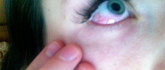
Red spot on a squirrel
The whites of the eyes may become covered with red spots - this occurs when a small blood vessel ruptures. Red spots that appear 1-2 times a year do not cause harm to health, especially if the organ does not hurt and there are no other accompanying symptoms. If blood vessels burst regularly, then it is necessary to contact a specialist as soon as possible. This may signal the development of infectious and viral diseases in the body.
Other symptoms
In 90% of cases, bleeding in the eye is a symptom of neurological problems and abnormalities. If the patient immediately begins to sound the alarm and seeks a complete comprehensive diagnosis, he will be able to prevent extremely negative consequences. If the presence of a red spot is accompanied by other neurological symptoms, this is already grounds for an urgent mandatory visit to the hospital.
These symptoms are:
- Lethargy, disorientation in space, confusion;
- Severe headache, dizziness;
- Complete or partial paralysis;
- Tremor of the limbs;
- Speech disorders;
- Noise, pounding in the ears;
- Heart rhythm disturbances;
- Memory losses;
- Difficulty swallowing, breathing.
If red spots in the eyes are accompanied by any of the above symptoms, immediate medical attention is needed. When a stroke is diagnosed, the patient is prescribed immediate resuscitation measures.
Treatment of red whites symptom
Red spots on the sclera indicate minor hemorrhage. Before you begin to treat the pathology, it is necessary to find out the cause of the rupture of blood vessels. The capillary can burst due to overexertion, heavy physical activity and severe stress. In most cases, the clinical syndrome goes away on its own and does not require any specific treatment. In the first hours after a rupture, experts recommend applying ice.
If ice compresses do not reduce the area of the spot, you can use eye drops. Medications help expand the lumen of blood vessels, which minimizes the risk of their rupture.
When a visit to the doctor cannot be postponed
After the eyes turn red, accompanying signs may appear indicating the development of dangerous pathologies. If, simultaneously with hyperemia, a burning sensation, itching, pain on palpation, purulent discharge, or peeling of the skin around the eyes occur, you should immediately consult an ophthalmologist. All signs indicate the development of a viral or bacterial infection inside the visual organ.
Complex of therapeutic measures
Pathologies of the visual organs, accompanied by an inflammatory process, can be treated in two ways. Drug therapy includes the use of topical medications and drops. According to reviews, these are accessible and inexpensive medicines with a different spectrum of action. They come in 3 types:
- non-steroidal;
- combined;
- steroid.
Nonsteroidal drops can be used regardless of the stage of the disease. They help most well with mild degrees of inflammation. Steroid drops are used for severe illnesses and during the rehabilitation period after surgery. Combined drops can be used not only for inflammation, but also for allergic reactions. Main indications for use: conjunctivitis, barley, blepharitis.
Traditional medicine acts more gently, especially in the initial stages of eye inflammation. Pathologies are treated by brewing - soak a cotton pad or gauze folded in several layers in strong black tea and apply to the eyes for 10-15 minutes. After each procedure, used cotton pads must be disposed of; they cannot be reapplied to the organ.
For barley and conjunctivitis, wash the eyes with infusion of chamomile or dill. The drugs have a pronounced anti-inflammatory effect. Before using any medications, you must remember the possible contraindications that are indicated in the instructions for use.
What to do if the squirrels are red?
Transient redness, which occurs irregularly and disappears quickly, does not require special treatment. If a person constantly has red whites of the eyes, it is necessary to find out the cause and treatment is prescribed in accordance with it.
The main treatment for vision pathologies is the use of eye drops and ointments. If the cause is a general disease - infection, hypertension, gout - appropriate therapy is prescribed.
- Emoxipin drops help remove red blood vessels on the whites of the eyes. The drug has a vasoconstrictor effect and restores the capillary wall. It is used for conditions accompanied by rupture of blood vessels and the formation of hemorrhages.
- Antiviral and antibacterial agents are used to treat infectious conjunctivitis and keratitis. “Ophthalmoferon” and “Poludan” drops have an antiviral effect. Eye antibiotics - “Tsipromed”, “Normax”, “Tetracycline ointment”, “Oftocipro”. Such medications are prescribed by an ophthalmologist after taking the necessary tests.
- Treatment of allergic manifestations consists of the use of antihistamines - “Allergodil”, “Lecrolin”, “Cromohexal”. For severe inflammation and chronic allergies, ointments and drops with corticosteroids are prescribed - “Hydrocortisone”, “Dexamethasone”.
- Redness and hemorrhages in the white of the eye associated with fatigue can be treated with vasoconstrictor and moisturizing drops. These include “Vizin”, “Systane”, “Hypromellose”.
- Means for reducing intraocular pressure - “Azarga”, “Cosopt”, “Fotil”. Used to treat glaucoma. Medicines are dispensed from pharmacies only with a doctor's prescription.
If discomfort is felt in the morning, the red whites of the eyes are visible, the doctor prescribes a course of vitamins - “Pro-Visio”, “Lutein-complex”. Drops with a vitamin, regenerating effect - “Taufon”.
The use of folk remedies is permissible after consultation with an ophthalmologist. Decoctions of medicinal herbs are used in the form of lotions and rinses:
- chamomile;
- sage;
- Oak bark.
It is unacceptable to put any folk remedies into the eyes.
If the cause of red eyes lies in incorrectly selected vision correction products (glasses, lenses), they are replaced with others. If possible, laser correction surgery is performed.
We invite you to watch an educational video about the causes of red eyes and treatment:
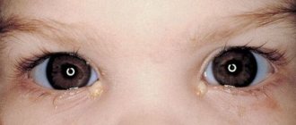
Problems in children
The whites of children's eyes change color for the same reasons as in adults. The main cause of hyperemia is chronic non-infectious diseases, allergic reactions, viral and bacterial pathologies. Most diseases are transmitted through contact. If a child attends school or kindergarten, the risk of developing diseases of this nature increases.
Stress and emotional overstrain play a big role. An unstable emotional state of a child can be caused by a number of reasons, including puberty.
Regular mood swings lead to thinning of the vascular walls, which increases the risk of rupture of large and small blood vessels.
Redness of the eye in a child and infant
Many mothers are interested in the answer to the question why the whites of the eyes turn red in newborns and children of primary school age. Experts identify several causes of red eyes, which include:
- entry of a foreign body into the organ of vision;
- allergic reactions to incorrectly selected baby food or complementary foods;
- infectious viral eye infection;
- congenital form of dacryocystitis.
Infants in the first months of life are restless and often capricious. Capillaries can burst due to the crying of a newborn baby.
Prevention measures
Few people know why the eyes are red and hurt, what to do in this situation and how to improve their overall well-being. Experts recommend preventing the appearance of unpleasant symptoms in the form of inflammatory processes in the organs of vision. Preventive measures include regular personal hygiene. Hands should be washed as often as possible, especially after visiting the restroom and being in crowded places, at least 2-3 times a day.
It is necessary to avoid stressful situations, completely give up bad habits, including alcohol and tobacco products (the latter reduce the vascular lumen and increase the risk of vascular rupture). Physical activity should be alternated with rest.