What is emerging scotoma of the eye?
Human vision is binocular (seeing with both eyes). It is limited by its field, that is, by the space that we can see with motionless eyes and head. When a dark area appears on it, like a drop of acid in a photograph, this is an eye scotoma.
It is formed when some structure of the visual apparatus (nerve, retina, receptors) is affected. The picture cannot be correctly formed and analyzed by the cerebral cortex. Such an area falls out of the general field of vision.
Watch a video about the features of the disease:
Possible complications
One of the complications may be chronic or transformed migraine. In this case, headache occurs more than 15 days a month, the frequency of attacks gradually increases and the intensity of pain decreases.
Nausea and sensitivity to bright lights and loud noises may be an accompanying factor, but are not required.
Another common complication is status migraine. We are talking about a condition when a migraine attack lasts longer than 3 days and meets all the criteria for migraine without aura.
It may also happen that aura symptoms last for more than one week. Sometimes a migraine can trigger an epilepsy attack. Most often this is characteristic of migraine with aura, and the attack occurs during or immediately after it.
As a rule, migraine has a favorable prognosis; only in rare cases can potentially dangerous complications arise. The main burden of migraine is its impact on a person's life and environment.
Classification
There are many varieties of cattle, we will try to bring order and figure out what they are.
By function:
- physiological;
- pathological
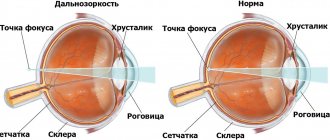
Physiological scotoma exists normally in every person. It is caused by the optic nerve (its disc) or vessels projecting onto the retina, forming a blind spot.
This zone does not have the visual receptors necessary to perceive the picture. However, we do not feel this, since the visual fields overlap each other from each eye, compensating for the physiological defect.
This area is called Mariotte's spot and is located in the temporal region of the field; angioscotomas are located next to or around it. They are caused by the presence of retinal vessels; there are also no visual receptors in these places.
The pathological form occurs due to pathological processes in the structures of the eye (retina, optic nerve, visual structures in the brain, choroid). When a scotoma is present in only one eye, the visual structures of the brain (chiasm) are not damaged.
The nature:
- positive – the one noted by the patient himself. Looks like a gray or dark spot;
- negative - a person does not feel it on his own, it is detected only instrumentally.
By density:
- absolute – the scotoma area is a blind spot, objects are not visible;
- relative - a defect area with partially reduced visibility; silhouettes can be distinguished.
According to the depth of perception, scotomas are divided into 1st and 2nd order scotomas, which differ depending on the strength of the decrease in light and color sensitivity of the eyes.
By location:
- central scotoma – located in the center of the visual field. A common cause is retinal pathology;
- peripheral - it is detected at the periphery of the visual field;
- paracentral - scotoma of the eye, found near the central point of fixation of the eye and adjacent to it;
- pericentral - it surrounds the fixing point without touching it, it can be ring-shaped. A special type of scotoma is Bjerrum's scotoma. Initially, it consists of separate blind islands, which later merge to form an arch. One part of it is associated with the blind spot, and the other extends to the nasal part of the visual field. Characteristic of the onset of glaucoma.
Separately, there is atrial scotoma, or ocular migraine. It refers to a neurological disease. Deterioration of vision develops due to vascular disorders during a migraine attack, and after its end it is completely restored.
Inhibition scotoma (functional) is a defect that occurs in people with strabismus. The developmental mechanism is to prevent double vision. The brain suppresses (inhibits) the image coming from a squinting eye. As soon as the healthy eye is closed, the scotoma will disappear.
Risk factors and causes of development
Given that the causes of ophthalmoplegic migraine are not fully known, triggers are highly specific to each individual. The provoking factors are twofold.
Uncontrollable factors include, for example, the menstrual cycle, weather changes and other triggers that are beyond our control.
Conversely, controllable triggers such as stress, lack of sleep, active or passive smoking, certain types of foods, bright lights, loud sounds, strong odors, etc. can be avoided.
Thus, every person prone to migraines should identify their manageable risk factors and try to avoid them in everyday life.
Abuse of certain medications, in particular barbiturates and opiates, can also provoke an attack of Charcot's syndrome and aggravate its course. Often, stopping hormonal contraception is enough to completely get rid of migraines.
Causes of the defect
The main reasons for the appearance of scotomas on the eye may be the following:
- Demyelinating diseases, such as multiple sclerosis, damage the myelin (the sheath that conducts nerve impulses) of the optic nerve.
- Retinal vascular thrombosis - the nutrition of the eye tissues along with the visual receptors (rods and cones) is disrupted.
- Glaucoma is a disease of increased pressure within the structures of the eye, which destroys the cells of the optic nerve. Its frequent manifestation is a change in the visual field - scotoma with further narrowing of the field.
- Ocular migraine, the classic form of which is atrial scotoma. It can be considered an atypical migraine. Severe headaches cause spots and flickering highlights to appear, which is why such a scotoma was called atrial scotoma.
- Age-related changes in the structure of the eyes (macula, optic nerve, retinal receptors).
- Vegetovascular dystonia. With it, visual field defects can move from one eye to the other and can be felt even when the eyes are closed.
- Hypertension - increased blood pressure leads to damage to the visual cortex and retina.
- Diabetes mellitus – its complication is diabetic retinopathy. The retinal vessels become inflamed, and microthrombi may form, causing the death of the eye receptors.
- Eye injuries – those that penetrate the retina are especially dangerous.
With cataracts (clouding of the lens), a gradual decrease in objective vision occurs, but scotomas do not occur.
Types of migraine and its phases
The disease can be divided into two main types, namely classic and general migraine. There are other types, but they are relatively rare and are more common in children.
Ophthalmic migraine manifests itself at the age of 6-12 years and, in addition to headache, is characterized by weakness of one of the eye muscles. At the same time, basilar migraine tends to cause dizziness and speech difficulties.
Another rare form of migraine causes weakness in one part of the body but is mostly hereditary. Also, only the aura may appear, without a headache, or the aura appears during pain.
The difference between classic and common migraine is very small. In general attacks, the second stage, called aura, is absent. An aura occurs due to decreased blood flow to certain parts of the brain.
If this decrease is not noticeable enough, the aura does not appear, and the migraine is classified as classic. General migraine is more widespread, and in approximately 80% of cases the headache comes without a preliminary aura.
Having gone through all the phases...
Classic migraine has four phases of development:
- The first one is called prodromal , and most people don't even know about it. It can last from one hour to the whole day. Characterized by decreased dexterity, yawning, feeling tired, stiff neck, thirst, increased sensitivity to light or sound, irritability, cravings for sweets.
- The second phase is the aura , which usually lasts 10-60 minutes. The most common symptom is visual inability, resulting in sparkling, flashing, double vision, or temporary blindness. There may be a decrease in sensitivity in the hands, and sometimes speech difficulties.
- , a moderate to severe headache occurs , which is felt on one side of the head. It is usually accompanied by sensitivity to light and noise, fatigue and drowsiness. Nausea and/or vomiting may be present.
- The fourth phase is the final one, during which deep sleep occurs . After it, a person feels tired, he has no appetite, but there is a feeling of relief.
Symptoms of the defect
The distinctive signs of scotoma are:
- a blind spot or a spot with a pronounced decrease in visibility in the field of vision is a classic sign of absolute (which a person notices) scotoma;
- impaired perception of surrounding objects, visual images - decreased color, transparency, brightness;
- the appearance of dark spots in the eyes;
- signs characteristic of atrial scotoma are flickering highlights, spots, floaters, a luminous halo of objects associated with a migraine attack.
When there are many scotomas, they can merge, then such a defect is characterized by complete loss of vision in one eye.
Diagnostics
In order to identify blind spots, it is necessary to undergo an examination by an ophthalmologist. Depending on the cause of the disease, he may suggest the following methods:
- perimetry – examination of the visual field using a perimeter (an arc with degree markings). A colored (to determine the loss of color vision) pointer is led along an arc, determining by degrees where the person has stopped seeing it. An analogue is computer perimetry;
- examination method on a flat surface - campimetry. The examination is carried out with a campimeter. This method is more narrowly focused. It is used to detect central or near-central blind spots;
- computed tomography of the brain - performed to exclude vascular pathology (for example, stroke), neoplasms;
- fundus examination - to assess the condition of the retina;
- Ultrasound examination of the eyeball – detects foreign bodies;
- measurement of intraocular pressure;
- neurological examination - to distinguish flickering scotoma associated with vasospasm from true ophthalmological one.
Notes
- ↑ 1 2 3 Scotoma
- article from the Great Soviet Encyclopedia. - "Possible Roles of Vertebrate Neuroglia in Potassium Dynamics, Spreading Depression, and Migraine", Gardner-Medwin, J. Exp.
Biology (1981), 95, pages 111–127 (Figure 4). - Skoromets A. A. Topical diagnosis of diseases of the nervous system: A guide for doctors. 1st ed. L.: Medicine, 1989. - P. 91-92 - 5000 copies. — ISBN 5-225-01582-4
Treatment
Therapeutic measures to cure vision loss will depend on the cause of the disease. This is where you should focus your attention. If this is atrial scotoma, then its treatment consists of prescribing vascular and anti-migraine drugs.
If blood flow through the vessels is disrupted due to a blood clot, antiplatelet (blood thinning) therapy is required, and visual disturbances in diabetes require correction of sugars, without which treatment is ineffective.
Age-related changes in the retina, early manifestations of cataracts and glaucoma, which lead to blind spots, are reduced with the help of eye drops (vitamins that reduce intraocular pressure).
The modern arsenal includes magnetic stimulation of the optic nerve itself or areas of the cerebral cortex. As well as stimulation of affected areas with peptide drugs that help tissue restoration.
When the cause of a visual field defect is a tumor or retinal detachment, then surgery or laser correction cannot be ruled out.
Prevention
The main prevention of any visual field impairment is as follows:
- constant monitoring of blood pressure levels;
- normalization of the patient’s psycho-emotional state;
- maintaining visual hygiene,
- If any ophthalmological abnormalities occur, immediate contact with an ophthalmologist is recommended.
It should be noted that any changes in visual fields are a consequence of some pathological process. But if detected, you need to undergo a number of examination methods prescribed by an ophthalmologist.
In rare cases, it is necessary to involve related specialists in assistance, for example, in the case of disorders and neuropsychiatric diseases.
When changing the field of view, treatment should completely depend on the nature of the main pathological process.
Preventive measures
Any disease is easier to prevent than to overcome. In order for it to bypass you, you need to carefully monitor your health, namely:
- undergo routine preventive examinations with an ophthalmologist;
- Monitor your blood pressure and blood glucose levels;
- exclude alcoholic drinks, nicotine;
- get enough sleep;
- do not neglect safety measures, wear safety glasses where the situation requires it;
- maintain a daily routine and rest schedule;
- wear properly fitted glasses or contact lenses, especially for myopia (a high degree of myopia is dangerous for retinal detachment).
A blind spot in the eye is a complex and serious disease; do not let the disease take its course. If you find yourself with similar symptoms, contact a specialist.
Don’t forget to share the article with your friends so that even more people learn about scotoma and can see a doctor on time.
Clinical picture step by step
Ocular migraine, like other forms of the disease, has several phases of development:
- Prodromal phase . This phase is an unobtrusive warning moment immediately preceding the disease. It usually manifests itself as mood swings, anxiety, nervousness, fatigue, depression, irritability, loss of appetite, etc. Often, however, these symptoms are so minor that they go unnoticed by the person.
- Aura . This phase is not always present. It occurs in only about 20% of migraine patients and usually lasts from 5 minutes to 1 hour.
- Directly, headache . This phase is manifested by moderate or unbearably severe headaches, which, as a rule, are of a vivid nature and are localized unilaterally, usually around the eye. The attack may be accompanied by vomiting and nausea. This phase of migraine is the most terrible and painful. At the moment a person tries to find a quiet, dark corner. Common symptoms of ophthalmoplegic migraine include abnormal sensitivity to auditory, visual, olfactory, and tactile stimuli. The person may also experience internal confusion, drowsiness, irritability, dizziness, loss of vision, or double vision. More severe migraines are accompanied by vomiting, which is characterized by the fact that it brings relief.
- Residual phase . In this final phase, recovery occurs and the headaches gradually subside. It can take from several hours to several days and is usually accompanied by fatigue and loss of appetite.