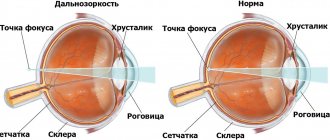
Hypertelorism is a pathological condition in which the distance between paired organs is abnormally large. This disorder usually affects the organs of vision. In this case we are talking about visual, or ocular hypertelorism.
Hypertelorism is a congenital pathology, but it is not a separate disease, but is formed as a result of other disorders. The development of pathology occurs in utero. It is often observed against the background of various syndromes (Down, Apert, Edwards), neurofibromatosis and other diseases associated with genetic abnormalities.
Causes:
- genetic predisposition (the presence of diseases of a similar nature in one of the parents or close blood relatives);
- taking strong medications during pregnancy;
- severe infectious diseases suffered by the mother during fetal development;
- abuse of alcohol, drugs and other harmful substances by the expectant mother.
All these reasons are hypothetical. Often the cause of hypertelorism is heredity.
Cause
Most often, pathology occurs during the embryonic development of a child. Laying of leaves is disrupted in the first three months after conception. A damaging factor can be the use of alcoholic beverages, psychotropic substances, and medications by the mother. The reasons for the formation of a bone defect are:
- improper fusion of cranial sutures due to increased arterial and intracranial pressure;
- hernias located in the central nervous system, localized near the nose and cranial fossae;
- clefts that form in the facial area, disrupting the location of the orbits, accompanied by a violation of the structure of the eyelid, macrostomia (excessively large gap for the mouth);
- the formation of genetic mutations that affect the shape of the skull, disrupting it;
- the formation of tumors in the face or brain, which leads to deformation of the skull,
- an acquired form of the disease that develops as a result of mechanical damage, which leads to improper fusion of bone processes (an increase in the distance between bones is formed due to the formation of callus, the application of metal prostheses, or their improper implantation).
The congenital form of the disease is much more common. The doctor must identify the root cause to eliminate the risk of recurrence of the disease after treatment.
Prevention
Since ocular hypertelorism is caused by a genetic abnormality, there is no specific prevention of the defect. At the stage of pregnancy planning, you should collect a hereditary history and study the presence of similar diseases in relatives.
If a pathology has been identified, then consultation with geneticists, careful monitoring of the condition of the fetus and genetic tests for early diagnosis are necessary.
To prevent congenital pathologies, it is necessary to undergo a comprehensive study at the stage of pregnancy planning. The mother should avoid drinking alcohol, smoking, using embryotoxic drugs that pass through the placenta, and avoid exposure to radiation and other harmful environmental factors.
Hypertelorism is a pathology that is rare and is diagnosed in childhood. It should be operated on both for medical reasons and to improve the patient’s quality of life.
Author: Albina Neiman, doctor, especially for Okulist.pro
Risk group
The risk group includes the following categories of patients:
- children whose mother suffers from alcoholism or drug addiction or was irradiated during pregnancy;
- patients who have a history of serious mechanical damage to the brain or facial part of the skull;
- persons who have undergone implantation of metal prostheses in the facial area.
If a person is at risk, he needs to consult with doctors, especially a plastic surgeon, who can help correct cosmetic defects.
Hypertelorism - what is it: the eye of the fetus on ultrasound, the nipple, the eye of the child
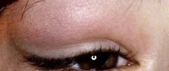
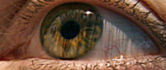
Hypertelorism, which is a symptom and not an independent pathology, most often manifests itself as an abnormal position of the eyes. Visually, the increased distance between their inner corners and the pupils is clearly noticeable.
Hypertelorism accompanies genetic diseases that affect the area of the skull and face, leading not only to cosmetic defects, but also to deterioration of vision, impaired sense of smell, and even problems in terms of mental development. The distances between the inner corners of the eye sockets widen when the wings of the sphenoid bone are too enlarged.
What is orbital hypertelorism?
The degree of the defect depends on the location of the visual organ, the distance between the eye sockets and the age of the person. With ocular hypertelorism, the oval of the face changes and the nasal fissures are deformed.
Scientists distinguish two types of this anomaly. With false epicanthus, the distance between the corners of the visual organ increases. True hypertelorism is characterized by expansion of bone formations. It begins when the front part of the skull is damaged, when a tumor appears in the nose.
A defect is considered a pathology if the value of the indicator in the form of the orbital-circumferential index exceeds 6.8. The angle at which the axes of the eye sockets diverge is sometimes 60 degrees, which is twice the norm.
How to treat viral conjunctivitis in children is described in detail in the article.
Deformation of the skull in the orbital form of the disease
Find out how to treat internal stye on the eye here.
Reasons for the appearance of a baby
The position of a child's eyes changes when the ethmoid bone, which is located at the base of the nose, widens and lowers. Problems in the development of the facial part of the skull, which lead to hypertelorism, arise from pathologies of a genetic nature, which are accompanied by a large number of defects:
- With Apert syndrome, the bones of the skull fuse much earlier, and the baby is severely retarded in mental development.
- With neurofibromatosis, a tumor forms in the brain, growths appear on the skin and various organs.
- For a chromosomal disorder, which is typical for Edwards syndrome, the child has deformed feet, low muscle tone, and a reduced size of the lower jaw. Small eye sockets are located at a great distance from each other.
- Hypertelorism occurs with craniostenosis, when bone sutures heal prematurely. It accompanies hernias of the skull and brain, congenital defects of the structure of the facial area.
Negative effects lead to disruption of intrauterine development of the fetus:
- prenatal infection;
- gene mutations at conception;
- hereditary predisposition;
- medications taken by a pregnant woman.
Hypertelorism occurs in Down syndrome, manifests itself in Crouzon and Toast syndromes, and develops when the facial bone is damaged due to trauma. It usually does not begin independently, but is combined with other pathologies.
A proven antiglaucoma agent - instructions for Ganfort eye drops.
A genetic test will help reduce the risk of developing such problems
Weakness of the blood vessels in the eyes is the cause of hemophthalmos.
Forms and symptoms
Features of the disorder in orbital hypertelorism are determined by the distance between the internal angles of the visual organ. In the first degree it is less than 34 mm, in the second – 39, in the third it exceeds 40. The norm for this indicator is 3 cm.
It happens that a child has very small eye sockets and slits. Sometimes a baby is born without them at all. This type of pathology, called microorbitism, affects the bones of the skull. They thicken and become deformed.
Rare congenital pathology
With a symmetrical appearance of the anomaly, the distance between the eyes widens. With asymmetric hypertelorism, they are located at unequal heights. The pupils look in different directions. Over time, heterotropia occurs.
Because the structure of the bones in the facial area changes, a person may have a bridge of the nose that is too wide or flat.
Hypertelorism accompanies many genetic pathologies and manifests itself in the form of:
- underdeveloped jaw;
- deformed feet and eyebrows;
- optic nerve atrophy;
- fusion of fingers and toes;
- cheekbone displacement;
- abnormal joint mobility.
Children who have this syndrome are usually severely retarded in mental development; adults have problems with intelligence.
If hypertelorism occurs together with Toast's disease, eyelash dysplasia is present, the child suffers from chronic conjunctivitis. With Greig's syndrome, paralysis of the abducens nerves of the eyes and astigmatism may occur.
Antimicrobial therapy – Gentamicin sulfate eye drops.
Diagnostic methods
To determine the type of lesion, they begin with an external examination. The doctor will find out what the person is complaining about, whether he has relatives with an abnormal face shape or wide-set eyes. The neurologist evaluates the patient's intellectual abilities and checks the condition of the muscles.
Using MRI and fluoroscopy, defects are found in the skull and brain. This method helps to find out why the facial bones are deformed. The distance between the eye sockets must be measured, which makes it possible to determine the degree of hypertelorism. To confirm the presence of pathology, a computed tomography is performed.
Farsightedness is not a death sentence – mild hyperopia is what it is.
Use of the medicine for other than its intended purpose - heparin ointment against bruises under the eyes.
Features of treatment
The article is for informational purposes only; a doctor who specializes in such syndromes must establish a diagnosis and prescribe a method of therapy. A consultation with an otolaryngologist is required.
Hypertelorism cannot be cured with medications and physiotherapeutic procedures. The only way to get rid of cosmetic defects and unpleasant symptoms is through surgery.
Before the operation, the type of which depends on the severity of the symptoms, the patient is examined by an ophthalmologist. The most favorable time for its implementation is no older than 8 years. Young children need intervention if heterotropia develops and the lacrimal canal narrows.
It is advisable to perform the operation on preschoolers, since then teeth will begin to form in the upper jaw, which will interfere with the procedure. If intervention is started at an earlier age, there is a risk that the baby’s cheekbones will shift.
If the problem is just beginning to appear, the shape of the skull is corrected by inserting special staples. In the first degree of violation, the wall of the orbits is cut, moved to the required distance, and cartilaginous tissue is restored.
When hypertelorism enters the second stage, they resort to rhinoplasty using part of the parietal bone. With the third degree of damage, it is necessary to change the shape of the forehead and model the base of the skull.
If microorbitism or false hypertelorism is diagnosed, bone tissue is dissected, nasal deformities are eliminated, and the appearance of the visual organ is restored.
Usually, cosmetic defects in children are corrected quite well. If the baby suffers from Down's disease, the corners of the eye sockets will be repeatedly adjusted and the skin on the bridge of the nose will be removed.
Hormonal therapy for the treatment of inflammation - hydrocartized eye ointment.
Will Gilan Comfort eye drops help you cope with dryness? Read here.
Preventive measures
Hypertelorism is often inherited by the baby from the mother or father. It is difficult to prevent its occurrence. If your relatives have experienced symptoms of this syndrome, you should consult a genetics specialist before having children.
Following simple rules helps prevent congenital pathology. Those who are planning to conceive a baby need to give up alcoholic beverages and forget about smoking. A pregnant woman should try not to be exposed to the aggressive influence of the external environment, not to become infected with infectious diseases, and to use medications only as a last resort.
You can get rid of hypertelorism, which manifests itself in the form of defects that disfigure the face, in the first years of the baby’s life. It is undesirable to postpone the operation, since with age the deformities will begin to increase, vision will deteriorate, speech will suffer, and this will affect the psycho-emotional state of both the baby and the parents. See also information about anisocoria.
Classification
There are three degrees of development of the disease depending on the distance between the main orbits:
- first degree: 3-3.5 cm;
- second degree: 3.5-4 cm;
- third degree: 4.1 cm or more.
The disease is divided depending on the correspondence between the right and left sides of the face:
- symmetrical - the right and left parts correspond to each other, are located equally;
- asymmetrical - the right and left parts do not correspond to each other, the location of the eye socket, wings of the nose, and cheekbones changes.
Based on the degree of development of bone defects, two types of the disease are distinguished.
- A true violation. The pathology develops due to the expansion of the bones that are located between the eyes. The bridge of the nose widens, the bridge of the nose becomes flat.
- False violation. Only the distance between the inner corners of the eyes increases, but the location between the orbits corresponds to normal parameters. The bone tissue has a normal location and is fully formed.
The doctor classifies the disease in order to correctly register it in the medical history and select treatment.
Hypertelorism
The term “ocular hypertelorism” has been used to refer to the increased bony distance between the orbits.
It was first proposed by LG Greig in 1924 and implied a delay in the displacement of the orbits in the medial direction during fetal development, their fixation in the prenatal state and the resulting resulting increased distance between their medial walls.
Subsequently, P. Tessier proposed a term that more correctly characterizes this condition - orbital hypertelorism. It should be differentiated, first of all, from telecanthus - an increase in the distance between the medial canthus of the palpebral fissures without changing the bone distance, which also imitates hypertelorism.
Therefore, we can highlight
- true hypertelorism , when the eye sockets are widely set due to abnormally wide bone formations between them,
- false (epicanthus) , when the distance between the medial corners of the palpebral fissures is increased, despite the fact that the bone distance between the orbits is within normal limits.
The increased distance between the inner edges of the orbits is usually a consequence of excessive development of the lesser wings of the sphenoid bone. At the same time, the eyes are widely spaced, the bridge of the nose is wide, and the bridge of the nose is flat.
Orbital hypertelorism is not an independent disease and accompanies such anomalies as craniostenosis of the sutures of the anterior cranial fossa, anterior and basal cranial hernias, clefts of the craniofacial skeleton, craniofacial dysplasia, as well as craniofacial tumors and post-traumatic deformities.
Hypertelorism is considered pathological if the interorbital-circumferential index (IOCI) is more than 6.8. MOI is the result of dividing the head circumference by the distance between the inner canthuses, multiplied by 100.
Classification of orbital hypertelorism
The main anatomical characteristic of the OG is an increase in the width of the ethmoid bone in the horizontal plane. The perforated plate in OH is often located at a lower level than is normal. An increase in the width of the cells of the ethmoidal labyrinth is more often observed in the anterior part of the sinus and less often in its posterior part.
In accordance with this, the angle of divergence of the sagittal axes of the orbits from the midline in OH can reach 60°, whereas normally it does not exceed 30°. These anatomical characteristics of the OG are of decisive importance when choosing a method for approximation of the orbits and surgical correction of the middle zone of the facial skeleton.
Tessier classification of orbital hypertelorism
Tessier's classification is based on measuring the interorbital distance (IOD) between the middle of the anterior lacrimal crests.
- with grade I OH, the MOR is 30-34 mm,
- with stage II OH, the MOR is 35-39 mm,
- with grade III OH, the MOR is 40 mm or more.
Tessier himself called this classification “artificial”, since the degree of OH does not indicate the depth of prolapse of the perforated plate of the ethmoid bone. And the latter is crucial in determining the method of surgical intervention.
Classification of orbital hypertelorism V.A. Belchenko
Classification by V.A. Belchenko is based on the difference in measuring the interorbital distance (IOR) at the beginning of the upper third of the orbits, i.e. in the area of the junction of the frontal, lacrimal and maxillary bones and MO in the standard measurement location - between the middle of the anterior lacrimal ridges.
- OG l degree 1st type - MOR between the middle of the anterior lacrimal ridges - 30-34 mm, MOR between the inner edges of the orbits at the level of the beginning of the upper third of the orbits - no more than 34 mm.
- OG l degree of the 2nd type - MOR between the middle of the anterior lacrimal ridges - 30-34 mm, and at the level of the beginning of the upper third of the orbits - 35-39 mm.
- OGII degree, type 1 - MOR between the middle of the anterior lacrimal ridges - 35-39 mm, MOR at the level of the beginning of the upper third of the orbits - no more than 39 mm.
- OG II degree 2nd type - MOR between the middle of the anterior lacrimal ridges - 35-39 mm, and at the level of the beginning of the upper third of the orbits - 40-44 mm.
- OH III degree, type 1 - MOR between the middle of the anterior lacrimal ridges is 40 mm or more, and MOR in the area of the beginning of the upper third of the orbits exceeds this figure by no more than 4 mm.
- OH III degree of the 2nd type - MOR between the middle of the anterior lacrimal ridges is 40 mm or more, and MOR at the beginning of the upper third of the orbits exceeds the first one by 5 mm or more.
The type of OG is determined by the difference in measurements between the MOR at the beginning of the upper third of the orbits and the MOR between the middle of the anterior lacrimal ridges. For type 1, this difference does not exceed 4 mm; for type 2, it is 5 mm or more.
In addition, there are asymmetrical variants of hypertelorism, when in addition to the wide distance between the orbits, rotation of the orbits and differences in height are possible.
Etiology
The intrauterine development of hypertelorism occurs when some kind of failure occurs in the process of bringing the eye sockets together. The reason for this may be the adverse effects on the body of a pregnant woman and the fetus of various factors.
Conventionally, there are several groups of causes that lead to the occurrence of hypertelorism:
- Premature fusion of the sutures of the skull (craniostenosis), namely the sutures of the anterior cranial fossa
- Cranial hernia
- Craniofacial clefts
- Craniofacial malformations (cranial bone dysplasia, Crouzon syndrome, Apert syndrome, etc.)
- Other conditions (craniofacial tumors, post-traumatic changes in the skull bones, etc.)
False forms of orbital hypertelorism
Among the causes leading to the occurrence of false forms of orbital hypertelorism, the most common are trauma to the nasal-orbital-ethmoidal complex with an increase in the interorbital distance and various neoplasms occupying the space of the ethmoid and nasal cavity. And although these manifestations are in most cases regarded as false orbital hypertelorism, the methods for eliminating it can be the same as for eliminating true OH.
Crouzon syndrome
Crouzon syndrome is a genetic disorder known as branchial arch syndrome. Specifically, this syndrome affects the first branchial (or pharyngeal) arch, which is the precursor to the jaw and mandible.
It is primarily formed by premature closure of the coronal and sagittal sutures. Heredity is autosomal dominant, but in 25% of cases there may be a fresh mutation.
Ocular manifestations:
- Exophthalmos due to a shallow orbit is the most noticeable sign. Develops secondarily due to growth retardation of the upper jaw and cheekbone. In severe cases, the eyeballs are dislocated and lie in front of the eyelids.
- Hypertelorism (wide distance between orbits).
- V-shaped exotropia and hypertropia.
- Vision-threatening complications include exposure keratopathy and optic neuropathy due to compression of the optic nerve at the optic foramen.
Pathology of the eyeball: aniridia, blue sclera, cataract, lens subluxation, glaucoma, coloboma, megalocornea and optic nerve hypoplasia.
Greig's syndrome (familial hypertelorism)
This autosomal dominant syndrome was described in 1928 by Scottish surgeon David Middleton Greig. In this disease, the development of the skull is disrupted at an early embryonic stage.
Common manifestations include: brachycephaly, wide nasal bridge, high palate, dwarfism, cryptorchidism.
Mental retardation, convulsive seizures, abnormalities of the jaws and teeth, umbilical hernia, clubfoot, and clinodactyly are often observed.
Ocular manifestations:
- hypertelorism,
- epicanthus,
- deformation of eyebrows and eye slits,
- enophthalmos,
- weakness of convergence,
- high degree of astigmatism,
- bilateral abducens nerve palsy,
- atrophy of the optic nerve as a result of narrowing of the optical canal, in some cases - pigmentary degeneration of the retina.
Diagnostics
A preliminary diagnosis is made based on a primary external examination. The distance between the inner corners of the eyes is measured. The presence of maxillofacial deformities is established. Attention is drawn to the shape of the skull. Possible visual and olfactory impairments are identified.
The diagnosis is confirmed by instrumental examination data: X-ray examination and computed tomography of the head, and in some cases MRI to exclude combined anomalies of brain development (cranial hernia, etc.).
An x-ray of the skull bones determines the presence or absence of hypertelorism and the degree of asymmetry of the orbits.
Computed tomography gives a more detailed picture of the existing pathology and allows you to more accurately determine the amount of treatment.
The complex of diagnostic measures must necessarily include an examination by an ophthalmologist, ENT specialist, neurologist, as well as other specialists, depending on the cause of the development of hypertelorism (cranial hernia, craniostenosis, etc.).
Stereolithographic biomodeling
The creation of a real 3-dimensional model significantly increases the possibility of planning surgical intervention. A real operation plan, performed on a model, greatly facilitates the surgeon’s orientation and helps to predict the course of the operation and the detailed implementation of individual stages.
Symptoms
There are the following clinical symptoms during the development of the disease:
- proximity of the eye sockets to the temporal areas;
- changing the correct shape of the orbital region;
- the hairline on the forehead, which is called the “widow’s peak”;
- incorrect anatomical structure of the nose: the base is excessively wide, the tip has folds;
- the nasal septum is curved, making normal breathing difficult;
- the function of the visual organs is reduced: myopia or farsightedness develops, objects double, and an irregular shape of the eyeballs forms (astigmatism);
- exophthalmos (bulging eyes) develops only in some cases.
Based on clinical symptoms, the doctor suggests a diagnosis. But it can only be confirmed using diagnostic tests.
Diagnostics
If the disease is hereditary, consult a geneticist. If the form is acquired, it is recommended to consult a traumatologist. In all cases, surgical treatment is recommended, so contact a plastic surgeon for consultation.
Diagnosis of the patient’s condition consists of several stages:
- Anamnesis collection. This is data obtained from the words of the patient or his close relatives. Based on them, the doctor suggests a diagnosis and prescribes additional research methods.
- General inspection. It shows the characteristic symptoms of hypertelorism.
- Assessment of visual acuity. For this purpose, ophthalmic tables are used, which depict symbols, pictures, and letters. The longer the patient sees, the better the quality of his vision.
- X-ray. The image will show bone tissue and its deformation, orbital distance, and asymmetry of the orbits. Using the method, the doctor detects extensive bone tissue disorders.
- MRI, CT. The technique allows us to identify the layer-by-layer structure of the brain, eyeballs, and bone tissue. The study is prescribed before surgery to eliminate the risk of complications, damage to blood vessels and nerves.
Using diagnostic tests, the doctor makes a reliable diagnosis and prescribes treatment.
Treatment
Treatment of the defect is only surgical. Surgery is performed at ages 5 years and older to ensure maximum jaw development and prevent damage to developing teeth. Before this age, the operation is performed if the child has eyelid abnormalities, narrowing of the tear ducts, or strabismus.
In case of I degree of orbital hypertelorism, the doctor may recommend extracranial correction, i.e. reducing the distance between the bones of the inner parts of the nose and eye socket. In this case, the surgeon does not affect intracranial formations.
In severe cases, combined intracranial and extracranial surgery may be necessary, performed by a plastic surgeon and a neurosurgeon. During surgery, an incision is made from ear to ear, the frontal bone is temporarily moved back, and excess bone between the eyes is removed.
The eye sockets are moved closer to each other, and the soft tissues are returned to their place. At the same time, you can change the position of the nose and upper jaw.
Complications
With prolonged absence of therapy, the following complications arise from the development of the disease:
- inflammatory condition of nervous tissue;
- corneal deformation;
- decreased visual acuity up to complete blindness;
- atrophy of nervous tissue, if the cause is mechanical damage;
- chronic conjunctivitis;
- replacing binocular vision with monocular vision, that is, a person sees with only one eye.
To avoid the risk of complications, you must consult a doctor promptly.
conclusions
Ocular hypertelorism is a common symptom that is characteristic of many genetic diseases. Due to the fact that there are no effective ways to treat and prevent the disease, you should approach pregnancy responsibly, starting from the planning stage. And if pathology still cannot be avoided, it is necessary to raise the question of surgical correction as early as possible, which will reduce the risk of complications and facilitate the child’s social adaptation.
Also read about what aphakia of the eye is (congenital or acquired absence of the lens) and whether color blindness can be cured.