What is xanthelasma of the eyelids, and how to treat it.
Xanthelasma of the eyelids, one of the types of xanthoma, is a skin growth that forms on the periorbital area of the face. The neoplasms have a benign structure, and the disease itself does not pose a serious health hazard, since not a single case of xanthoma degeneration into a malignant tumor is known. But damage to the skin can be an alarming signal of the development of a chronic disease, and the best option is to immediately consult a doctor.
You can quickly and easily get rid of xanthelasma!
9 most powerful remedies!
These simple remedies are gentle and effective!
Only those who have xanthelasma know how annoying it is.
It is a slightly yellowish, flat, benign growth that appears on the top of the eyelids.
This is quite unsightly in appearance and affects a person's self-esteem.
To determine that the resulting plaque is actually xanthelasma, you should study the lipid spectrum of the blood.
If the test is positive, steps should be taken to reduce blood lipids and cholesterol levels to normal levels.
Women are more likely to suffer from this evil, and the older they are, the more common it can be.
Some practitioners believe that xantholasma could be considered a symptom of severe atherosclerosis or would indicate a high risk that the patient is suffering from heart attacks.
However, the reasons for this problem are not known.
Xanthelasma is often observed in patients with obesity, diabetes, myxedema, nephrosis lipoids, pancreatitis, liver cirrhosis and high blood cholesterol. It may be hereditary. In these cases, a genetically determined disorder of fat metabolism develops.
As a rule, xanthelasma appears on both eyelids. Sometimes it merges into a solid yellow stripe with an uneven contour, passing through the entire upper eyelid.
Its development occurs gradually and slowly, without subjective sensations in the patient. It can be the size of a small pea to the size of a bean.
To eliminate it, you can try the following natural remedies:
1. Garlic
The enzymes that can be found in garlic will help you reduce these small pockets of fat. Garlic also lowers cholesterol levels, which is more important.
Garlic extract is an effective and completely safe alternative to high cholesterol pills and blood pressure medications.
Grind one clove of garlic to make a paste. Apply to the affected area and cover with a cotton pad to prevent garlic from getting into your eyes.
Leave on for ten minutes and then rinse with warm water.
You can also consume a couple of cloves of raw garlic before breakfast every day.
If you decide to use a garlic supplement, consult your doctor.
Warning: Do not use garlic on the skin for long periods of time as it may burn the epidermis.
And if you have sensitive skin, never use it.
2. Castor oil
Castor oil contains ricinoleic acid, which has the ability to lower cholesterol.
To treat, simply place a few drops of oil on a cotton swab and place on the affected area, massaging gently. Make a bandage to secure the disc to the skin and leave it on overnight. The next morning, rinse.
The best choice is organic cold-pressed castor oil.
3. Apple cider vinegar
This ingredient will help reduce bad cholesterol and at the same time remove toxins from the body. Vinegar contains acetic acid, which can reduce and eliminate these fatty plaques.
To use it, simply soak a cotton pad in vinegar and place it on the affected area.
Secure with a bandage and rinse with warm water after 1-2 hours. Do this twice a day.
4. Fenugreek seeds
They are rich in fiber and great for lowering cholesterol.
Mix two tablespoons of seeds in water and let steep overnight.
The next morning, drink the liquid on an empty stomach. You can also use this liquid as a compress. The ingredients penetrate into the deep layers of the skin and help dissolve fat.
5. Onion
Roast the onion until soft, then chop to form a paste.
After this, take a small piece of neutral soap and mix with chopped onion.
Apply the mixture to the affected area, avoiding contact with the mucous membrane of the eye. Let the mixture sit for five to ten minutes, apply once or twice a day.
After removing the compressor, rinse with warm water.
6. Banana peel
Banana peels can dissolve fat deposits due to the powerful enzymes they contain. Being an excellent antioxidant, banana peel is also beneficial for treating various skin problems.
Place a piece of ripe peel on the affected area, secure it with a bandage and leave overnight. Remove in the morning and rinse.
Repeat until you get the desired results.
7. Coriander
Coriander is very rich in various antioxidant enzymes that can dissolve bad cholesterol. To treat xanthelasma, simply boil a tablespoon of coriander in a glass of water for 1-2 minutes. Leave for 15 minutes. Then drink.
It is recommended to drink 1 glass three times a day until cholesterol levels decrease and plaques around the eyelids disappear.
8. Orange juice
Orange juice contains vitamin C, flavonoids and folates, which are effective in increasing high-density lipoprotein levels (known as good cholesterol) and reducing bad cholesterol.
Drinking fresh orange juice regularly will help reduce bad cholesterol.
100-150 ml of freshly squeezed orange juice every day is enough to maintain normal cholesterol levels. You can also eat an orange every day if you don't want to juice it.
9. Almonds
Almonds contain a lot of fiber and have the ability to quickly reduce bad cholesterol levels in the body. Apart from this, it is also rich in certain enzymes that dissolve the fatty deposits created around the eyes.
All you need to do is grind 1 nut into powder. After this, add a couple of drops of milk to the powder to make a paste.
Apply this paste on the affected area. When the paste dries naturally, remove with your fingers and rinse with clean water.
Do this every day until your plaques are completely gone.
In addition to the recipes, follow these instructions:
— Limit consumption of saturated fats
— Add organic coconut oil to your diet, just a spoonful of oil in cocoa or coffee
- Avoid excess sugar and processed carbohydrates
- Limit your intake of foods that increase bad cholesterol levels, including red meat, high-fat dairy, and processed foods.
— Eat more whole grains, fresh vegetables and fruits.
- Consume foods high in fiber, such as legumes
- Always try to maintain a healthy weight
— Avoid heavy alcoholic drinks and smoking
- physical activity: half an hour for five days - this facilitates blood circulation and prevents fat accumulation
— Check your cholesterol and lipid levels regularly.
Surgical treatment of the adnexal apparatus of the eye
Reconstructive and plastic surgery on the eyelids
Even minor changes in the shape of the eyelids and their position in relation to the eyeball can disrupt the functions of the eye.
In addition, eyelid surgery is of great cosmetic importance. In all cases, the existing defect affects the psycho-emotional sphere, leading to isolation, depressed mood, and a sharp decrease in the effectiveness of family, professional, and social functions. MC "OPTICS-Surgery" can help you correct the following eyelid defects:
- Entropion, eversion of the eyelid (Entropion); - Blepharochalasis; - Pterygium (pterygoid hymen); - Papilloma; — Lipoma (fat on the eye); - Atheroma; - Chalazion; — Neoplasms of the conjunctiva; — Neoplasms of the skin of the eyelids; - Xanthelasma of the century;
Inversion of the century (Entropion)
An abnormality in the position of the eyelids, in which the edge of the eyelid with the eyelashes growing on it is turned towards the eyeball. Inversion of a small part of the eyelid or the entire eyelid may occur. Sometimes this condition can be severe and the entire eyelid curls inward along with the eyelashes. In this case, severe pain occurs, since the eyelashes irritate the cornea and can cause minor damage, scratches of the cornea, which can result in dystrophic, ulcerative processes in the cornea and its clouding.
Entropion of the eyelid often occurs after burns, after inflammatory processes of the eyes (diphtheria, trachoma). In this case, the volvulus is cicatricial due to the formation of scars in the eyelids and conjunctiva. With severe spasm of the eyelids, spastic inversion of the eyelid may occur. More often it occurs on the lower eyelid. And quite a lot of cases of spastic inversion of the eyelid occur in elderly patients, due to the fact that the skin of the eyelids is flabby and stretched.
Eversion of the edge of the eyelid
In this case, the ciliary edge of the eyelid is not adjacent to the eyeball, but is turned outward. Eversion of the eyelid can be of a minor degree, when the eyelid is simply loosely attached to the eyeball or droops somewhat; with a more significant degree, the mucous membrane (conjunctiva) turns outward in a small area or throughout the entire eyelid. The conjunctiva is visible above the ciliary edge. Eversion can develop with inflammatory diseases of the eyelids and conjunctiva - spastic inversion. Paralytic eversion of the eyelid occurs in diseases of the facial nerve (facial nerve neuropathy) due to weakness of the orbicularis oculi muscle. Atonic eversion of the eyelid occurs in old age due to loss of tone of the orbicularis oculi muscle and stretching and atrophic changes in the skin. Cicatricial eversion of the eyelid is a consequence of traumatic lesions of the eyelid, burns. Any degree of eversion of the eyelids is always accompanied by profuse lacrimation and damage to the skin due to the fact that it is constantly wet. The conjunctiva dries out and thickens. Various infectious processes may develop. Ultimately, the cornea may become damaged and corneal ulceration (keratitis) may occur.
Blepharochalasis
Blepharochalasis is hypertrophy, an increase in the number of tissues of the upper eyelid, as a result of which the upper eyelid folds and hangs over the eye in the form of a bag.
Blepharochalasis is a disease caused by repeated swelling of the eyelids, leading to overhanging atrophic skin folds. Swelling leads to thinning of the skin, like tissue paper. A skin fold forms on the upper eyelid, which hangs over the palpebral fissure, hangs over the eyelashes and eye, causing a cosmetic defect and limiting the field of vision from above.
Main clinical manifestations: “bag-like” drooping of the upper eyelids, blurred vision, conjunctival hyperemia, increased tearing.
The disease can create the impression of drooping of the upper eyelid.
Treatment consists of removing excess skin and, if necessary, performing repair of the tendon of the levator palpebrae superioris muscle.
Pterygium (pterygoid hymen)
The pterygoid hymen is a triangular-shaped duplicate of the mucous membrane of the eyeball, growing into the superficial layers of the cornea most often from the nasal side, and is found in older people. Residents of areas with high solar insolation and those who spend a lot of time outdoors are most susceptible to the growth of this film; the presence of mechanical and chemical irritants also plays a role in the development of pterygium. Pterygium is visible to the naked eye. Symptoms may include decreased vision due to astigmatism, as well as eye irritation, redness, and watery eyes. The part of the pterygium tightly fused with the cornea is called the head, the rest, penetrated by vessels, is called the body. The non-progressive form of the pterygoid hymen, which has barely crossed the limbus, does not require surgical treatment. With a progressive form, the pterygoid hymen moves along the cornea to its center, reducing visual acuity. In such a situation, surgery to remove the pterygium becomes inevitable. This operation is performed under local anesthesia. If you follow your doctor's instructions and instill anti-inflammatory drops as prescribed by your doctor, full recovery will occur within two weeks.
Papilloma
The papilloma virus that has entered the human body almost always provokes the formation of warts. The location of these warts depends on the type of virus and the place where contact with the patient with papillomas occurred. Papillomas themselves are safe for humans, but they cause physical discomfort, and primarily cosmetic discomfort. Papilloma on the skin of the eyelids signals reduced body resistance or digestive disorders.
There are different methods for removing papilloma: laser excision, cryodestruction and electrocoagulation, the traditional one is surgical.
With the traditional method, the operation is performed on an outpatient basis under local anesthesia. The manipulation does not take much time, but requires the care and accuracy of the surgeon.
Lipoma (wen on the eye)
Lipoma is a subcutaneous neoplasm that consists of overgrown adipose tissue. A lipoma on the eye causes discomfort and, first of all, an unpleasant cosmetic defect. Lipoma is a benign tumor, a capsule filled with yellowish fat.
The location of Lipoma on the eye is most often the upper eyelid, less often the lower eyelid. Often, over the course of many years, a lipoma does not change in size, and sometimes it can disappear on its own.
In some sources, lipoma is mistakenly called atheroma. Atheroma is also an accumulation of fat, but only due to blockage of the sebaceous glands.
There are several ways to remove lipoma: surgical, laser, electrocoagulation. The simplest is surgical. On an outpatient basis, under local anesthesia, an incision is made and the capsule along with its contents is removed. There are usually no relapses after surgery. Subtle scars may remain.
Atheroma
Atheroma is a blockage of the sebaceous glands, in which their functions are lost, and a capsule filled with fat (cyst) is formed at the site of accumulation of sebum. Wen is localized on any part of the body where there are sebaceous glands (head, neck, back and other parts of the body)
The reason for the development of atheroma is a violation of metabolic processes in the body, chronic tissue injury, excess weight, increased sweating, and hormonal changes. Atheroma does not cause pain. The appearance of atheroma is not dangerous, but if infected, it can lead to suppuration, sepsis and blood poisoning. Therefore, when atheroma appears, it must be removed. To do this, you need to consult a specialist doctor.
Types of operations: surgical, laser, radio wave method.
The simplest is the classic surgical method, performed on an outpatient basis under local anesthesia using a scalpel and further removing the capsule with its contents. The duration of the operation is on average 15-20 minutes.
Chalazion
A chalazion is a round, dense formation located on the edge of the eyelid and formed due to a chronic inflammatory process of the meibomian gland. The disease appears when the exit channel of the gland is blocked and secretory fluid accumulates in it.
Chalazion can occur in both adults and children. The cause of the disease is mainly poor hygiene - frequent rubbing of the eyes with dirty hands, a person forgetting to wash, or improper use of contact lenses. There may also be a weakened immune system, oily skin, or endocrine diseases (such as diabetes). Chalazion can often appear as a complication after a stye.
If a chalazion becomes infected, an abscess may develop (swelling and redness of the skin around the nodule). A fistula can form between the eyelid and the chalazion cavity. Often the fistula opens and pus begins to flow. In this case, it is necessary to perform surgery to remove the pyogenic capsule, since the abscess may inevitably recur.
Treatment of chalazion can be: - medication, in the early stages of the disease (antibacterial eye drops, ointments, physiotherapy, steroid hormones); - surgical, with a nodule diameter of more than 5 millimeters. The operation is performed on an outpatient basis, under local anesthesia, within a few minutes.
Xanthelasma of the eyelids
Xanthelasma of the eyelids is a benign formation that is localized on the skin around the eyes. The appearance of a tumor is associated with an imbalance of lipids in the patient’s body, which can lead to the development of atherosclerosis and other serious complications. Therefore, when a neoplasm is detected, patients are advised to undergo examination and also find out what it is.
The tumor differs in color from the surrounding skin - it has a yellowish tint. The formation has a soft consistency; when pressed, the patient does not feel pain. Xanthelasmas can be single or multiple. The tumors usually affect the eyelids of both eyes. Multiple xanthelasmas can merge into single plaques that have fuzzy edges and a bumpy surface. Often the neoplasms form a continuous thin line that runs along the entire surface of the eyelid. Xanthelasma grows very slowly and gradually spreads to the entire surface of the eyelid. Xanthelasmas usually grow to the size of a small pea; in rare cases, they grow to the size of a bean (approximately 4-5 cm). Tumors do not cause any unpleasant sensations to the patient, with the exception of psychological discomfort from their appearance. However, tumors significantly affect the patient’s appearance and prevent them from feeling healthy, so they are considered a rather large cosmetic problem.
In addition to tumors on the eyelids, patients often develop tumors on other parts of the body - face, neck, limbs, joint surfaces. Such formations are called xanthomas. Their appearance indicates that the patient has a specific disease - xanthomatosis. After treating the underlying pathology, the patient can remove the tumor. Treatment usually takes place on an outpatient basis.
Surgical correction/treatment of eyelid defects (Reconstructive and plastic surgery on the eyelids) is carried out both on a commercial basis and under the program of the territorial compulsory insurance fund of the city of Sevastopol (TFIF) - free of charge.
To make an appointment with a doctor, please contact the Medical Registration Office.
Sevastopol, st. Novorossiyskaya, 43A Mob. tel.+7(978)202-53-70 Daily 8:00-17:00 Saturday, Sunday - closed
PATIENT NOTE:
- Memo Preoperative preparation for surgery
- Memo List of tests and examinations
- Methods of drug administration
Removing xanthelasma at home
Removal of xanthelasma at home can be carried out with the help of medications, diet food, as well as recipes from traditional healers. The pathology is characterized by the appearance of neoplasms on the eyelids that have a yellow tint. These growths can be convex or flat and are often localized near the inner corner of the visual organs.
Removal of xanthelasma of the eyelids
One of the benign skin neoplasms that arise as a result of lipid metabolism disorders is xanthelasma. These are yellow, flat, slightly raised plaques that form symmetrically, most often around the eyelids. Quite often, this disease can be found in people over 50 years of age, but there are known cases of its earlier development. Professionals from the State Research Center for Laser Medicine will remove xanthelasmas in the shortest possible time at the most attractive prices. Although these growths do not affect health in any way, they look rather unattractive. Today, this problem can be solved quite simply: it is enough to use the services of specialized centers.
Causes and symptoms
The exact factors that provoke the development of pathology have not been identified. However, doctors note that neoplasms can arise due to a failure in fat metabolism. Diagnosis of patients who suffer from xanthelasma often shows that they have diabetes and hypertension. Fat metabolism disorders often occur in old age or are inherited. In such a situation, xanthelasmas of the eyelids form in the first year of the child’s life.
The occurrence of xanthoma can also be caused by nephrosis and cirrhosis, chronic pancreatitis and an increase in the amount of cholesterol in the blood. However, the appearance of growths on the eyelids does not always indicate the presence of pathologies. Such a defect may also be cosmetic. In this situation, we are talking about a local accumulation of skin fats, while there is no general metabolic disorder. Mostly the occurrence of growths is caused by non-compliance with personal hygiene rules.
If we talk about symptoms, then this pathology is characterized by symmetrical damage to two eyelids or localization of growths on one eye. Often, neoplasms appear near the inner corner of the eye. If there is a serious disorder of fat metabolism, xanthelasma spreads to the outer corner and can sometimes affect the cheeks. In exceptional situations, growths form on the oral mucosa. At the same time, the neoplasms do not cause discomfort to the patient, do not itch or itch.
Navigation:
- Methods for removing xanthelasma
Another name used is the term “planar eyelid xanthoma”, since the disease is considered a type of xanthoma. And this, by the way, is the only form of xanthoma, the cause of which is not always a violation of lipid metabolism or a multiple excess of cholesterol in the blood.
Externally, xanthelasmas look like plaques that rise slightly above the surface of the skin. Mostly they “sit” on the inner corners of the upper eyelids. It is noteworthy that xanthelasmas are more often observed in women in old age; in men, cholesterol plaques on the eyelids form much less frequently.
Xanthelasmas can be single or multiple. Sometimes plaques on the eyelids indicate xanthomatosis of the skin, which is characterized by the general process of formation of plaques in different places of the body. In individual patients (or rather, female patients), multiple plaques can merge with each other and create solid lumpy elements. Doctors say that these tumors are correctly assessed as a kind of marker that determines the level of risk of developing severe atherosclerosis or the onset of myocardial infarction.
Structurally, xanthelasmas are local deposits of adipose tissue formed in the papillary dermal layer. Palpation of these tumors does not bring any painful sensations. As for the sizes, there is a wide variation here - from a tiny pea to a bean.
Xanthelasmas appear “suddenly,” suddenly, without any prerequisites or preliminary symptoms. In general, they do not pose a threat to health, do not become malignant, and therefore are perceived only as a cosmic defect in appearance. Sometimes very significant.
Treatment at home
Medicines
Xanthelasma is often removed with a laser, but medications are often used. If the problem arose due to an increase in the amount of cholesterol in the blood, therapy involves normalizing fat metabolism. First of all, treatment is aimed at eliminating the concomitant disease, which is responsible for the appearance of growths on the eyelids. Patients are then prescribed topical medications that target localized fat accumulation. Zinc-ichthyol or hydrocortisone ointment is prescribed. Medicines will need to be applied to the affected areas of the skin and rubbed in with massage movements. You need to use the ointment 2-3 times a day. You can remove xanthelasma at home using this method in 3 weeks.
During therapy, it is important to avoid contact of the product with the mucous membranes of the visual organs.
Treatment with folk remedies
Compresses
Therapy with unconventional methods can be carried out only after consultation with a doctor. You can get rid of xanthelasma with the following folk remedies:
- Honey. You will need to prepare an egg, 1 tablespoon of flour and 1 teaspoon of honey. Prepare the dough from the ingredients and make a small cake out of it. Apply to the affected eyelid and wait 10 minutes. After removing the compress, the visual organs will need to be washed with running water without using soap. Use the product 2 times a day until the xanthoma is completely removed.
- Onion. You need to peel the onion and place it in the oven until it becomes soft. Grind the vegetable using a blender and grate 5 grams of laundry soap, mix the ingredients. Wrap with gauze and apply to the eyelid, avoiding contact with the mucous membrane. Leave for 10 minutes, then rinse with water. Apply a compress twice a day.
- Chestnut. The fruits in the amount of 5 pieces need to be baked in the oven and then chopped. Mix with 1 tablespoon of honey and half a small aloe leaf, which has also been previously crushed. Apply the resulting mixture to the affected area of the eyelids and wait 15 minutes. Rinse off with running water at room temperature.
Return to contents
Effective decoctions
Diet food
With the help of diet it is possible to normalize lipid metabolism. Doctors from Dr. Shilova’s clinic note that it is important for patients to limit animal fats in their diet, replacing them with vegetable ones. You need to stop eating fatty meats. Vegetable, olive and flaxseed oil will also be beneficial. White rice, semolina, sweets and pasta should also be excluded from the menu.
The diet should contain a sufficient amount of vegetables and fruits. Doctors recommend consuming them fresh. This food contains the required microelements and vitamins that improve fat metabolism and the general condition of the body. In addition, you can get useful substances from vitamin-mineral complexes, which are sold in pharmacy chains. It is permissible to take them after consultation with a specialist. The menu should also contain fiber, which is found in cereals and legumes. It is important for the patient to stop drinking alcohol and smoking.
Source: etoholesterin.ru
Xanthelasma on the eyelids
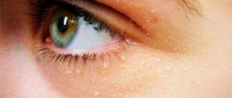
Xanthelasma of the eyelids, or flat xanthoma of the eyelids, is a benign formation in the form of yellowish plaques that rise above the skin. Location: inner corner of the upper eyelid. Sizes can vary from the size of a grain to a bean; plaques can be single or multiple. In this case, they can merge with each other and extend onto the bridge of the nose. The surface of xanthelasma is smooth, the plaques are soft to the touch and do not cause any pain upon palpation. Such formations are not prone to degeneration. But since they look quite unaesthetic, many would like to get rid of them.
For information: The term “xanthelasma” itself comes from the Greek language. "Xanthos" literally translates as "golden yellow", and "elasma" means plate or plaque. Such formations are observed in people all over the planet, regardless of race, age and gender. But it is noted that they occur more often in mature women, less often in men, and extremely rarely in children. It is noted that xanthelasma usually occurs in a pre-infarction state or signals developing atherosclerosis.
Xanthelasmas
In medicine, there are two similar concepts - xanthelasma and skin xanthoma. In fact, these are two names for one formation, but the first one usually means plaques on the eyelids, and the second one means all other localizations. Xanthelasma itself is not dangerous; they are not neoplasms or tumors, but belong to disorders of metabolic processes in the body. Xanthelasma disease develops when lipid metabolism is disrupted, resulting in fat deposition in the papillary layer of the dermis.
Xanthelasma is a benign neoplasm that looks very much like a flat plaque, soft and painless. There may be several such plaques, and when there is a large accumulation of them, they gather into one smooth tumor, causing a lot of inconvenience. Firstly, it all looks rather unpresentable, and secondly, it can interfere with the eyelid. Therefore, it is advisable to remove such neoplasms, even if they do not carry any physical harm.
Xanthelasma does not degenerate into cancer, but a person may suffer from a significant cosmetic defect. Sometimes, xanthelasma can signal serious problems in the body. You can get rid of xanthelasma only by removal - there is no other way to remove the formation from the skin. But since there are lipid metabolism disorders in the body, complex therapy must be prescribed. It includes treatment with a specific diet and medications that will help normalize metabolism and reduce cholesterol.
Such tumors progress slowly, but do not develop into malignant ones. There are various methods for treating xanthelasma, but in order to completely and permanently get rid of them, the only way to get rid of them is through surgery.
The most effective and aesthetic method of treatment is surgical laser removal. It leaves almost no marks on the face and body, the procedure is painless due to the preliminary administration of anesthetic medication (anesthetic).
The formation is evaporated layer by layer, while the doctor strictly controls the depth and area of influence of the laser beam. Healing occurs in 1-2 weeks depending on the depth of the plaque. Within 3-4 months, the area of formation removal will become invisible
Laser removal of xanthelasma. It is considered one of the best and most popular methods today. Its popularity is due to the fact that this procedure has many advantages and virtually no disadvantages.
Risk factors:
- diabetes;
- obesity;
- high blood cholesterol;
- thyroid diseases;
- lipid metabolism disorder - dislipoproteinemia.
- Hereditary factors may also play a role
ADVANTAGES OF LASER COAGULATION Laser removal of xanthelasma has a number of advantages compared to other methods:
- After laser exposure, the risk of new formations is reduced several times;
- Cholesterol deposits are evaporated using a CO2 laser, so this procedure does not have any mechanical or contact effect;
- In most cases, only one procedure is enough to achieve the desired effect;
- Exposure to a laser beam leaves virtually no marks on the skin, and therefore is considered the most effective procedure in aesthetic and cosmetological terms;
- After such procedures, virtually no recovery time is required.
Symptoms and causes
Xanthelasma at the initial stage can go unnoticed for a long time. People perceive small yellow spots on the eyelids as natural age-related skin pigmentation. Usually women disguise them with powder and eye shadow. If the plaques are large, many people mistake them for wen and try to remove them using various folk remedies.
Xanthelasma can be recognized by the following characteristic signs:
- formations from light yellow to light orange on the outer surface of the upper or lower eyelid;
- the size of the spots varies from 0.5 to 1.5 cm;
- may be single, or may merge and move to the bridge of the nose;
- the plaques do not itch, do not hurt, and do not cause any discomfort other than moral discomfort.
What is
Xanthelasma is a benign formation that occurs on the upper eyelids. It develops extremely slowly and is practically asymptomatic. Outwardly, it resembles small nodules (5 mm-1.5 cm) with a golden color. Xanthelasmas are soft to the touch. Their surface is smooth. In rare cases, uneven. They do not cause any inconvenience to humans, other than aesthetic ones.
As a rule, neoplasms appear only in one place, but it happens that they spread over the surface of both one and the other eyelid. When a second plaque appears next to the first one, the second one immediately merges. Multiple xanthelasmas can only be dealt with surgically.
As a rule, yellow plaques appear in older women. They occur extremely rarely in children and men.
Is cloudiness in front of one or both eyes dangerous - treatment of eye cataracts.
Diagnostic methods
Since plaques on the eyelids are caused by metabolic disorders, if such a phenomenon is detected, you should contact a dermatologist and endocrinologist. Diagnosis is based primarily on visual examination of the patient. Diascopy is also performed. The doctor presses on the plaque with a glass slide so that the blood drains out and it becomes possible to determine the true color of the formation.
The patient receives a referral for a detailed blood test to determine the level of cholesterol and lipid proteins, and a full examination to determine the quality of fat metabolism in the body.
It is extremely important to carry out a differential diagnosis to exclude pseudoxanthoma, a tumor of the sweat glands or the skin. After making an accurate diagnosis, it is determined what treatment will be and whether it is required.
Diagnosis of the disease
If the skin changes described above appear, you should consult a dermatologist. The diagnosis is made by visual examination. The diagnostic search consists of identifying lipid and cholesterol levels when examining blood from a vein, as well as investigating the cause of lipid metabolism disorders. Often, the patient requires consultation with a therapist, cardiologist, nutritionist, as well as instrumental studies (ultrasound of the liver, gastroscopy, ECG, echocardiography, etc.). Xanthomas are an indirect marker of an increase in cholesterol levels of more than 6.24 mmol/l.
Treatment methods
There are no specific treatments for yellow plaques on the eyelids. It is necessary to treat the underlying disease - obesity, diabetes or hypertension. The main goal is to restore normal fat metabolism in the body. For this purpose, a therapeutic diet is mandatory.
Its main points are as follows:
- Replacing animal fats with vegetable ones.
- Minimum consumption of salt and spicy seasonings, or better yet, eliminate them completely.
- Exclusion of fatty and fried foods, priority is given to vegetables and fruits, cereals.
- Complete cessation of nicotine and alcohol.
- Limit calories consumed per day to no more than 2000.
In addition, the doctor selects lipotropic drugs, these can be:
There are herbal preparations that have the same effect. As a rule, they contain extracts of plantain, birch buds, rose hips, and dandelion root.
To support the liver, it is recommended to take hepatoprotectors, for example, Essentiale. What else can a doctor prescribe to get rid of xanthelasma faster:
- choline chloride;
- ascorbic acid;
- a nicotinic acid;
- pyridoxine;
- cyanocobalamin.
Usually, doctors immediately warn the patient that treatment for xanthoma will take a long time and is not always successful. Your health will certainly improve, but there is no guarantee that the plaques will disappear. For this reason, the patient is informed about the various options for removing the lesions. Various methods can be used, and the optimal one is determined first of all by the doctor individually, since each of them has its own side effects and contraindications.
- Electrocoagulation. The plaques are cauterized by electric current, which may leave scars. This method is suitable for removing only small plaques.
- Cryodestruction. The formations are cauterized with liquid nitrogen; there is also a risk of scarring after the procedure.
- Surgical excision. The plaques are removed under local anesthesia using scissors or tweezers and then cauterized.
- Laser removal. A directed laser beam destroys the tissue of formations; it is considered the most modern method. But it is not applicable when the eyelid hangs over the plaque; in this case, surgical removal is indicated.
For information: removing xanthelasma does not mean a complete cure, so that it does not form again, you should continue the treatment prescribed by your doctor, remember a balanced diet and a healthy lifestyle.
Treatment of the disease
Xanthelasma has no specific treatment. If xanthelasma or xanthomatosis occurs against the background of a disease that may cause a disorder of fat metabolism, treatment of this disease is necessary. According to indications, insulin and thyroidin may be prescribed.
Patients with identified blood lipid disorders or increased cholesterol should adhere to a diet low in animal fats. To do this, animal fats are replaced with vegetable fats, for example, sunflower and olive oil. Such patients with xanthelasma are prescribed lipotropic drugs and drugs that reduce blood cholesterol. These include: cytamifene, parmidine, linetol, lipoic acid, lipamide, diosponin, clofibrate. Among herbal preparations, the following have a lipotropic effect: birch buds, dandelion root, rose hips, corn silk, plantain juice, immortelle flowers. It should be remembered that these drugs have a choleretic effect and their use is contraindicated in case of violations of bile drainage along the biliary tract.
In the treatment of xanthelasma, nicotinic and ascorbic acids, cyanocobalamin, pyridoxine, calcium pangamate, choline chloride, and essentiale are used.
Surgical treatment of xanthelasma is indicated for cosmetic reasons. It is carried out by excision of xanthelasma, laser removal, electrocoagulation, cryotherapy or radio wave destruction. Removal in most cases is performed under local anesthesia on an outpatient basis. Small xanthelasmas are usually removed using diathermocoagulation. Larger plaques are separated with scissors and tweezers. The edges of the wound are brought together and lubricated with iron sesquichloride, which forms a strong scab and allows the wound to heal by primary intention within 1-1.5 weeks. After separating xanthelasmas with a wide base, the edges of the wound are cauterized by diothermocoagulation. When xanthelasmas are combined with an overhanging skin fold on the eyelid, they are surgically excised together with excess skin of the upper eyelid.
To prevent redivision of xanthelasma after surgery, the patient is recommended to follow a dairy-vegetable diet with the exclusion of animal fats and limitation of carbohydrates. The daily intake of butter should not exceed 25g, sunflower oil - 75g.
Traditional medicine for xanthoma on the eyelids
Many patients are prejudiced against the removal of any formations on the skin or cannot undergo it due to contraindications. In this case, treating xanthelasma of the eyelids with folk remedies can help. Professional doctors do not recognize the effectiveness of traditional medicine methods, moreover, they do not recommend them. Patients often violate the recipe and dosage of the proposed products and, as a result, only harm their health and add new problems.
However, many are looking for just such recipes to get rid of formations on the eyelids without removal. Below are the most popular and acceptable ones.
- Honey cake. Combine a tablespoon of flour and natural honey, beat in one egg and stir until a fairly dense dough is obtained. Apply the mixture to the affected eyelid for 10 minutes, then rinse with warm water, but without soap. The procedure is carried out every other day until the xanthoma completely disappears. After this, a medicinal ointment can be applied to the eyelid.
- Ointment. Twice a day for 14 days, a thin layer of 1% mercury ointment or zinc-ichthyol ointment is applied to the eyelid. Both drugs have contraindications, so before starting a course of treatment it is recommended to consult with your doctor.
- Herbal collection for oral administration. Mix 100 g of rose hips, 100 g of mint leaves, 75 g of immortelle. Pour the resulting collection with water at the rate of one glass of boiling water for each tablespoon of collection. Close tightly and leave for at least 4 hours. The strained decoction is taken 4 times a day, 150 ml, thirty minutes before meals. The course of treatment lasts thirty days, then a break is taken for sixty days, after which the course can be repeated.
- Infusion of yarrow. Two tablespoons of herbs, dry or fresh, are poured with a glass of boiling water, covered and left for one hour. Then filter and take a quarter glass three times a day before meals.
- In the postoperative period, to eliminate scars and scars, traditional medicine recommends applying 0.5% hydrocortisone ointment to the eyelid.
Whether to try these recipes or not, everyone decides for themselves. Doctors do not deny their benefits in the postoperative period for rapid recovery, as well as for the prevention of relapses. But it is still not recommended as the main method of treatment.
Laser treatment – excellent results
Xanthelasmas are considered benign formations and have the appearance of flat yellow plaques, the size of which varies from 2 mm to 5 cm. Mostly such growths are located around the eyelids, much less often they can appear on other areas of the skin of the face. If you discover signs of this disease and decide to get rid of cosmetic defects on your face, you must first contact our doctors for examinations and medical history. Among the many ways to remove such formations, laser treatment is the most effective. In many cases, one procedure is sufficient to completely get rid of skin plaques. The State Research Center for Laser Medicine can offer its patients several methods using such devices:
- Surgical lasers “Atkus 15-3” and “Crystal”. Capable of solving many problems related to aesthetic medicine. When exposed to this technique, no burns or scars are formed on the skin. The natural pigmentation of the skin is preserved;
- device for radio wave surgery "Surgitron". It has proven itself best in dermatology. It can get rid of any skin tumors without pain or injury. When using this device, healing occurs very quickly;
- carbon dioxide laser (or CO2 laser) “Lancet”. Serve to remove warts, papillomas and other unwanted formations. After surgery using this equipment, patients do not experience any residual marks. The number of relapses is practically reduced to zero.
Methods of laser and radiosurgical treatment of skin pathologies and neoplasms are one of the most modern and effective methods in aesthetic cosmetology and dermatology. They are of interest in connection with an effective, fast and virtually painless method of treating benign tumors on the face and body. they reduce the number of relapses and complications, shorten wound healing time, allow for a one-stage procedure and provide a good cosmetic effect. The removal procedure is carried out under local anesthesia (applying an anesthetic cream or injection into the base of the formation) and is well tolerated. When removed, the tumor tissue is taken for histological examination to determine the degree of aggressiveness of the tumor. Healing of the wound defect occurs under a formed crust, which disappears within 4-14 days.
Reviews
Summary: Xanthelasma is not a malignant tumor; it can not be treated, but only perceived as a signal of certain changes in the body. If the plaques grow and cause moral discomfort to the patient, there are various methods for removing xanthoma. The use of folk remedies in this case is inappropriate due to low efficiency, but a rather high risk of injury to the organs of vision. Reviews recommend finding a good clinic and getting rid of plaques in a safe and modern way. There is no way to prevent the formation of xanthelasma.
Source: gsproekt.ru
How to treat
To get rid of golden plaques forever, going under the knife to a specialist who will remove them will not be enough. First of all, you need to determine the cause of their appearance and try to eliminate it. In addition, it should be prevented from occurring in the future. Otherwise, some time after the operation, the tumors will appear again.
Read why your child has red circles under his eyes here.
Recipes from our grandmothers to combat skin defects
Often, in order to cope with pathology, doctors offer their patients to use folk remedies.
Causes and symptoms
It is difficult to say why the yellowish plaques shown in the photo appear on the body. Experts identify several patterns and risk factors:
- metabolic disorders, namely problems associated with lipid metabolism;
- obesity;
- diabetes;
- liver diseases;
- pathology of the pancreas.
According to some studies, the formation of xanthelasma may indicate problems with the cardiovascular system. Thus, atherosclerosis was found in some patients.
All plaques are straw-yellow in color . They are soft to the touch and their surface is quite smooth. Sometimes several formations located nearby merge into one. The main places of localization are considered to be the upper and lower eyelids.
Xanthelasma is recognized as a benign neoplasm. Science knows of no cases in which a plaque became malignant. In addition, pain and any discomfort are excluded.
As a rule, growths occur in older women . Although men also sometimes encounter such a cosmetic defect. Education develops gradually. First, a tiny nodule appears on the eyelid, which gradually reaches the size of a large bean.
Description of the disease and its symptoms
“Flat xanthomas of the eyelids” is another medical name for the disease, due to the appearance in the form of flat plaques of beige, straw or yellow color. Growths protrude above the skin, on the border of the mobile and fixed zones of the eyelids, at the inner corner in the paraorbital region.
They are usually soft to the touch and do not cause a person pain or discomfort (inflammation, redness, itching, burning). They do not interfere with the full functioning of the eyelids, but as a cosmetic defect they do not look aesthetically pleasing. But this information will help you understand what kind of eye diseases a person has.
Xanthomas most often have a smooth surface and can be located either in a single form or, merging with neighboring neoplasms, transform into a group of tuberous elements running along the upper eyelid. Less commonly, skin growths appear on the lower eyelid, extending onto the bridge of the nose or closer to the temporal part. From the moment of appearance there is a tendency to grow slowly, and if appropriate measures are not taken, the plaques reach the size of a large bean.
Often, xanthomatous formations can be combined with growths located in other parts of the body, but modern medicine has not fully elucidated this reason.
The main risk group for eyelid disease is older women. The disease occurs less frequently in men. In children they are expressed in isolated cases and are often associated with metabolic disorders or a hereditary form of hypercholesterolemic xanthomatosis.
It is also worth paying attention to what dirofilariasis looks like in humans.
The video shows a description of xanthelasma of the eyelids:
Causes of the disease
Modern medicine considers the main reason for the appearance of eyelid xanthelasma to be a violation of the lipid metabolism of the human body, which is of a general or local nature. One of the primary manifestations of disorders, at the examination stage, is an increase in blood cholesterol, or certain fractions of triglycerides.
First of all, emphasis should be placed on curing the patient’s underlying disease, if present.
Patients most susceptible to the appearance of flat xanthomas of the eyelids are:
- with severe chronic liver damage (hepatitis of various forms, cirrhosis, Wilson-Konovalov disease);
- with obstruction of the bile ducts (inflammation of the pancreas, acute pancreatitis, gallstones);
- with developing atherosclerosis;
- with diabetes mellitus;
- with dysfunction of the thyroid gland;
- congenital deficiency of lipoprotein lipase (changes in biochemical blood parameters).
Sometimes the cause of flat plaques is alcohol abuse.
You may also find it useful to learn about how upper eyelid ptosis is treated.
The video shows the reasons for the appearance:
When hyperlipidemia occurs, cells oversaturated with lipids are localized in the upper dermal layers, granulating around small vessels. But there is an idiopathic form in which xanthoma appears. In this case, lipid metabolism, according to laboratory results, is normal, and at the initial stage of examination no initial pathology is detected.
It is also worth learning more about why the corners of your eyes and eyelids itch, and what can be done about this problem.
Xanthelasma removal
Such neoplasms never disappear on their own . Moreover, their number may increase. To avoid this, you should identify possible causes and eliminate them, but this does not always help.
Most often, growths are removed. Although such neoplasms are completely harmless, they spoil the appearance .
- Cryodestruction involves applying liquid nitrogen to the damaged area of the skin. The exposure time is determined by the doctor depending on the size of the plaque and some other factors.
- Laser removal is considered gentle, because during the procedure there is no risk of injury to healthy tissue. This method is completely painless and safe. Immediately after removal, the patient can go home. There will be no scars at the site of xanthelasma.
- The radio wave method is also safe because it is non-contact. Thanks to high-frequency waves, the cells of the growth heat up and begin to evaporate, while the patient does not feel pain or discomfort.
- Surgery involves local anesthesia. Scissors and tweezers are used to remove plaques. The rehabilitation period lasts 1–2 weeks. During this time, the wound tightens and heals. The disadvantage of the procedure is the possible formation of a scar.
- Electrocoagulation is sometimes used in conjunction with surgical excision. So, after removing the growth, the edges of the wound are connected and then cauterized with an electrode. A crust forms at this place, after which no traces remain.
Diagnostics
The diagnosis is made based on external examination. A patient who has characteristic neoplasms on the upper eyelid should undergo a consultation with two specialists:
First, a diascopy must be performed. This procedure involves pressing on the plaque with a glass slide. During this, it is bled, thanks to which it is possible to accurately determine the color of the neoplasm.
Xanthelasma should be distinguished from xanthoma. The first type of skin defect occurs only on the upper eyelid, and the second - on any part of the body.
A proven drug to combat the symptoms of glaucoma is Cosopt eye drops.
The endocrinologist should refer the patient for examination of lipid metabolism. If a violation is detected, the doctor will prescribe appropriate therapy.
This article will help you determine the causes and methods of treating red whites of the eyes.
Treatment at home
You can try to get rid of xanthelasma at home. However, this must be done carefully, since even folk remedies have their contraindications. It is best to consult your doctor first.
It is worth giving preference to herbal compositions that will improve lipid metabolism and facilitate the functioning of the pancreas and liver.
- In 1 tbsp. boiling water you need to brew 20 g of birch buds. Drink the finished infusion 2 tbsp. l. three times a day.
- 2 tsp. dry crushed yarrow is poured into 1 tbsp. boiling water and leave for an hour. The liquid is filtered and divided into 4 doses.
- You can take a decoction of dandelion root. To prepare this product you will need 1 tbsp. water and 1 tsp. dried herb.
- An infusion of mint, immortelle and rose hips provides a good effect. Take 100 g of all ingredients and pour 3 tbsp. boiling water The mixture should be infused for 4 hours.
Dietary recommendations
is to some extent , you should reconsider your diet.
- It is important to limit the amount of animal fats. We are talking about offal and fatty meats. It is better to use vegetable oil instead of butter.
- You should add at least 300 g of vegetables, except potatoes, and 200 g of fruit to your daily diet.
- You should consume fiber and avoid flour products made from premium flour. It is important to add bran bread, brown rice, oats, buckwheat, lentils and beans to your diet.
- Be aware of omega-3 fatty acids, which are found in flaxseeds, fish and nuts.
about 1.5 liters of water per day and giving up bad habits, including alcohol and smoking.
Preventive measures
To minimize the likelihood of developing xanthelasma, it is worth taking measures to prevent disorders that can lead to the formation of plaques.
- It is important to monitor your weight to avoid obesity.
- It is worth reviewing your diet, reducing the amount of fatty foods and adding fresh fruits and vegetables.
- Possible skin injury should be avoided.
- If desired, you can take decoctions aimed at normalizing lipid metabolism.
Yellow plaques that form on the eyelids are considered benign. Xanthelasmas do not disappear on their own, which means that, if necessary, they will have to be removed using a laser or another method. To avoid reappearance of the growth, it is worth adjusting lipid metabolism.
Source: dermatolog.guru
Prevention
Preventive measures for xanthelasma of the eyelids are individual in nature and are prescribed by a medical specialist in accordance with the results of a complete diagnosis of the patient.
For example, if a xanthoma is present, the patient does not suffer from a chronic disease, he needs to refrain from eating fatty foods. It is also advisable to drink up to 2 liters of water, and, if possible, replace the liquid with herbal infusions. Include fiber-rich foods in your diet, and increase the amount of fruits and vegetables you consume daily.
Patients with high cholesterol or impaired lipid metabolism should limit the consumption of animal fats, replacing them with vegetable fats. If this diet is ineffective, experts prescribe lipotropic or lipid-lowering drugs.
It will also be useful to learn about what to do when a speck gets into the eye in the upper eyelid.
Video shows xanthelasma removal:
Lipotropic agents promote the oxidation and breakdown of fats in the liver tissue. Lipid-lowering drugs must be used with caution, as their effects can negatively affect the condition of the liver and choleretic system.
If xanthomas have formed, the above preventive measures can limit the appearance of neoplasms, but are not able to clear the skin from resorption of the bumpy growths that have already appeared. This also applies to the use of various cosmetics (medical gels (Vidisik eye gel), creams and other procedures).
