Emmetropia is a term that describes a visual condition in which parallel rays coming from a distant object are focused by refraction precisely on the retina when the eye is relaxed. In other words, this is a normal state of refraction in which a person clearly sees distant objects.
Emmetropia is achieved when the refractive power of the cornea and the axial length of the eyeball are balanced, allowing light rays to focus accurately on the retina.
Definition
Emmetropia is a vision condition characterized by the fact that passing light rays are focused by refraction precisely on the retina of the eye, provided that it is completely relaxed. This is a completely normal manifestation, so a person sees very clearly even very distant objects.
Emmetropia of the eye occurs when the refractive power of the cornea and the axis of the eyeball are stabilized, allowing light to be accurately focused on the retina. With this phenomenon, a person has the clearest vision.
He sees equally well near and far from himself. When reading, if the object is located at a distance of approximately 30-33 cm, you need to make a minimum of effort, as a result of which the eyes get tired much later than in any other condition.
Emmetropia is a proportionate refraction in which the ratio of refractive power and the axis of the eye is optimal. When the indicators deviate in any direction, ametropia occurs.
Emmetropization
Many patients who have been diagnosed with this are interested in what it is - eye emmetropia. The description of this condition suggests that this condition is considered normal, but requires medical supervision.
Emmetropization is the process of emmetropia, which is controlled by incoming signals. The mechanisms that control this process are not yet precisely understood. Presumably, it is influenced by a genetic factor.
Emmetropization occurs as a result of ongoing active and passive processes. Passive processes involve enlarging the eyes to the required size as the child grows. The active process implies mechanisms for regulating the length of the axis of the eyeball.
Do you need glasses?
People who have just learned that they have -1 vision are concerned about one pressing question: do they need to wear glasses? With accommodative myopia, this is not necessary and even harmful.
There is a popular belief that wearing glasses for vision problems only makes them worse. In the presence of this type of myopia, this statement is one hundred percent true.
Muscles that are not strong enough to give the lens the desired shape will atrophy even more when wearing glasses, and vision will only worsen.
But with anatomical myopia, glasses are not only harmless, but also necessary.
The muscles that regulate the lens in this type of myopia work normally, but their excessive strain can further compress the eyeball, which is already degrading.
Therefore, neglecting glasses or contact lenses for such myopia is a sure way to rapid deterioration of vision.
However, wearing glasses may not be enough to correct anatomical myopia. Medical correction may be needed.
She has several types. The most modern and safe correction is laser vision correction. Its essence is that it cauterizes the cornea, making its shape flatter.
Because of this, the distance from the lens to the cornea is reduced, and the rays hit the reading plane in a focused manner.
Early correction method
A method of early correction is wearing special glasses that adapt the eyes to normal functioning.
They only need to be worn a few hours a day for several months. This method does not always help.
But with severe distortions of the eyeball, laser correction alone is not enough. Complex surgical repair is required. But it is better not to bring your eyes to this state by doing laser correction in the early stages of myopia.
Treatment of accommodative myopia, in turn, is simpler and does not require serious medical intervention. It is enough to develop muscle tone.
To do this, you need to perform visual exercises, use special eye drops, play sports and normalize blood circulation.
Emmetropic disorders
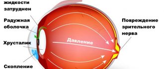
Disorders of emmetropia are called ametropia. In this case, light rays do not reach the retina. Ametropia is also called refractive error and emmetropia. Such disorders include farsightedness, nearsightedness and astigmatism. The ability of the eyes to focus light on the retina is based on such anatomical features as:
- eyeball length;
- curvature of the lens;
- curvature of the cornea.
If the eye has an excessively elongated axis, then the light flux is focused in front of the retina, which provokes the occurrence of myopia. If the light reaches the retina without focusing, then this is farsightedness.
The curvature of the cornea is also of great importance, since if it does not have an ideal spherical surface, then the light flux is refracted with disturbances and is focused rather unevenly, which provokes the occurrence of astigmatism. If the lens has an excessively curved shape, then this becomes the main cause of myopia, and if it is flat, then farsightedness.
Causes
There are two main reasons for this state of affairs: accommodative and anatomical. They differ greatly from each other, and it is the identified cause that determines how bad vision -1 is and what the further course of myopia will be.
Accommodative myopia indicates weakness of the muscles that control the movement of the lens. That is, this natural lens simply cannot take the desired shape for the correct refraction of incoming light.
This is typical of young children whose muscle development (including the eye) does not keep up with the growth of the body, which is why it becomes too weak to ensure its full functioning.
Anatomical myopia is a more serious disease. It is associated with changes in the structure and proportions of the eyeball.
It stretches, the location of the retina in relation to the lens changes and, therefore, the rays reach it in a defocused form.
Genetic predisposition plays a huge role in the occurrence of this type of disease, while the influence of external factors is not so significant.
To make a diagnosis, you need to undergo an ultrasound of the eyeball, during which accurate data on its structure and shape will be obtained.
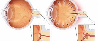
How does light pass through the eye?
When light passes through water or a lens, it changes direction. Some structures in the eye have refractive powers, similar to water and lenses, that bend light rays so that they converge at a specific point called a focus. This ensures clear vision.
Most of the eyeball's refraction occurs when light passes through the curved, transparent cornea. The natural lens of the eye, the crystalline lens, also plays an important role in focusing light on the retina. The aqueous humor and vitreous humor also have refractive abilities.
Nature has endowed the human eye with the ability to focus images of objects located at various distances. This ability is called accommodation and is carried out by changing the curvature of the lens. In the emmetropic eye, accommodation is needed only when viewing a close object.
What pathologies are there?
Emmetropia is higher visual acuity, which is considered normal. A person with such indicators has very clear vision and sees objects well within 1.5 m without eye strain. However, it is worth noting that in some cases various types of violations may occur, which include:
- physiological astigmatism;
- ametropia;
- amblyopia;
- myopia;
- farsightedness.
Astigmatism is an anomaly, which consists in the fact that the rays of the light flux are collected at one point, as a result of which a circle is formed on the retina. The larger it is in diameter, the less visual acuity. The depth of the lesion directly depends on the width of the pupil.
Ametropia is a disproportionate refraction in which the focus of the rays does not coincide with the retina. Emmetropia is a proportional refraction, and it belongs to the most perfect and harmless variety. Visual acuity in this case is 1.0 or more.
Myopia - the light flux is concentrated in front of the retina, resulting in the image being somewhat blurred. Such a person sees poorly at a distance but well up close. With farsightedness, the picture is blurry, as if in a fog.
Amblyopia is a serious pathology in which light does not reach the retina, which can occur with cataracts, cataracts on the cornea and irreversible disorders in the vitreous body.
Specifics of determining the quality of vision in children
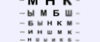
The Sivtsev vision test table has black letters printed on a white background of various sizes, the purpose of which is to determine visual acuity. The table contains various options of 7 letters of the same height and width, but having different sizes. According to the benchmarks, 2 equidistant points with an angular resolution of 1/60 degree are well visualized.
Sivtsev’s table helps determine the severity in the interval from 0.1 to 0.5 at a distance of five meters. The rows are presented as follows:
- the first ten rows differ in increments of 0.1 (V = 0.1 – 1.0);
- two rows have a difference of 0.5 (V = 3.0 – 5.0);
- three rows in 0.1. (V = 3.0 – 5.0).
There is additional information. The left column shows the distance, expressed in meters, from which a patient with 100% vision can see, and the right column shows the reference values when viewing from 5 m.
To get the most accurate results, you need to follow certain rules. The table cloth is placed five meters from the patient’s eyes. It must be illuminated by 2 fluorescent lamps. The light flux is directed onto the canvas, and not into the eyes of the subject.
Each eye should be checked separately, and they must be covered with the palm of your hand or taken an object for this. There is no need to put too much pressure on your closed eye, nor should you close your eyes. It is permissible to make no more than two or three mistakes. No more than three seconds are allotted for everything. The digital expression is in the last line where errors were made that exceeded the norm.
At the bottom there is a special column starting with “D = ...”, in this area they indicate the distance, expressed in meters, from which a person with ideal visual acuity is able to distinguish elements. Another column “V = …” numerical indicators when reading from five meters.
Golovin's vision test table consists of several optotypes, with the help of which the quality of vision is determined. It contains four rings of equal height and width with a gap. In accordance with the established norm, a patient with 100% vision visualizes 2 separate lying points located at an equidistant distance with an angular resolution of one minute, which is 1/60 of a degree.
Optotypes are placed as follows:
- 10 rows have a difference of 0.1 steps;
- two rows – 0.5 steps;
- three additional – 0.1 steps.
To decipher the results, additional columns are used: “D =” and “V =”. The first column suggests the distance, expressed in meters, from which a person with perfect vision can see, and the second column gives diagnostic results corresponding to the ability to visualize objects located up to five meters away.
The table will need to be illuminated using two fluorescent lamps with an illumination of 700 lux. Under no circumstances should the light beam be directed at the subject's face.
Reading results
With ideal vision quality, all characters in each line will be named accurately. If the sharpness is incomplete, it is permissible to make a mistake once in the rows from 0.3 to 0.6 and twice in the rows from 0.7 to 1.0. Visual acuity of 1.0 is considered normal. To determine visual function within less than 0.1, the patient must be gradually moved closer to the table every half meter and marks made on the floor.
The most common method for diagnosing vision is the Snellen vision test table, which was compiled in 1862 by the Dutch ophthalmologist Hermann Snellen. It looks like this: letters are placed in several lines, and their size decreases with each subsequent line, starting from the top.
The largest letters are located at the top. A person with perfect vision can easily read them from a 60-meter distance. The lower lines are read from 36, 24, 18, 12, 9, 6, 5 meters.
If it is planned to conduct the examination on a computer, then the subject is seated six meters from the screen. One eye is closed without putting pressure on it, and the signs begin to be read in order with the other. If a person managed to count the very last row, this indicates an excellent level of vision. Normally, you need to read one of the last lines while being at a distance of six meters (6/6) from it, some manage to do this from five meters.
Orlova's table is used to determine visual acuity in children under seven years of age. The lines contain pictures that decrease from line to line, starting from the top. On the left and right sides, normal indicators are indicated: D – distance, expressed in meters, from which a child with perfect vision can visualize all lines, the following readings are taken as the standard:
Ideal vision is fixed at 1.0 - this means that a preschooler can clearly distinguish the 10th line from five meters. If from a standard distance the child cannot see the first row, he should be placed closer to the table every half meter until he will not be able to see the items printed at the very top.
Before starting the procedure, the baby needs to be brought to a sign and asked to name several images. You should make sure that the child understands what is drawn in the pictures, at the same time, he must understand exactly what is required of him, only in this case can the study begin.
The younger the child, the faster he will get tired, so you should not show every picture. It is enough to invite him to name two or three images in each line. But if the baby in one of the rows did not name the picture, he is asked to look and try to name all the objects that are in this line. The problem series will indicate the quality of vision.
Rabkin's color tables for testing vision allow you to determine the presence of a disease called color blindness. This means that a person cannot adequately perceive colors and makes mistakes in their definition. Using this table, the ophthalmologist identifies this diagnosis, and also divides patients into three types:
- trichromant is an indicator of the norm;
- protoanope – inability to accurately perceive the color red;
- Deuteranope – inability to accurately perceive the color green.
Before the examination, you need to prepare, the patient must be in a good mood. Poor health can affect and distort the results. One image must be viewed for 10 seconds. Then they begin to examine another picture. The pictures show numbers on a polychromatic background, which makes it difficult to visualize the numbers.
Bailey-Lovey system
The technique looks like this: Latin letters are arranged in a table in a geometric progression. There are different intervals between the signs, which complicates perception. In the first row, the letters are large in size, they are located at a great distance from each other. In the next row, the size of the letters decreases, and the spaces between them become smaller. This diagnostic option is often used to certify pilots and drivers.
RORBA table
This technique is very similar to Sivtsev’s table, with the difference that it contains not 7, but 11 letter icons. They use not only the Cyrillic alphabet, but also the Latin alphabet. The system is placed on two sheets, on one - four rows, with capital letters, on the second - 12 lines. Thus, visual acuity can be detected in the range from 0.25 to 2.0.
There are a huge number of developments and programs on the Internet that can be used to test your vision. After undergoing an examination and discovering a deviation from the norm, you can consult a doctor, but this method should not replace examination by a specialist. It is not always possible to identify deviations from the norm in this way, which means missing the onset of the disease. To prevent this from happening, you need to regularly visit an ophthalmologist for preventive purposes.
In cases where the subject cannot accurately visualize the optotypes while being two meters away from them, the study is carried out by moving the fingers in front of the patient's face. The symbol for the meter is taken to be 0.2. The person is asked to name what he sees. If he cannot do this, they suggest determining whether a movement is being made to the right or left of him, while the ophthalmologist waves his arms alternately on each side. If the patient fails to do this, then use a light source. When a person does not react to light, they are diagnosed with zero vision.
Some people who have insufficiently good eyesight want to memorize the data from vision testing tables in order to undergo diagnostics and receive a certificate authorizing this or that activity. All the methods that doctors use in their practice are freely available on the Internet, they have a standard form. Anyone can easily learn and remember letters or table symbols and use them at their own discretion.
| Despite great advances in the field of recognition of eye features, test diagnostics using sign tables remains popular today. |
Kinds
Source: glazalik.ru Experts distinguish 4 forms of ametropia:
- Myopia or nearsightedness. Light rays are fixed in front of the retina. A person has difficulty seeing objects located in the distance. Myopia is most often diagnosed in school-age children. This is due to intense visual stress.
- Hypermetropia or farsightedness. The rays are fixed behind the retina, which makes it difficult to perceive objects that are close, while a person sees well into the distance.
- Astigmatism is a visual defect in which the rays lose the ability to converge at one point. As a result, all surrounding objects appear blurry or deformed.
- Presbyopia or age-related farsightedness. This type is mainly diagnosed in older patients. Presbyopia is associated with a decrease in the elasticity of the lens, when it cannot fully perform its functions.
For all of the listed forms of ametropia, weak, medium, and strong stages of the disease are distinguished - depending on how many diopters it is necessary to reduce or increase the refractive power of the eyes in order to correct vision.
Farsightedness and myopia are divided into several degrees of ametropia, depending on the number of diopters: weak - no higher than 3; average – no higher than 6; strong – above 6.
Astigmatism is measured by slightly different indicators: weak – up to 2; medium – up to 4; strong – more than 4. There is complicated and uncomplicated ametropia of the eye.
In the second case, the disease manifests itself in the form of a decrease in uncorrected perceptual acuity, but the ability to correct it remains.
If a pathological condition develops, then the disease becomes complicated - the visual analyzer changes. Such pathological processes occur when strabismus and asthenopia are diagnosed - the retina and optic nerve may change.
There are stationary and progressive ametropia, the latter includes myopia, which can be aggravated due to stretching of the scleral membrane and an increase in the length of the anterior-posterior axis.
Astigmatism
This is another type of ametropia. It can be caused by the refraction of the eye, which makes it difficult for the image to focus on the retina. Similar to myopia and farsightedness, three degrees of the disease are distinguished, only the division occurs along +4D and +2D.
Astigmatism is not cured, but corrected. But for this, its timely detection is important. Otherwise, visual acuity may sharply decrease, and even strabismus may occur. In children, astigmatism can be observed even in infants up to one year old. There are known cases of congenital forms.
Reference
It should be noted that a similar defect causing astigmatism is observed in all people, but it is within the normal range - 0.5D.
With this disease, the eyes become red and watery. A headache may occur. In children, this problem can be identified by observing their behavior. With astigmatism, the child will squint to see objects.
Contact lenses and glasses are used for treatment. Laser correction can be used.
Forms and degrees
There are the following forms of the disease: Mixed - the values of the optical axis and refractive power are outside the normal range. Combined - the indicators are normal, but their combination has a negative effect on refraction. Refractive – normally only the length of the optical axis.
Axial - on the contrary, only the magnitude of the refractive force is normal.
There are three degrees:
- more than -6D – strong;
- up to -6D – average;
- up to -3D – weak.
As a rule, myopia results in an enlarged eyeball.
How to test drivers' eyesight
A vision test for drivers is a mandatory procedure; it is done in order to issue a medical certificate to the driver. Only with such a certificate can you obtain a driving license. For diagnostics, they offer three tabular options: the Sivtsev and Golovin methods and the color perception test for drivers according to the Rabkin table.
The procedure is standard. The driver is asked to look at the signs alternately with one eye and the other. The doctor then interprets the results.
In cases where a deviation from the norm has been recorded, the driver will have to take measures to improve the quality of vision. You will need to undergo a course of vision correction, or you will need to purchase contact lenses or glasses, and possibly undergo surgery - this will be decided by the doctor.
When the problem is resolved, a re-check will follow, and if the indicators are good, the driver will be issued a certificate. If you have poor eyesight, you will not be given a certificate; such a driver is a danger on the road and can cause an accident.
Carrying out diagnostics
Refraction can be determined after using special means in the form of drops, since only in this case is it possible to obtain the most accurate information without distorting the result. In addition, deviations can be detected using special lenses. If objective methods for determining refraction are additionally required, then skiascopy and refractometry are used.
It is very difficult to recognize the presence of abnormalities in a newborn child, so the examination is limited only to detecting the presence of visual functions.
How to maintain good vision
Emmetropia is a normal state of refraction, with the clearest image and the greatest range of vision, tending to infinity. However, it is worth remembering that refraction may deteriorate over time, which is why you need to follow the recommendations of an ophthalmologist and undergo periodic examinations.
To maintain healthy vision for a long time, you must take into account such aspects as:
- nutrition;
- visual stress;
- stress;
- mental stress;
- vitamins.
Nutrition is very important, since the daily diet must contain many foods rich in vitamins and nutrients. It is necessary to minimize the amount of stress, reduce visual and mental stress.
How to prevent violations
To achieve this goal, there are several basic activities carried out, they need to be followed as closely as possible:
- visit a doctor once a year to check accommodative ability and vision in general;
- choose the optimal lighting mode, especially with visual stress, do not use fluorescent lamps;
- so that emmeotropic disease does not bother you, it is recommended to create a rational exercise regime, giving the eyes temporary rest;
- learn popular gymnastic exercises for the eyes, which are aimed at strengthening muscles and relaxing them;
- Together with a specialist, select the optimal vision correction, wear glasses exclusively for your refraction;
- Creating a balanced and nutritious diet will normalize the balance of nutrients and create normal vision.
An integrated approach to eye refractive disorders will provide high-quality prevention of all disorders and will stabilize visual functions.
Possible reasons for the appearance of nystagmus Causes of a disease such as conjunctivitis What diseases cause parasites to appear in the eyes Is bulging eyes disease treated?
What does visual acuity depend on?
Nowadays, many people have various vision problems. This is facilitated, first of all, by the fascination with digital devices - computers and smartphones, the constant use of which worsens visual acuity. But what does it depend on? Let's look at it in detail in this material.
What factors does vision depend on? What it will be like is influenced by many reasons. The most common cause of decreased visual acuity is poor heredity. Research by scientists has shown that if children are born to nearsighted parents, they will also suffer from vision problems. Other factors also influence the deterioration of vision clarity. For example, the most important ones are a sedentary lifestyle, regular viewing of the computer and TV, and incorrect posture, which children do not control while studying. All this leads to excessive overstrain of the eye muscles and, accordingly, a decrease in vision.
Also deteriorating the clarity of vision contribute to:
- eye diseases (especially dangerous diseases of the retina, cornea and lens);
- poor nutrition;
- refractive errors (myopia, hypermetropia, astigmatism);
- diabetes mellitus, thyroid disease;
- constant exposure to irritating and toxic substances.
All these factors lead to impaired visual acuity and, as a result, deterioration in clarity of vision. But there is also another parameter - the refraction of the eye, which determines how the image is projected onto the retina. What does it depend on? The most important factor is age. For example, newborns often experience farsightedness. As we grow older, the refraction of the eye shifts towards its intensification. In old age, everything happens exactly the opposite: the refraction of the eye changes in the direction of its weakening due to changes in the lens. So what needs to be done to maintain normal vision? We have prepared a list of tips for you below that will help you maintain clear and stable vision for many years.
- First of all, you need to reduce eye strain. If you work a lot at the computer, then every hour you need to rest your eyes for a few minutes. During this time, you can do several eye exercises. Eye gymnastics will be given below.
- Adjust your diet. You need to add to it those products that contain beneficial components for the eyes. This applies to carrots, blueberries, apricots, oranges, spinach, fish, and milk.
- Do eye exercises. During a short break from the computer, look left and right, up and down. Mentally draw geometric shapes with your gaze - a circle, a triangle, a square, a rectangle. At the end of charging, you can blink intensely.
- Get examined by an ophthalmologist. Even if nothing bothers you, you need to have your eyes checked at least once a year. This will help you notice impending vision problems in time and take appropriate measures.
MagazinLinz.ru team