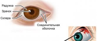
Causes of red whites of the eyes in a newborn
Immediately after birth
Redness that occurs immediately after the birth of a child can be caused by conjunctivitis, which the baby contracted in the womb from the mother. Conjunctivitis is an inflammation of the mucous membrane. The infectious form of the disease manifests itself immediately after birth, since the immune system is imperfect.
Dacryocystitis is another cause of red eyes immediately after childbirth. Inflammation of the lacrimal organs is considered a borderline condition between a congenital anomaly and an acquired disease. The congenital disease manifests itself only as hyperemia of the mucous membrane; other symptoms appear 2–3 days after the birth of the baby.
Other diseases that can develop in the womb and lead to hyperemia immediately after birth:
- Blepharitis - the causative agent of the disease is Staphylococcus aureus. The cause is infectious or allergic diseases during pregnancy in the mother. The development of blepharitis in a child is caused by a lack of vitamins, anemia, dry keratoconjunctivitis in a pregnant woman, which are not treated in a timely manner.
- Uveitis is an inflammatory process of the uveal tract. Its development in a newborn is provoked by the herpes virus, mycobacteria, Koch's bacillus or toxoplasmosis, which were discovered in the mother during pregnancy. The baby's optic tract becomes infected, and hyperemia appears at birth.
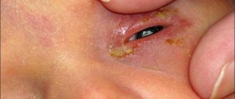
Conjunctivitis manifests itself in a newborn as a viscous, opaque or yellowish discharge from the eyes. The baby's body temperature rises.
If the birth was difficult and long, the baby could develop oxygen starvation. It appears as a result of prolonged presence of the head in the birth canal and compression. Due to the increase in blood pressure, the baby’s blood pressure rises, the vessels cannot withstand it and burst. The result is reddened whites of the eyes.
Sometimes oxygen starvation manifests itself as one small spot. This redness is considered physiological and does not entail complications or threaten the life of the newborn. The redness goes away within 2–3 days, even before the baby is discharged from the maternity hospital. Hyperemia may be a consequence of severe hemorrhage in the fetus or the result of passage through the birth canal. The integrity of the blood vessel of the eyeball is possible for the following reasons:
- Using medical forceps during the birth of a baby. They are designed to extract the fetus by the head in strict accordance with the natural biomechanism of childbirth. They are used for weak labor, acute intrauterine fetal hypoxia, and prolapse of umbilical cord loops. The fetus may experience more than just reddening of the whites, complications such as compression of the brain and hemorrhage into the cranial cavity are possible; in the latter case, the eye is very red and filled with blood.
- The use of labor stimulants - Dinoprostone and Oxytocin. Medicines affect the fetus, one of the side effects is eye hyperemia.
- Improper breathing of a woman during labor.
Causes of hemorrhages in the eye
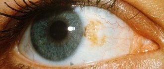
There are many reasons that cause hemorrhage. This is mainly due to the fragility of the capillaries in the eyeball. In this case, the vessel may burst after childbirth, as well as during strong crying of the child.
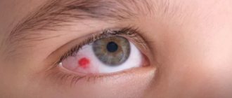
In newborns, the capillaries in the eye are so weak and fragile that they can break during emotional outbursts. That is why, if a young mother notices a small red spot on the baby’s sclera, this should not be a reason for much concern.
In children after one year, capillaries can burst as a result of various diseases (conjunctivitis) or injuries, viral diseases, such as the flu. In a child, hemorrhage may occur due to eye strain during frequent use of gadgets, so parents need to closely monitor the child, monitoring his regime.
Hemophthalmos
The cause of burst capillaries is high blood pressure. This can happen due to illness, during changes in weather, and also when swimming. When the question arises why a baby’s capillaries are so thin that hemorrhages appear in the eyes, not all parents immediately think that a possible cause may be a metabolic disorder.
New parents are afraid of the presence of toxins in the child’s body that accumulate as a result of some disease, hypovitaminosis, or the presence of diabetes. In this case, it is recommended to go with your baby to an ophthalmologist, who can accurately determine what the problem is and prescribe the appropriate treatment.
Sometimes hemorrhage in the eye occurs due to the fact that the baby rubs the eyes vigorously. This may be due to a foreign body entering the eye, too much light, or dry air.
Whatever the reason for the destruction of blood vessels in the child’s eye, parents should consult a doctor in any case.
Parents' actions
If a symptom has not gone away by the evening or morning of the next day, you should not ignore it. Even if there is a physiological problem, you should consult an ophthalmologist or pediatrician to prevent complications.
The child should be seen by a doctor as soon as possible. Before visiting the doctor, you can take the following measures to alleviate the baby’s condition:
- observe the rules of personal hygiene of the child;
- change bed linen (boil dirty ones);
- disinfect toys so that the child does not introduce infection into an already inflamed eye;
- ventilate the room more often, humidify the air in the baby’s room so that the eyes do not dry out;
- walk at least 2 hours a day;
- cut your nails and keep them clean;
- do not allow rubbing of eyes;
- do not allow watching cartoons for a long time, this will worsen the redness;
- make the light in the room less bright.
Before visiting a doctor, it is recommended to rinse your eyes with boiled, cooled water at least 2 times a day. Each visual organ is treated with a separate cotton pad. You should rinse from the outer corner to the inner one, this will prevent the development of an infectious process if the baby has conjunctivitis, blepharitis or uveitis.
If you have a runny nose, sneezing, lacrimation and hyperemia, this is an allergic reaction. Write down on paper all the foods your child ate that day. This will help the doctor identify the allergen. If an allergic reaction occurs, protect the child from flowers and pets, monitor the condition before visiting a doctor and write everything down in a notebook.
Description of the causes of eye hyperemia
The body of a newborn is very sensitive, so the use of any medications should be preceded by consultation with a specialist.
If parents notice red eyes in a newborn, they need to be wary, these are signs of an abnormal condition.
They can be caused by various reasons:
- Blockage in the nasolacrimal ducts. An ophthalmologist determines the signs, and treatment of dacryocystitis is carried out by massage and probing.
- Redness is often accompanied by purulent formations. The justification is the penetration of dust and dirt onto the mucous membrane. Because the eyes are sticking together, the baby begins to rub them. Mom needs to help him lift his eyelids, rinse carefully using warm boiled water. Removal of debris is carried out with a clean damp cloth. After these initial measures, be sure to contact the clinic.
- Intraocular pressure provokes redness; an ophthalmologist will help identify the source of the formation.
Cleanliness and proper care are necessary for all children in the first and subsequent periods of life. Young parents should be consulted about all preventive procedures during the mother’s pregnancy stage, then there will be no problems.
What not to do
If redness is detected, you should not begin self-treatment. Put eye drops, ointments and other medications aside. Do not allow other family members to treat the child themselves.
You cannot do the following:
- apply compresses - moisture will aggravate the situation;
- do not use home remedies (even washing with furatsilin or chamomile decoctions);
- do not touch the baby’s eyes unnecessarily - this creates additional conditions for injury to blood vessels;
- Don’t try to get the speck out if you can’t wash it off with water.
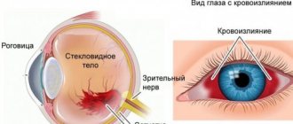
Forms of hemorrhage
Depending on the location of the burst vessel, ocular hemorrhages can occur in the sclera, anterior chamber, retina, or vitreous body. They can be distinguished visually upon inspection.
Into the retina
When the retinal capillaries are damaged, red “streams” appear, folding into a mesh.
Into the eye socket
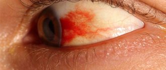
In this case, the size of the lesion can range from minor to global. The sclera may become covered with small red dots or hemorrhagic spots. In severe cases, the eyeball may become completely engorged with blood. Such damage is usually accompanied by limited eye mobility and decreased visual acuity. This situation most often occurs either as a result of injury or during surgery.
Into the vitreous
This hemorrhage looks like a bump on the sclera. If such damage is detected, you should immediately seek medical attention. Otherwise, the consequences can be very serious - up to partial or complete loss of vision.
To the anterior chamber
This hemorrhage differs from others in the mobility of the spot. It changes its position depending on whether the child is sitting or lying down. In most cases, it goes away without consequences within 7-10 days. If a speck of blood does not resolve for more than ten days, you should consult an ophthalmologist.
Treatment by a doctor
The cause of redness is determined by your family doctor or ophthalmologist. It is better to contact a specialist in a narrow field. Drops and ointments and other therapeutic and preventive measures are used.
Hyperemia caused by the allergen is eliminated with antihistamines. The most common method of treatment is taking second-generation antihistamines - Claritin, Kestin, Zyrtec. Immunostimulating medications are prescribed.
Dacryocystitis is treated by massaging the lacrimal sac, washing the eyes with Furacilin solution and Levomycetin eye drops. Treatment lasts 2 weeks. The regimen for using medications and rinses varies depending on the severity of the condition. If conservative therapy does not help, the doctor prescribes lavage of the lacrimal ducts.
The procedure is performed under local anesthesia; a probe is inserted into the lacrimal canaliculus and passed through the lacrimal ducts.
For ophthalmological pathologies of an infectious nature, treatment is long-term. Blepharitis in a newborn is treated with lotions of medicinal plants, eye washes and treatment of the eyelids with special gels.
For conjunctivitis, rinsing with a solution of potassium permanganate or furatsilin is prescribed. Both products have antibacterial and antiseptic properties. Next, antibacterial eye drops are used. In severe cases, medications are given by injection.
Uveitis is treated with anesthetics, glucocorticosteroids, antiviral and antimicrobial medications. Medicines are selected exclusively by the attending physician. The type of drugs chosen depends on the origin of the inflammatory disease.
Causes
Blood can only flow from the vessels, so the immediate cause of hemorrhage in the retina is always damage to the choroid that feeds it - a rupture or pathological permeability of the vascular walls (in the latter case, the volume of the accumulating effusion is much less). In turn, the main causes of vascular damage are:
- injuries (including those received during ophthalmic surgery) are statistically the most common factor, the share of which is 75-85%;
- vascular pathology (angiopathy) and the resulting degenerative processes in the retina (retinopathy) as an independent, primary disease;
- secondary angio- and retinopathy that developed as a result of other, more general diseases (diabetes mellitus, arterial hypertension, blood diseases, infections, atherosclerosis, etc.).
The direct consequences of retinal hemorrhage may be infiltrates, swelling, inflammation, etc., but the most likely and dangerous threat (especially when fluid leaks between the retina and the choroid) is retinal detachment, partial or total - which means a sharp deterioration in vision or irreversible blindness.
Separately, as a special form, retinal hemorrhage in newborns is considered, which has a clear cause-and-effect, clinical and prognostic specificity in contrast to “adult” hemorrhages.
Thus, with a worldwide trend toward an increase in the frequency and severity of retinopathy in newborns (especially premature infants), the frequency of postpartum retinal hemorrhages in infants reaches 20-30%. Important statistical patterns were revealed: firstly, such hemorrhages are extremely rare during cesarean section births, and secondly, their probability is much higher (about one and a half times) if a woman gives birth for the first time.
Subsequently, it was confirmed that the main cause of retinal hemorrhage in newborns is the birth itself, or more precisely, the nature of the birth process and methods of delivery. Difficult, complicated, protracted labor associated with compression of the fetal head, as well as mechanical obstetric and gynecological techniques (forceps, vacuum) sharply increase the likelihood of retinal hemorrhage, which is usually noted at 1-2 weeks of life. There were alarming concerns about a possible connection between retinal and brain hemorrhages, but subsequent in-depth studies using tomography fortunately did not find such a connection.