Diplopia is a visual impairment in which objects appear double and images overlap each other. Double vision usually occurs as a result of deviation of the eyeball. In this case, the object does not fall on the central region of the retina, but on the adjacent areas.
In all cases, diplopia leads to impaired binocular vision. If you close one eye, then most often the diplopia disappears. Monocular visual impairment occurs quite rarely, for example, when the root of the iris is torn off, subluxation of the lens, etc. with monocular diplopia, closing one eye does not lead to a positive result.
1.Why do I see double?
Double vision is a symptom that needs to be treated very carefully. Some causes of double vision may be relatively minor, but others require urgent medical attention. Let's try to figure out why it can cause double vision and what to do about it.
Why do I see double?
When you open your eyes and see a clear image of the object you are looking at, this is normal. But this seemingly simple and automatic process depends on the coordinated work of several areas of the vision system.
- The cornea
is the “window” of the eyes. The cornea helps focus the light entering the eyes. - The lens behind the pupil
also helps focus light on the retina. - The muscles
of the eyeball allow the eye to turn and look to the sides. - Nerves
are responsible for transmitting visual information from the eyes to the brain. - The brain
processes information received from the eyes.
When you see double, the cause may be problems with any element of this system.
A must read! Help with treatment and hospitalization!
Surgery
If the pathology is caused by disturbances in the functioning of the visual organs after injuries or in other cases, then surgical intervention is used to radically solve the problem. There are two ways:
- Recession of the eye muscle is its weakening through movement.
- Resection of a muscle is a reduction in its length.
The same operations are performed in the treatment of strabismus. They have been practiced in ophthalmology for several decades and show effective results.
2. Possible causes of this problem
When you see double, the cause may be problems with any element of this system.
Cornea problems
Problems with the cornea often cause double vision in only one eye. If you close it, you won't see double. Damage to the cornea distorts the incoming light and this becomes the main cause of diplopia. Infection can cause damage to the cornea (for example, shingles can cause distortion of the cornea). Scars on the cornea or dry cornea are other possible causes of double vision.
Problem with the lens of the eye
Cataracts are one of the most common lens problems that cause double vision. If there are cataracts in both eyes, perception will be impaired in each of them. Cataracts can usually be cured with simple surgery.
Eye muscle problems
If the muscles in one eye are weakened, that eye cannot move as smoothly as the healthy eye. And looking in a direction controlled by weak muscles causes double vision. Weakness of the eye muscles may be associated, for example, with myasthenia gravis, an autoimmune disease that blocks the muscles from stimulating the nerves inside the head. Early signs of myasthenia gravis are double vision and drooping eyelids. Graves' disease is another possible cause of eye muscle weakness. Graves' disease usually causes vertical diplopia, where double vision causes one image to be higher than the other.
Nerve problems
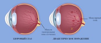
The causes of damage to the nerves that control the eye muscles vary. Multiple sclerosis can damage nerves anywhere in the brain or spinal cord. This means you will see double. Diabetes can affect the nerves connected to the muscles that move the eyeball. And this will also lead to diplopia.
Brain problems
The nerves from the eyes are directly connected to the brain. In addition, the entire process of processing visual information from the eyes also occurs in the brain. Therefore, there can be quite a few causes of double vision - stroke, aneurysm, increased pressure in the brain due to injury, bleeding or infection, as well as brain tumors, headaches and migraines.
What else could it be?
Double vision may begin on its own, without other symptoms. But depending on the causes, other symptoms may be present simultaneously with double vision:
- Strabismus;
- Pain when moving the eyes in one or both eyes;
- Pain around the eyes, for example in the eyebrow area;
- Headache;
- Nausea;
- Weakness in the eye muscles and general weakness;
- Drooping eyelids.
Visit our Neurology page
Visual gymnastics after a stroke
To restore vision, it is recommended to perform a set of special exercises. Gymnastics helps maintain tone, strengthen muscles and brain function. All types of gymnastics are based on relaxing the eyes, reducing intraocular pressure, combating irritation and visual tension. Exercises can include any eye movements, focusing the gaze at different distances, drawing objects and images in the air. Long-term therapy makes the picture clearer and eliminates distortions.
If you experience partial vision loss after a stroke, exercise can help retrain your brain. Doctors include exercises in physical therapy. The simplest exercise is with a pencil. You need to hold the pencil at a distance of 45 cm and follow it with your eyes, moving the pencil up, down and to the sides. You can't move your head. Also, a pencil is placed in front of the patient’s face and moved to the sides.
After a stroke, it is recommended to work with puzzles and drawings. Patients can complete drawings of objects and shapes, play word games, and solve puzzles. Exercises like this help retrain the brain and help it identify objects using vision.
3.Diagnostics of double vision
If for some unknown reason you begin to see double, you should immediately consult a doctor. With so many potentially serious causes of double vision, you need to find the cause as soon as possible.
Your doctor will likely use a variety of techniques to determine the cause of double vision. Blood tests, a physical examination, and possibly imaging tests such as a CT scan or MRI are the most commonly used.
General information is very important for diagnosing double vision. Here are some questions your doctor might ask:
- When did the double vision start?
- Have you hit your head, fallen, or been unconscious?
- Have you ever been in an accident?
- Does double vision get worse towards the end of the day or when you are tired?
- Do you have any other symptoms besides double vision?
- Do you have a habit of tilting your head to the side when you look somewhere?
Now focus your vision on some stationary object in your field of vision, for example, a window or a tree behind it.
- Are the two objects that appear as a result of double vision near each other? Or is one higher than the other? Or maybe they are located slightly diagonally? Are any of them larger or smaller?
- Are both images clear? Or is one clear and the other blurry?
- Close one eye and then open it and close the other. Does double vision go away when you look with one eye?
- Imagine that you are looking at a watch dial. Move your eyes in a circle, according to the numbers. Does double vision get worse at certain clock positions? Or is it getting better in some area?
- Tilt your head to the right and then to the left. Does double vision get worse/better in any of these positions?
About our clinic Chistye Prudy metro station Medintercom page!
Restoring vision after a stroke - basic principles
In order to be able to restore vision in the future, if you have a stroke, you need to urgently consult a doctor and get qualified help. An ophthalmologist treats visual impairment after a stroke. All stages of recovery must be approved by a doctor. It is very important to analyze the results and adjust the rehabilitation scheme in a timely manner.
Principles of vision restoration after stroke
- moisturizing the eyes with drops and gels;
- restoration of affected visual functions with the help of medications;
- regular and correct exercise;
- diet with vitamins, especially vitamin A;
- taking nutritional supplements.
After a stroke that causes vision loss, you need to seriously engage in general rehabilitation and restoration of visual function. Without making an effort, the patient risks remaining blind for life, because medications are not able to restore vision completely. Rehabilitation may include exercises, medications, and even surgery.
4.Treatment
If you see double, the most important thing is to find and treat the cause. In some cases this really helps.
- Eye muscle weakness or injury is treated with eye surgery;
- Myasthenia gravis is treatable with medications;
- Blood sugar levels in diabetes can be controlled with medications or insulin.
If a way to cure double vision has not been found, there are measures that can help reduce this symptom. This sometimes requires wearing an eye patch or special prism glasses to minimize the effect of double vision.
Species and types
- Monocular. When the healthy eye is closed, the patient continues to see a double image;
- Binocular. Double vision occurs when the patient looks at an object with both eyes at the same time.
This disease is also divided according to the type of plane in which the split image occurs:
- Vertical - distortion occurs vertically due to refraction created by the oblique eye muscles.
- Horizontal – image distortion occurs horizontally. The cause is a disruption of the normal functioning of the rectus ocular muscles.
- Cross - the projection of the image occurs crosswise: the image from the right eye appears on the left and vice versa. This causes great discomfort for the patient.
- Temporary. Occurs after a traumatic brain injury, overwork, during severe headaches, as a side effect of taking certain medications, due to severe poisoning and after alcohol intoxication.
- Congenital.
- Strabogenic. Occurs along with strabismus.
- Paralytic. The cause is paralysis of the extraocular muscles.
Symptom Description
The eyes see the same object from slightly different angles, but this difference is not noticeable to a healthy person. The brain processes the signals sent by the organs of vision and forms one accurate image from two pictures. With diplopia, a person sees both of these images at once, the brain does not process anything - this is the first and most important symptom of the disease.
You can verify that each eye has a different angle of view by closing one eye and looking at your finger relative to the background. Then, closing the other eye, you can see that the finger has “moved” relative to the objects in the background. With diplopia, a person observes two images of one object, not only horizontally, but also vertically, and also inverted crosswise.
At risk are patients who have been injured or suffer from diseases that may weaken the eye muscles.
Methods of traditional and folk medicine
The goal of therapy is to treat the specific cause of double vision. Main methods of therapy:
- Relief of volumetric processes in the orbit - puncture of the hematoma, removal of the tumor.
- Therapy of stroke, neurological and neuropathic disorders.
- Therapy of diseases of an inflammatory or infectious nature , which is carried out using diuretics, antibiotics and anti-inflammatory drugs.
- Therapy of the main disease . Correction is carried out to restore the functioning of the optic nerve.
- Prismatic correction to reduce pathology . In this case, it is prescribed to wear special glasses, made individually, which have a lens offset in the center. It is also common to use six prismatic diopters for each eye.
- Visual gymnastics . This is a set of exercises aimed at expanding the field of vision and normalizing binocular vision. These exercises can be done at home. To do this, you need to fix a sheet of paper with a marked strip on the wall. Then focus your gaze on this strip and turn your head in different directions. The goal of the exercise is to keep the image intact. The exercise is effective for incomplete diplopia. For sensory diplopia, the patient should combine a pair of distant strips into one.
- Surgical intervention . The operation can be performed by moving the muscle slightly back, after which the transected tendon is sutured to the sclera. Or the muscle is shortened to compensate for the work of another muscle.
Folk remedies
Therapy does not only consist of traditional methods. Treatment of diplopia with folk remedies is very effective:
- Prepare a tincture of crushed lavender leaves, valerian powder and a liter of white wine . The ingredients must be mixed and infused for three days. The container with the possessed must be shaken from time to time. After the product has infused, it needs to be strained. Take the tincture half an hour before meals, a tablespoon every day.
- Flower pollen is known for its healing properties . For a month you need to take a small spoon in the morning and evening.
- Vitamin collection . You need to drink regularly. The tincture can be prepared from the fruits of viburnum and rose hips. Pour the ingredients in equal quantities with water and boil for 15 minutes over low heat. After cooling, the tincture must be strained. Take 100 grams in the morning and evening before meals.
Diagnosis of the disorder
Diagnostics are carried out to determine the localization of the disorder to identify the causes of the disease and further prescribe treatment. Pathology affects:
- eye;
- optic nerve;
- auxiliary eye apparatus;
- brain.
Diagnostics involves traditional procedures - ophthalmoscopy, viziometry. The conjunctiva, visual acuity, color perception and light beam refraction are also examined. Next, a test is carried out containing questions that the patient must answer.
Additional studies are carried out using CT, MRI, ultrasound. If necessary, the doctor can refer the patient for examination to other specialists: neurologist, neuro-ophthalmologist, oncologist, psychiatrist, rheumatologist, endocrinologist, dermatovenerologist,
In the case of strabismus, the disease is diagnosed using coordimetry and diplopic provocation.
Such methods represent the implementation of test vision control. A man looks at a moving light source. The resulting image is transferred to a coordinate map. In this way, damage to any of the muscles is determined.


