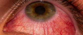
Conjunctival hyperemia is redness of the mucous membrane, which occurs against the background of various eye diseases. It appears due to blood flow, which is caused by infection or mechanical damage. In this material we will look in detail at the causes of hyperemia, symptoms and methods of treating it.
The main function of the mucous membrane of the eye is the natural protection of the visual apparatus from mechanical stress and injury, viruses, bacteria and other pathogens.
Causes of hyperemia, or red eye syndrome. a bunch of. Most often, redness indicates the appearance of conjunctivitis. But other reasons can provoke the development of this disease:
- blepharitis, phlegmon, stye (chalazion), iritis, etc.;
- various eye injuries;
- allergy;
- diseases of the cardiovascular system;
- pathologies of cerebral vessels;
- diabetes mellitus and other diseases of the endocrine system;
- influence of external factors (ionizing radiation, chronic stress and lack of sleep, high visual load, etc.).
Conjunctival hyperemia: what is it?
Basically, this condition develops against the background of an inflammatory process on the mucous membrane of the organ of vision, or in the layer of blood vessels. The reason for this phenomenon is the impact of negative factors. Hyperemia was also observed against the background of eye injuries, diseases of the iris, and eyelids.
There are two forms of the pathological condition: acute, chronic. In the first option, redness occurs very quickly, first on one side, then on the other.
Sometimes hyperemia is accompanied by the separation of purulent contents, and a change in the shade of the eyeball and cartilage is recorded.
The chronic course of the pathology is often accompanied by severe itching, the feeling of a foreign object in the eyes, and constant tension in the organ of vision. The purulent discharge is not profuse, redness extends only to the upper part of the cartilage.
Treatment of conjunctival hyperemia
Inflammatory diseases of the conjunctiva require complex treatment. The patient may be prescribed the following medications:
- Levomycetin drops;
- Tobrex;
- Phloxal;
- Tetracycline ointment.
In addition, it is recommended to frequently rinse your eyes with Furacilin solution. You can prepare it yourself or buy it ready-made at the pharmacy. Wash the eyes from the outer eye to the inner one, take a separate cotton swab for each visual organ.
If the inflammation is severe or caused by eye injury, then systemic antibiotics are prescribed. They should be taken for at least 5 days. If the drug Azithromycin is prescribed, then it is taken for only 3 days, which is associated with a prolonged effect.
Traditional methods also help well. It is recommended to frequently wash your eyes with a decoction of chamomile or sage. In some cases, tea bags can also help reduce inflammation.
Diagnostics
To identify the causes of red eyes, your doctor will need to perform several diagnostic tests:
- Questioning the patient. The doctor identifies disturbing symptoms; people often complain of dryness and pain in the eyes.
- A general examination, during which the doctor may use a slit lamp to view details on the surface structures of the eye. When the organs of vision are red, purulent exudate may be released, increased or decreased production of tear fluid, damage to the mucous membrane, foreign objects and other formations.
- Fundus examination. This technique allows the doctor to evaluate all intraocular elements. The person is first instilled with Atropine or similar drugs that disrupt the process of pupil accommodation. Using a slit lamp, the doctor can evaluate the entire internal structure.
- Assessment of visual acuity using diagnostic tables. The more lines a person sees, the better the quality of his vision. The method is used to eliminate the possibility of decreased visual acuity in many diseases that cause redness of the mucous membrane.
- Measurement of intraocular pressure to eliminate the possibility of glaucoma formation.
- Bacterial culture to detect the infection and the antibiotic to which it is sensitive.
- PCR. This is a virological method with which the exact type of pathogen is determined.
- Ultrasound of the eyeball. Using directed ultrasonic waves, the position and condition of the intraocular elements and the eyeball itself are assessed.
- MRI, CT. Such testing is often performed when it is impossible to accurately identify the cause of the condition using other methods. On the screen, the doctor will see a layer-by-layer image of the visual organs, nerve fibers, and brain. Using the test, you can accurately determine the cause of redness.
Based on genetic tests, the doctor determines the exact cause of the disease, and only then can treatment begin.
Diagnosis and treatment
Therapy is prescribed only after consultation with a competent ophthalmologist. The conjunctiva is examined using a special magnification lens (ophthalmoscopy). During this procedure, a violation of the microcirculation of the eye is detected (overflow of blood vessels and dilated lymphatic ducts). Stagnation of fluid in the eyeballs is detected.
Scrapings from the conjunctival membrane and purulent discharge from the eyes (if any) are also taken to examine and identify the infectious agent that caused the inflammatory process.
Treatment is prescribed after identifying the cause that provoked the appearance of hyperemia. If the redness of the membrane is caused by conjunctivitis, then treatment is carried out using eye drops that contain local anesthetics, such as Trimecaine, Lidocaine, Pyromecaine. In addition, potassium permanganate, Furacilin, brilliant green, Dimexide, Oxycyanate are usually prescribed, which are used to wash the eyelids and eyelashes.
Important! Solutions for washing the conjunctival membrane using such antiseptics are prepared exclusively in the dosage prescribed by the doctor, otherwise an eye burn is possible.
After eliminating the discomfort and washing the eye, instillation of antibacterial, antiviral, sulfonamides, and antiallergic medications into the conjunctival sac is carried out.
Read in a separate article: Eye strain: symptoms and eye drops for tension and fatigue
Bacterial damage to the conjunctiva can be eliminated using antibacterial drugs or sulfonamides (Albucid). If the conjunctiva is inflamed due to viral penetration, the disease is eliminated with the help of Kerecid, Florenal and other antiviral agents. The allergic form is treated with antihistamine drops, which contain Dibazol and Diphenhydramine.
An ophthalmologist may prescribe the following medications:
- Ointments with antibiotics (Tetracycline, Erythromycin, Gentamicin), with antiseptics (Yellow mercury ointment).
- Antiseptic drops (Sulfacyl Sodium, Piloxidin), antibacterial (Levomycetin), anti-inflammatory (Diclofenac, Olopatodin, Dexamethasone), antihistamine (Fenistil, Suprastin).
Treatment is carried out until all clinical signs of the underlying disease completely disappear.
Important! In no case is it practiced to apply various blindfolds to the eyes, as this procedure promotes the rapid proliferation of bacteria, which leads to complications of the eye disease.
Causes
A common cause of the development of conjunctival hyperemia is ignoring the rules of personal hygiene. In this case, the mucous membrane gets infected, and the disease develops acutely.
The following varieties are found:
- Adenoviral. It develops during mass epidemics and is distinguished not only by obvious redness of the mucous membrane, but also by the appearance of small watery blisters on it. The disease is accompanied by an increase in the behind-the-ear and submandibular lymph nodes;
- Follicular (chlamydial). The cause of this pathology is an untreated sexually transmitted infection. The disease is not too severe and complications occur rarely;
- Gonorrheal. One of the most severe infections, the spread of which through the mucous membrane sometimes leads to loss of vision. Treatment of pathology in this situation should be carried out only in a hospital;
- Dry. Hyperemia can develop when exposed to hot conditions or in older patients. The main sign of pathology is the feeling of a foreign object;
- Viral. It is considered a childhood infection, since the maximum number of people affected are children of school and preschool organizations. If you consult a doctor in a timely manner, it goes away completely without a trace.
In addition, hyperemia of the mucous membrane can be of an allergic nature. It develops during the flowering season of plants, or is associated with household allergens (dust). The disease is accompanied by lacrimation, swelling, and hyperemia.
Chronic conjunctivitis occurs for other reasons. The most common are:
- Mechanical irritations. Occurs when tiny foreign bodies come into contact with the mucosa. Workers in flour mills, textile factories, construction and ceramic shops often suffer from chronic conjunctivitis;
- Chemical exposure. The disease develops when being in smoky, gas-polluted rooms, when in contact with toxic fumes from acids and alkalis;
- Pathological abnormalities in the functioning of other organs. Problems with the gastrointestinal system, vitamin deficiency, diseases of the sinuses, and helminthic pathologies can cause hyperemia;
- Poor eyesight. Avoiding glasses can also lead to redness of the conjunctiva.
Redness of the eyes is also observed in the following diseases of the organs of vision:
- Scleritis;
- Barley;
- Phlegmon;
- Glaucoma;
- Blepharitis;
- Chalazion.
Regardless of what caused the development of the pathology, you must immediately consult a doctor.
Competent treatment is the key to a speedy recovery. Self-medication in this situation is strictly prohibited, as it can lead to even greater complications.
What is the cause of the disease?
Most often, conjunctival hyperemia appears due to failure to properly comply with hygiene rules and infection. It is the conjunctiva that first encounters inflammatory agents. A person can get an infection in the eye through a handshake, or by involuntarily rubbing the eye.
Red eye syndrome can be a symptom of the following pathologies:
- scleritis;
- uevitis;
- barley;
- phlegmon;
- chalazion;
- iridocyclitis;
- acute attack of glaucoma;
- tumor process;
- diseases of the eyelids
Transient hyperemia of the conjunctiva is not a disease, but only a symptom
The cause can also be cardiovascular diseases, cerebral vascular pathologies, and endocrine disorders.
The chronic process can be caused by the following reasons:
- dust or smoke in production;
- contact with acids, alkalis, fumes of toxic substances;
- hypovitaminosis;
- anemia;
- sinusitis;
- helminthic infestations;
- chronic gastrointestinal diseases.
Causes of hyperemia
For your information! One of the common causes of hyperemia is failure to comply with personal hygiene rules (for example, using someone else's towel, rubbing the eyes with dirty hands, improper care of lenses, etc.).
Other reasons experts include:
- Uveitis (inflammation of the choroid of the eyeball).
- Scleritis (acute inflammation of the deep layers of the sclera - the white membrane of the eye).
- Barley (purulent inflammation of the hair follicle of the eyelash).
- Iridocyclitis (inflammation of the iris and ciliary body).
- Purulent inflammation of soft tissues as a result of infections in the mucous membrane of the eye .
- Acute glaucoma (sudden increase in intraocular pressure).
- Mechanical damage and trauma to the conjunctiva.
- Allergic reaction to cosmetics, household chemicals, pollen, etc.
- Blepharitis (inflammation of the edges of the eyelids).
- Chalazion (chronic inflammation of the eyelid margin).
- Stress and chronic fatigue.
- Radioactive radiation.
- Constant lack of sleep.
- Increased visual load (especially when working at a computer).
Know! In addition to infectious and inflammatory eye diseases, the cause of hyperemia can be cardiovascular pathologies, diabetes mellitus and other endocrine disorders, cerebral vascular diseases, etc.
Causes
Most often, redness of the mucous membrane is observed with conjunctivitis. This ophthalmological disease can be provoked by pathogenic bacteria, viruses and allergens. In some cases, pathogenic fungi lead to conjunctivitis. This disease is usually accompanied by burning and pain in the eyes. The eyelids become swollen, and pus leaks from the corners of the eyes. In the mornings, the patient cannot open his eyes normally, as the eyelids are strongly stuck together with purulent masses.
Other diseases of different etiologies can also lead to eye hyperemia. Redness occurs most often in the following cases:
- For serious ophthalmological diseases - barley, chalazion, blepharitis, iritis and a number of others;
- Injuries to the visual organs, in which the integrity of the membranes or structures is disrupted;
- Allergy;
- Diseases of the heart and blood vessels;
- Endocrinological diseases, most often diabetes mellitus;
- Diseases of the brain in which cerebral circulation is impaired.
Some external factors can also provoke redness of the mucous membrane. This may be too dry or dusty air, some chemicals and work in hazardous industries. In some people, the conjunctiva turns red in the frosty air, in this case they speak of an allergy to the cold.
Chronic fatigue and lack of sleep can trigger redness. The cause of this problem may also be working at the computer for too long.
Types of hyperemia
Depending on how deep the infection has penetrated, redness is divided into two types: ciliary and mixed. Ciliary hyperemia is diagnosed when the infection spreads deep into the eye. In this case, the vessels are not the standard red, but more blue in color. This condition indicates that deep-lying arteries are affected.
Mixed hyperemia is called hyperemia, in which infection occurs not only of the conjunctiva, but also of the ciliary body. In this case, the infection penetrates the organ of vision through the blood.
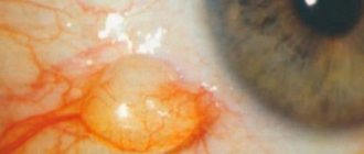
Focal hyperemia
Focal redness is usually caused by bleeding or the presence of tissue elements. The most common cause of subconjunctival bleeding is trauma. Although the bleeding may seem heavy, no treatment is required unless all other structures are damaged. If there is no history of trauma, then the possibility of systemic coagulation disorders should be investigated. Prolapse of the third eyelid gland is the main cause of red masses. The gland is returned to its place by attaching it to the eye socket or holding it in place using a special pocket. Conjunctival neoplasia is rare, but a biopsy of any questionable lesions is always indicated. Episcleritis and nodular granulomatous episcleritis are inflammatory diseases that often affect the lateral aspects of the corneal limbus and respond to topical and/or systemic treatment with prednisone and azathioprine.
Treatment of hyperemia
Treatment is selected depending on the cause (or pathogen) of the disease. Drops, ointments, topical antibiotics, etc. are used as therapy.
Thus, patients are prescribed the following types of medications :
- Drops and ointments with an antibacterial effect (Tobrex drops, Sulfacyl sodium drops, Tsipromed drops, Levomycetin ointment, Oftocipro ointment, etc.). They are used for the development of bacterial infections in the eye area.
- Antiviral drops and ointments (Ophthalmoferon drops, Actipol drops, Dexamethasone drops, Zovirax ointment, Acyclovir ointment, etc.). The drugs are indicated for viral etiology of the disease.
- Antihistamines (Allergodil drops, Claritin tablets, Cortisone tablets, etc.). They are recommended in case of an acute reaction to a particular allergen.
It is worth noting! It is recommended to instill drops into the conjunctival sac, slightly pulling the lower eyelid down. Ointments are placed there (in small quantities).
The products must be used 4-5 times a day. The course of treatment is determined individually.
In case of hyperemia, it is not recommended to make compresses or apply bandages to the eyes , as this can only aggravate the disease.
Eye hyperemia: treatment
Eye hyperemia can be a symptom of a number of serious diseases, so if it occurs, you should make an appointment with an ophthalmologist. The doctor will conduct a thorough examination, make a diagnosis and prescribe treatment.
Depending on the symptom of which disease is hyperemia around the eyes, the following therapeutic measures may be prescribed:
- instillation of local anesthetics into the eyes (trimecaine, lidocaine);
- washing the mucous membrane of the eye and eyelids with an antiseptic;
- prescription of antiviral agents or antibiotics (depending on the type of pathogen);
- taking corticosteroids;
- prescribing antihistamines (if the cause of hyperemia is an allergy).
The selection of medications should be carried out by the attending physician, taking into account the diagnosis and the individual characteristics of the patient’s body. Self-medication is unacceptable!
In case of hyperemia, you should not apply bandages to your eyes; this can only worsen the patient’s condition.
In our online store we offer you the following drugs for the treatment of eye hyperemia, which can be used as part of complex therapy for this disease:
- Amino complex - amino acid complex;
- Complex B-50;
- Beta Carotene;
- L-Carnitine;
- OCU Support;
- Taurine;
- Blueberry complex.
Before using any drug, you should consult your doctor.
Symptoms
Depending on the form and type of disease with hyperemia, symptoms can vary significantly.
General signs of pathology are:
- Redness of the eyes;
- Burning;
- Swelling;
- Unbearable itching;
- Constant lacrimation;
- Discharge of pus;
- Pain in the eyes;
- Fear of bright light.
The disease can occur on one side or both. When one eye becomes infected, the inflammation often spreads to the other. Sometimes the pus-like discharge becomes so abundant that the patient cannot open his eyes in the morning.
With an allergic form of pathology, a cough may be added to the main symptoms, and with a viral form, enlargement of the lymph nodes may be possible. After exposure to toxic substances, hyperemia is accompanied by acute pain.
Risk group
The risk groups for acute or chronic eye redness include the following categories of patients:
- suffering from systemic diseases, for example, hepatitis, diabetes, hypertension;
- people whose work involves the use of chemicals and harmful substances;
- people who, on duty, spend a long time on the street, even despite bad weather conditions (road workers, traffic police officers);
- patients who sit in front of a computer for long periods of time, use phones, TVs, tablets and other electronic devices, and read books in poor lighting.
If a patient is at risk, he needs to have his vision examined by an ophthalmologist annually to prevent many diseases.
Hyperemia of the conjunctiva of the eyes: diagnosis
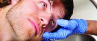
The patient’s quick and complete recovery directly depends on high-quality diagnosis and prescribed treatment. The visual examination takes only a few minutes. The doctor uses special instruments to examine the condition of the mucous membrane of the eyes, and then, based on the results obtained and the patient’s complaints, makes a diagnosis. Depending on what caused the disease, additional laboratory tests and consultations with other specialists may be required: a therapist, an allergist, a surgeon.
Etiology
Uncomfortable sensations in the eye area, their redness are phenomena that are familiar to many people. Constant strain on the organs of vision associated with working at computer monitors, lack of sleep, lack of vitamins, all this leads to the fact that the protective mucous membrane of the eye (conjunctiva) becomes red. This sign indicates the appearance of an inflammatory process and should not be ignored.
In most cases, eye hyperemia indicates the presence of a disease such as conjunctivitis - inflammation of the mucous membrane of the eye. But in some cases, redness of the conjunctival membrane can be caused by more serious eye problems, such as iritis, iridocyclitis. It can be a sign of eye injury, tumors of various etiologies, changes in the condition of blood vessels, problems with the brain, or heart disease. Due to the loss of activity of the walls of the blood vessels of the eye, they protrude, which contributes to the appearance of hyperemia.
Hyperemia of the conjunctival membrane has several varieties, differing in cause and symptoms:
- Ciliary. It is caused by penetration of an infectious agent into the iris of the eye, damaging its vessels.
- Mixed. The ciliary body and conjunctiva are involved in the inflammatory process. This process is caused by the penetration of the pathogen through the blood.
Hyperemia can be accompanied by the formation of barley on the eyelid, inflammation of the edges of the eyelids (blepharitis), chalazion (inflammation of the subcutaneous gland of the eye), and orbital phlegmon.
Types of inflammatory process
Conjunctivitis can be acute or chronic. An acute process is characterized by an acute onset. First, the inflammation affects one eye, and over time the second is also involved in the process. The discharge is mucous or even purulent in nature. They stick the eyelids together so tightly that after waking up it is difficult for a person to open his eyes. The conjunctiva is red, swollen and cloudy.
The chronic form lasts quite a long time. Patients complain of eye fatigue, heaviness of the eyelids, and itching. Conjunctival hyperemia is mild. Discharge from the eyes is scanty and mucopurulent in nature.
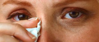
It is very easy to get an infection in the eye. Don't rub your eyes with dirty hands
Acute conjunctivitis, in turn, is divided into the following forms:
- adenoviral. It is characterized by the formation of pinpoint vesicles, enlargement of the submandibular and parotid lymph nodes, as well as the appearance of infiltrates on the cornea, which spoil vision;
- gonorrheal This is a serious form of inflammation, in which the outer shell of the eye is sharply hyperemic, swollen and bleeding. The process can spread, even moving to the cornea. The appearance of ulcers on it is fraught with loss of the eye;
- chlamydial Characterized by the formation of granular follicles. Develops against the background of urogenital chlamydia;
- viral keratoconjunctivitis is more often diagnosed in childhood;
- allergic. Most often occurs during the flowering period. Household allergens are also often the cause of increased sensitivity. In addition to hyperemia of the mucous membrane, swelling of the eyelids and profuse lacrimation appear;
- dry. It mainly occurs in older people, as well as in those living in hot climates. When moving the eyeballs, a feeling of the presence of a foreign body appears.
If hyperemia of the mucous membrane of the eye is accompanied by swelling and pain, this may indicate the presence of the following diseases:
How to treat viral conjunctivitis
- allergic reaction. Allergens can include household chemicals, food, medications and much more;
- blepharitis - inflammation of the eyelids, in which they thicken;
- chalazion - growth and inflammation of the meibomian gland;
- barley;
- orbital phlegmon.
Symptoms of conjunctivitis depend on its form. Still, experts identify common symptoms:
- edema;
- discharge;
- itching and burning;
- lacrimation;
- pain;
- photophobia.
Depending on the underlying cause, conjunctival hyperemia may be accompanied by pain, a sensation of a foreign body in the eye
Depending on the degree of damage to the mucous membrane of the eye, experts distinguish three main forms of the disease:
- Conjunctival injection. The mucous membrane that covers the eyelids turns red. Hyperemia of the transitional fold and eyeball also occurs. The pathological condition is accompanied by photophobia, lacrimation, pain and burning in the eyes.
- Ciliary injection. Redness affects the iris of the eye. Deeper tissues are involved in the process. Most often, this form occurs with iridocyclitis and iritis. In this case, the mucous membrane has a purple tint, it is smooth, and there are no blood vessels protruding above it, as is observed with a conjunctival injection. This is because the inflammatory response is caused by the blood vessels inside the eye.
- The mixed form occurs during acute infectious processes, which are characterized by a hematogenous transmission route. The iris changes its color and a purulent infiltrate is observed.
Determining the source of eye hyperemia
When the eyes of an animal are red, the localization of the pathological process is important (Fig. 1). Redness can be focal or generalized. Patchy redness may represent bleeding or the presence of tissue elements. Generalized redness is usually caused by conjunctivitis, corneal disease, or pathology of the deep ocular media. The most important and difficult step in localizing the redness is differentiating the vessels of the conjunctiva and sclera. True conjunctivitis causes little harm to vision, but some diseases that cause conjunctival/sclera hyperemia can lead to rapid blindness. If serious blinding diseases such as glaucoma or anterior uveitis are treated as simple conjunctivitis, the results will be disappointing. The redness of conjunctivitis mainly involves the capillaries of the conjunctiva. These superficial vessels lighten more quickly with topical phenylephrine or epinephrine than the deeper vessels surrounding the sclera. Conjunctival vessels are also more mobile than episcleral vessels. The veterinarian can rub the conjunctiva through closed eyelids with their fingers, or after local anesthesia, they can try to push the conjunctiva over the sclera using a damp cotton swab.
If the enlarged vessels are mobile, then they belong to the conjunctiva.
Superficial redness caused by underlying tissue pathology usually involves the episcleral vessels. Episcleral vessels are large and immobile. These vessels will not lighten quickly after topical application of phenylephrine or epinephrine. They may extend caudally from the corneal limbus, as seen in glaucoma, or they may extend around the limbus, as in anterior uveitis.
Chronic disease of the cornea can lead to vascularization with possible granulation of corneal tissue. Hyphema can fill the entire chamber, then the entire cornea will be red and completely cover the iris and pupillary opening. Bleeding in the vitreous can also cause red eyes (this should be distinguished from the normal red fundus reflection in albino dogs and cats). A retinal detachment may appear as blood vessels against the caudal side of the lens.
After localizing the redness, a full examination of the eye should be performed to diagnose concomitant diseases, give a prognosis and prescribe treatment. To do this, the pupillary reflex, blink reflex, intraocular pressure are determined, the intraocular fluid is examined, the cornea is stained with fluorescein, the Schirmer tear test, and the eyelids, cornea, iris, lens, vitreous body and fundus are carefully examined.
Treatment
The course of treatment for inflammation of the conjunctiva should be comprehensive. The use of antibacterial tablets and vitamin preparations is required. The patient will also be prescribed eye drops that contain hormones - corticosteroids and antihistamines to reduce swelling. In addition, the following are often prescribed:
- Local anesthetics;
- Washing with antiseptics;
- Virus remedies;
- Systemic pain medications.
During the period of therapy, the patient needs to protect the inflamed mucous membrane as much as possible. When you are outside, you should wear glasses or a blindfold, and at home strictly follow the rules of personal hygiene. It is also recommended to avoid places with large crowds of people to prevent secondary infection.
If the symptoms of the pathology do not go away after a 7-10 day course of therapy, the patient is prescribed inpatient treatment. In this case, all necessary procedures (washing) take place indoors, and the risk of re-infection is minimized.
In severe cases, surgery may be required. Surgery is necessary if the patient is diagnosed with damage to the main arteries, hemorrhage, or aneurysm. If malignant tumors are detected and the function of the organ of vision is completely impaired, its amputation is possible.
How to eliminate the symptom?
Treatment should begin after consulting an ophthalmologist. After the examination, the specialist will be able to prescribe appropriate treatment. Treatment methods largely depend on the underlying cause of the redness. Therapy should be aimed not only at relieving symptoms, but also at combating the root cause.
Don't overload your eyes
Conservative treatment methods include the use of the following drugs:
- antibacterial and antiviral agents;
- hormonal drops with corticosteroids;
- washing the eyes with antiseptics;
- For allergic reactions, antihistamine drops and tablets are prescribed.
Bandages and compresses will only make the situation worse. Do not self-medicate!
Surgery is performed in exceptional cases. The indication for surgery is hemorrhage in the eye, damage to the main arteries or the development of an aneurysm. Most often, vascular bypass surgery is performed. If the eye dies, amputation is performed.
As you know, it is easier to prevent a disease than to treat it. The following recommendations will help reduce the likelihood of the disease:
- undergo regular preventive examinations;
- promptly treat ophthalmological disorders;
- follow the correct daily routine, do not forget about rest;
- when working in hazardous industries, use protective equipment: goggles, mask, respirator, etc.;
- observe the rules of personal hygiene;
- do eye exercises.
You can use traditional medicine after consulting a specialist.
As a supplement, you can use non-traditional recipes:
- dill. The juice should be extracted from the grass; to do this, it should be finely chopped. The resulting juice is mixed in equal proportions with water. Soak the bandage in the resulting solution and apply to the affected eye for twenty minutes;
- honey. Take a spoonful of honey and two spoons of boiled water. The resulting mass should be used in the form of eye drops. Place two drops in your eyes twice a day;
- rose hip. A tablespoon of dry crushed berries is poured into a glass of boiling water. The solution should be boiled over medium heat for twenty minutes. After the product has cooled, it should be strained. Make a cotton swab and apply it to the eye.
Purpose
Important! The conjunctival sac is a space formed when the eyelids close and houses the eyeball.
The main functions of the body are:
- Treatment of eye diseases involves the complex use of eye drops and ointments, which are introduced into the container of the conjunctival sac. When used correctly, the drug is evenly distributed over the surface of the mucous membrane and promotes rapid recovery.
- The conjunctival sac contains glands, their activity is aimed at producing tears. Tears contain mucus and liquid. They provide systematic hydration of the mucous membrane of the eyeball, eliminating itching, burning, and inflammation of the conjunctiva. This symptomatology develops as a result of prolonged work at a personal computer.
- The protective ability of the eye sac is to activate tear production when small debris, dust, or lint gets into the eyes. Activation of tear production promotes rapid removal of the foreign body.
Prevention
Conjunctival hyperemia, like any other disease, is easier to prevent than to treat. If you follow medical recommendations, the likelihood of developing pathology is much lower.
- Monitor your eye health and regularly sign up for preventive examinations;
- When working in hazardous, hazardous industries and being in polluted and dusty rooms, wear goggles or a mask;
- When working in an office, you must periodically perform special eye exercises;
- Maintain your own hygiene rules;
- Treat eye diseases in a timely manner.
Forecast
In most cases, the prognosis for conjunctival hyperemia is positive. The development of complications occurs very rarely. When the first signs of the disease appear, you should immediately consult a doctor in order to begin the necessary therapy in a timely manner.
Sometimes patients begin to treat themselves at home, using traditional methods. However, this should not be done without visiting an ophthalmologist. Instillation of herbal decoctions and infusions, as well as rinsing, can lead to a worsening of the condition. Drug treatment is aimed at eliminating the cause of the disease, but folk remedies only eliminate the symptoms. Against the background of visible improvement in the condition, complications may develop.
Diagnosis and treatment of conjunctival hyperemia
Eliminating any symptom is inextricably linked with eliminating the cause that caused it. Treatment should be aimed not just at combating the external manifestation, but at the pathology itself. Usually, hyperemia and its root cause are treated medicinally - with antibiotics, antiviral, hormonal and antihistamine drugs, as well as by washing the eyes with antiseptics.
In rare, especially severe cases, the attending physician may resort to surgical methods.
If eye redness persists for a long time, we strongly recommend that you consult an ophthalmologist. At Dr. Belikova’s Eye Clinic, you can undergo a full examination of your vision organs using modern diagnostic equipment, as well as receive recommendations and effective treatment from our specialists.
Mixed injection
When a conjunctival injection is added to the ciliary injection, a mixed injection occurs. It is typical for acute inflammation, for example, when, in acute conjunctivitis, a ciliary injection is added to the conjunctival injection, or when, in acute iritis, ciliary hyperemia is complicated by a conjunctival injection. Anastomoses between the conjunctival vascular system and the ciliary system make it possible to understand the mechanism of development of mixed hyperemia. With ciliary injection, you should also look for other symptoms of iritis: discoloration and blurring of the pattern in the iris, as well as signs of exudate formation - precipitates, hypopyon, etc.
In an invivo study, the picture of the conjunctival vessels is different than in injectable preparations, because only part of them contains blood. When there is a disorder of fluid exchange in the eyeball or orbit, stagnation occurs. In acute glaucoma, along with hyperemia, chemosis and edema of the eyelid occur.
Sometimes on the conjunctiva there are dilations of lymphatic vessels, as well as blood entering there. This phenomenon is called hemorrhagic lymphangiectasia.
Changes caused by general diseases may also be visible on the vessels of the conjunctiva (for example, in diabetes, aneurysms are sometimes observed in large numbers). Aneurysms, for unknown reasons, also occur in hypertension.
In addition, an accumulation of red blood cells (the “sludge” phenomenon) is often observed in the vessels of the conjunctiva. This phenomenon is typical for general and local diseases. Although the mechanism of its development is very close to the pathomechanism of erythrocyte sedimentation, this study cannot replace ROE. However, the observed accumulation of red blood cells in the vessels of the conjunctiva with significant ROE may suggest the existence of chronic diseases or tumors.
Conjunctival hyperemia
Conjunctival hyperemia is also accompanied by various pathological changes in the orbit and eyelids, as well as congestive processes in the eyeball. Often, redness of the mucous membrane is a consequence of compression of the vessels by a space-occupying formation of the orbit. This clinical sign can also be observed in cases of an acute attack of glaucoma.
With inflammatory changes in the deeper tissues and membranes of the eye, in particular the choroid, redness of the conjunctiva is also observed. A similar situation develops with inflammatory changes in the iris and ciliary body of the eye.
Due to the fact that conjunctival hyperemia caused by various pathological processes has characteristic features, based on this feature it is possible to differentiate diseases from each other. And a correct diagnosis is an integral part of effective treatment.
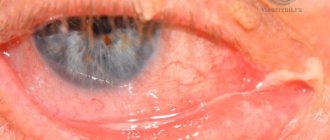
Conjunctival injection
With inflammatory changes in the conjunctiva, redness of the mucous membrane covering the eyelids is most often observed, but hyperemia of the eyeball and transitional fold is often observed. The degree of severity can vary significantly.
In the case of chronic conjunctivitis, there is only redness of the mucous membrane covering the cartilage. An acute process, on the contrary, is characterized by redness of the mucous membrane of the eyeball itself against the background of a change in the color of the cartilaginous surface.
When the conjunctiva is hyperemic, the mucous membrane covering the eyeball near the transitional fold turns especially bright red. In this area you can find small vessels protruding above the surface, which can be displaced along with the surface layer.
In addition to redness, which is the main manifestation of conjunctivitis, there is an excessive amount of liquid discharge (tears), swelling of the eyelids, a feeling of irritation or sand, and fear of bright sunlight.
With a pronounced inflammatory process, the infection can spread to the ciliary body and muscle, resulting in a combined infectious process.
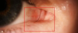
Ciliary injection
When inflammation spreads to deeper tissues, iridocyclitis or iritis occurs. This disease is accompanied by redness in the iris area. The hyperemia is not bright red in color, but a purple hue. The surface of the changed area is smooth and the vessels on it cannot be traced, since the cause of the color change is the deeply located arteries, which are visible through the semi-permeable sclera.
Mixed injection
If the infectious process spreads to both the conjunctiva and the ciliary body, then a mixed infection develops. This usually happens during acute inflammatory changes. Due to the fact that there is communication between the superficial and deep vessels of the eyeball, a hematogenous route of infection can be assumed. If we are talking about damage to the ciliary body, then the infectious process is accompanied by a change in color and blurring of the contours of the iris, as well as the formation of inflammatory exudate and hypopyon.
Diagnostics
When examining the vessels of the conjunctiva in vivo, a completely different picture is observed. This is due to the fact that not all of them are filled with blood. With congestion, there are disturbances in the exchange of the liquid fraction in the eyeball and orbit. The redness that accompanies congestive glaucoma will be discussed further. However, it should be noted that in acute glaucoma, in addition to hyperemia, chemosis or edema of the eyelid is present.
When examining the conjunctiva, dilated lymphatic vessels can also be detected, into which blood cells enter as a result of backflow. This condition is called hemorrhagic lymphangiectasia and occurs when the lymphatic ducts dilate.
The vessels of the mucous membrane of the eye can also change under the influence of systemic processes and diseases. For example, with diabetes mellitus and hypertension, a large number of aneurysmal dilatations are formed. Often the cause of such changes cannot be determined.
When examining the vessels of the conjunctiva under high magnification, it is possible to detect the so-called “sludge” phenomenon. In this case, red blood cells densely fill the capillaries, which impedes normal blood supply. Although the pathogenesis of this phenomenon is similar to the erythrocyte sedimentation reaction, this analysis cannot replace this study. Among other things, the “sludge” phenomenon is characteristic of tumor diseases and systemic chronic pathologies.
As already mentioned, the cause of hyperemia of the conjunctiva of the eye can be various eye diseases that require the participation of an ophthalmologist in the treatment. In this case, it is important to choose an eye clinic where they will really help you, and not to “deduct” money without solving the problem. Below is a rating of specialized ophthalmological institutions where you can undergo examination and treatment if you have conjunctival hyperemia.
Where to treat
Eye clinic of Dr. Belikova
4.6 reviews 54
- Moscow, Budyonny Avenue, 26, bldg. 2.
- Mon-Sun 08:00-20:00
- +7(495)-366-43-83 belikova.net