Causes of mydriasis
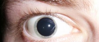
The shape and size of the pupil depend on the activity of two muscles, the radial and circular. The first causes the pupil to dilate, the second is responsible for its narrowing. If a patient has deformation of the circular muscle, its muscle tone is disturbed, then mydriasis occurs.
The activity of the circular muscle is influenced by external and internal factors. Normally, the pupil should dilate in low light conditions, when the sympathetic nervous system is excited (stressful situations, excitement or emotional upsurge).
There are provoking factors leading to the development of pathological mydriasis:
- blunt non-penetrating eye injury, which provokes the development of organ contusion;
- compression of the oculomotor nerves;
- trauma to the skull and its base, causing damage to nerve endings;
- intoxication with barbituric acid derivatives, acetylcholinesterase inhibitors, antihistamines, estrogens;
- infectious diseases that cause damage to intracranial and ocular nerve nodes and muscles (botulism, polio, meningitis);
- ophthalmological pathologies (glaucoma);
- severe hypoxia due to impaired cerebral blood flow;
- neoplasms in the brain (tumors, hematomas);
- drug use;
- surgical treatment of individual structures of the visual apparatus;
- progressive form of diabetes mellitus;
- palsy of the optic nerve due to tuberculosis, carbon monoxide intoxication.
Examination by an ophthalmologist
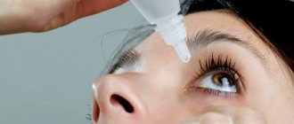
Ophthalmologists who examine the patient use mydriatics - eye drops that dilate the pupil (Mydriacyl, Cyclomed, Midrimax and atropine; Fig. 3). With a wide pupil, the periphery of the fundus of the eye is examined, where a specialist can identify dystrophic changes and even retinal detachment, and the structures of the eye are also examined using a slit lamp.
Rice. 3. Eye drops that dilate the pupil (from left to right): Midriacil, Cyclomed, Midrimax and atropine
Professor Yu. Z. Rosenblum recommends selecting glasses in four stages: I. Examination of the patient in natural conditions. Purpose: determination of the static refraction of each eye. II. Examination in conditions of cycloplegia. Goal: accurate determination of the static (clinical) refraction of each eye. III. Final examination in natural conditions. Objectives: assessment of the state of dynamic refraction and binocular functions and, based on the results obtained, selection of correction for distance and reading. IV. Examination of the patient wearing ready-made glasses. Purpose: checking the correctness of the glasses manufactured and the patient’s tolerance, if necessary, changing the prescribed correction. Let's take a closer look at stage II - examination under conditions of cycloplegia. Cycloplegia is a drug-induced paralysis of the ciliary muscle. The gold standard for cycloplegia is instillation of atropine drops in an age-specific dosage (atropine 0.1% in the first year of life, 0.5% at 5 years, 1% at 10 years and older). The drug is instilled 2 times a day, morning and evening, after meals, one drop for 3 days. Significantly more often used means of milder and short-term action: “Cyclomed” 1%, “Midriacyl” 1%. These solutions are instilled one drop at a time 2 times with an interval of 10 minutes. After cycloplegia, an ophthalmologist can perform retinoscopy (skiascopy), autorefractometry and obtain data on cycloplegic refraction and identify disorders of the optical system of the eye. The iris and ciliary body have the same innervation (oculomotor nerve), therefore, with cycloplegia, the pupil dilates (medicinal mydriasis).
Modern classification
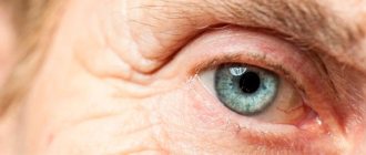
Depending on the causes, mydriasis can be physiological or pathological. Normally, pupil dilation occurs symmetrically, affecting both eyes.
Depending on the type of provoking factor, the following forms of pathological mydriasis are distinguished:
- Medication. Ophthalmologists create artificial mydriasis to assess the condition of the inner lining of the eye (retina), perform the necessary surgical procedures, and relieve muscle tension. For this purpose, medications are used: Atropine, Scopolamine, Ephedrine, Platiphylline. In such cases, pupil dilation persists for 10-24 hours;
- Traumatic. The condition develops in response to various damage to the visual apparatus and nervous system. After surgery, mydriasis can persist for 1-2 years;
- Spastic. It occurs against the background of dilator spasm due to irritation of the sympathetic nerves (cervical spine) and the use of adrenergic-type drugs. The occurrence of spastic mydriasis often indicates the development of severe pathologies (meningitis, poliomyelitis, syringomyelic syndrome, kidney disease, liver disease);
- Paralytic. The cause of the pathology is paralysis of the ocular sphincter due to diseases of the oculomotor nerves, central nervous system, hydrocephalus, epilepsy, botulism, Parkinson's disease, glaucoma, and drug use. In such situations, the pupil stops responding to light;
- Paradoxical. Disturbances in the functioning of the nervous system can cause the pupil to constrict in the dark and dilate in the light. This pathology is extremely rare.
Clinical picture
Pathological mydriasis is accompanied by the following symptoms:
- change in the shape of the pupil (oval or pear-shaped);
- absence or decreased reaction to light;
- impaired mobility of the eyeball;
- lack of reaction to approaching objects.
If mydriasis develops against the background of injury, then the pupil diameter varies from 8 to 10 mm. The pathological condition may disappear after the patient recovers or becomes permanent. Traumatic mydriasis occurs against the background of the following symptoms:
- lacrimation;
- pain and severe discomfort in the eye area;
- photophobia;
- increased fatigue while reading or working at the computer;
- decreased response to light;
- partial afferent failure.
The paralytic type of mydriasis provokes the occurrence of strabismus and drooping of the upper eyelid. In the spastic form, patients note a unilateral localization of the pathology, a decrease in the pupil's reaction to light and the movement of objects, and discomfort in bright lighting.
If mydriasis develops against the background of viral infections, it leads to loss of accommodation.
Diagnostic measures
To determine the causes of mydriasis, a comprehensive examination of the patient is carried out, which includes the following steps:
- careful history taking;
- conducting a visual ophthalmological examination to identify injuries, violations of the integrity of the tissues of the visual analyzer;
- prescribing an MRI or CT scan to identify pathological formations in brain tissue;
- biochemical blood test;
- conducting a general blood test to identify inflammatory processes;
- neurological examination, which consists of determining the presence of a reaction to light, strabismus, diplopia, and breathing problems;
Consultation with an ophthalmic surgeon, neurosurgeon or oncologist if necessary.
Diagnostics
Despite the fact that mydriasis has quite specific symptoms, only an integrated approach is required to identify the causes and establish the correct diagnosis.
First of all, the clinician needs to:
- get acquainted with the medical history to search for a pathological source;
- collect and analyze your life history - this will help diagnose drug-induced or traumatic mydriasis;
- conduct a thorough ophthalmological examination;
- interview the patient in detail to obtain complete clinical information regarding the course of such an illness, which sometimes helps to identify the underlying disease.
Among the instrumental procedures the following stand out:
- CT and MRI of the head;
- chest x-ray;
- ultrasonography of internal organs;
- ophthalmoscopy under mydriasis - to obtain more detailed information about the condition of the fundus.
Ophthalmoscopy
As for laboratory tests, they are limited to:
- general clinical blood test;
- hormonal tests;
- general urine analysis;
- blood biochemistry.
In addition to the ophthalmologist, the following persons take part in the diagnostic process:
- neurologist;
- traumatologist;
- endocrinologist;
- neurosurgeon.
Features of therapy
No special treatment is required to eliminate drug-induced mydriasis. But if pupil dilation persists throughout the day, then drugs of reverse action are prescribed - miotics (Salicylic acid physostigmine, Hydrochloric acid pilocarpine and Hydrobromic arecoline).
If pathological mydriasis occurs, medications are prescribed based on the established diagnosis.
To normalize the patient's condition, they usually use:
- M-cholinomimetics (pilocarpine hydrochloride, Aceclidine);
- N-cholinomimetics (Cytotin);
- Nootropic drugs (Piracetam, Lucetam, Phenotropil);
- Antiplatelet agents to restore cerebral blood flow;
- Hypoglycemic agents for patients with diabetes mellitus;
- Diuretics to relieve swelling in the brain area.
If a brain tumor or hematoma is detected, drug therapy does not bring results. The pathology can only be eliminated surgically.
Prognosis for mydriasis
Drug-induced mydriasis goes away on its own within 24 hours. In the traumatic form of the pathology, the prognosis is generally favorable. Mydriasis usually persists for 1-4 months. After eliminating the underlying disease, you can cause constriction of the pupil with Pilocarpine.
Paralytic and spastic types of mydriasis have a poor prognosis. The outcome directly depends on the form of the underlying disease and the effectiveness of its therapy.
There is currently no specific prevention that would eliminate the possibility of pupil dilation. Therefore, to prevent the development of mydriasis, you should regularly undergo preventive examinations with an ophthalmologist, eat a balanced diet, and promptly treat diseases of the eyes and nervous system.
Prevention and prognosis
Physiological mydriasis does not imply the use of any preventive recommendations, and to reduce the risks of developing such a disease, it is necessary to adhere to simple rules.
Prevention of this disease includes:
- complete abstinence from alcoholic beverages, nicotine and drugs;
- avoiding any eye injuries;
- rational use of medications, especially such as Atropine, Platiphylline and Scopolamine: any drug should not be taken without the approval of the attending physician;
- eating only high-quality foods;
- early detection and comprehensive treatment of any diseases that can lead to the development of the described disorder;
- regular visits to an ophthalmologist and other specialists, as well as laboratory and instrumental examinations in the clinic.
The prognosis of such a pathology will be favorable if the disease has developed against the background of provocateurs that do not have a pathological basis. Otherwise, the outcome will depend entirely on the etiological factors and their timely treatment.
If you think you have Mydriasis
and symptoms characteristic of this disease, then an ophthalmologist can help you.
Source
Did you like the article? Share with friends on social networks: