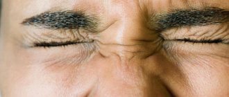
Floaters before the eyes are floating spots of various shapes and sizes (dashes, lines, nets, cobwebs), which are clearly visible when looking at light-colored objects or a bright background. They are especially visible against the background of the sky, a white wall or snow. Such floaters can appear in front of one or both eyes at the same time; occur periodically or be constant companions. Their distinctive feature is their floating nature. The flies seem to float following the movement of the eyes. When looking from object to object, they continuously repeat the same trajectory, only slightly delayed.
In most cases, the appearance of floaters before the eyes is not accompanied by unpleasant symptoms. But sometimes this phenomenon can be associated with a headache, flashes before the eyes or decreased vision.
What are black spots in the eyes
Such defects are associated with the occurrence of problems in the vitreous body and indicate its destruction (process disruption).
There are no clear causes of the disease. Predisposition to pathology is caused by: nervous shock, age, poor health. A common cause of blackheads is the death of cells that accumulate, reducing the transparency of the vitreous. Over time, too many dead cells appear in the eye, and they become clearly visible to a person.
The easiest way to identify them is to look at a light, monochromatic object (preferably white). It is not the cells themselves that block the view, but they cast a distinct or blurry shadow, which a person sees as a “dark midge.”
Black dots before the eyes, reasons:
Blackheads most often occur due to abnormalities in the structure of the vitreous body. It is located between the retina and the lens. All dying elements of these “details” of the visual apparatus are collected in the vitreous body and are perceived by the eyes as “interference” for normal and complete vision.
The main reasons for the development of pathology:
- Mechanical damage to the eye, causing cell death;
- Penetration of a foreign body onto the surface of the eye;
- Aging. This is the most popular reason for the appearance of floaters. It is most often observed after sixty years of age. After turning eighty, a person stops paying attention to them. However, if the points cause severe discomfort, consult a doctor;
- Osteochondrosis. Pathology can develop against the background of problems with the cervical spine. The points arise due to a disturbance in the pressure in the vascular system that supplies the brain;
- Detachment of the retina from the vitreous body;
- Exposure to harmful microorganisms that cause a variety of ophthalmic diseases;
- Eye strain. Occurs if a person is focused on one object for a long time (for example, staring at a computer monitor screen);
- Malfunction of the vascular system or problems with blood pressure. In this case, urgent therapy is required. If the pressure is low, then the processes of decomposition of the vitreous accelerate, since the vessels are filled with blood;
- Poisoning. If harmful substances enter the body, they negatively affect the central nervous system, the eyes “do not stand aside” and also react to deviations in the functioning of the system;
- Lack of vitamin A in the body. As a result, destruction and death of parts of the retina are observed;
- Degradation of the organ of vision. Most often diagnosed in patients over fifty years of age;
- Failure in the circulatory system. Pathology can be caused by increased physical activity or abuse of alcohol and tobacco products. All these factors lead to damage to blood vessels, the ejected blood is gradually transformed into dense clots;
- Head damage. If floaters appear after a traumatic brain injury, visit an eye doctor. Since this symptom may indicate retinal detachment.
In some situations, destructive processes in the vitreous body can be an “independent” ailment, i.e. not be a sign of any abnormality in the functioning of the body. In this case, the pathologies are hereditary.
Pregnant women periodically suffer from blackheads, this indicates the development of eclampsia. The disease is dangerous not only for the expectant mother, but also for the baby.
The problem is often diagnosed in people suffering from myopia. If the flies do not cause discomfort and do not interfere with leading a full life, there is no need to worry. Otherwise, consult a doctor. Return to contents
Causes of brown dots
The main reason for the appearance of black dots on the eye is the destruction of the vitreous body. It occurs due to problems with the lens, insufficient blood circulation, which contributes to the destruction of collagen fibers with age and in serious diseases affecting the eyeballs. Fiber fragments become concentrated at one or more points in the lens, obstructing the passage of light, creating spots. Bad habits stimulate the manifestation of this pathology. Reasons why black spots appear in the eyes:
- crystalline formations;
- the appearance of malignant tumors in the eye;
- diabetic retinopathy;
- exposure to aggressive substances (acids, alkalis, etc.);
- entry of foreign substances and dirt particles into the eye;
- occurrence of injuries;
- migraine;
- long exposure to bright light;
- depletion of visual organs;
- other damage to the eye shell.
If black lines fly before your eyes, this may be a consequence of more serious pathologies. Such problems are not always immediately noticeable, but due to the fragility of the blood vessels they progress quickly. Peripheral vision deteriorates, areas of clouding in the visual organs grow, sparks and flashes may occur. It is important to see a doctor immediately and surgery may be necessary. Such symptoms may be the result of diseases:
- fungal or viral infections;
- hemophthalmos (bleeding into the vitreous body);
- vascular pathologies;
- tumors;
- retinal detachment or other damage;
- vitreous detachment.
To independently identify vision problems, look at a white or light-colored surface so that it occupies 100% of the visual area. If, in addition to the white color, there are defects, spots, or the appearance of some particles flying before your eyes, consult a doctor. You may not have a pathology, but it’s worth being safe. The doctor will make a diagnosis and prescribe treatment.
Under no circumstances do doctors recommend self-medication. And all because this method of treatment can often lead to deterioration of health and serve as an impetus for a progressive process. Therefore, you should not try to diagnose yourself and prescribe treatment. The best way to solve this problem is to consult an ophthalmologist.
Almost all of us have experienced red eyes at least once. In particular, this phenomenon is often encountered by people who spend a lot of time at the computer. Redness can be partial or complete. To eliminate this problem, just give your eyes a little rest and everything will go away by itself.
Brown spot on the white of the eyes. What is this? There are cases when a person is faced with the appearance of a brown dot on the white of the eye. To understand how to deal with this, you need to observe additional manifestations of the stain. The color of the stain will also be important. A brown dot in the eye indicates the following things:
- Your blood pressure may be too high or, conversely, too low. This means that the capillary bursts and creates a small hematoma. This unpleasant moment cannot be treated, since there is no need for it. We must not lose sight of the very cause of what happened; it is worth thinking about treating the diseases that contributed to this.
- Fatigue or temporary stress on the body. For example, women during childbirth experience heavy stress, which leads to increased blood pressure. As a result, the capillaries burst. A person does not remain in this state for long, but the strictest treatment is required.
- A sharp increase in eye pressure. Only an ophthalmologist can help you solve this problem.
- There are also frequent cases of spots that are congenital, something like birthmarks. A brown spot of this kind will be completely harmless. It does not have any effect on vision function. If it bothers you only because of the beauty of your face, then you need to consult a doctor. He will be able to determine exactly what can be done.
- A more serious manifestation of a dark brown spot may be a floater on the eye. Such a spot does not appear every time, but only at the moment when a person moves his eyeball in any direction. This is a sign that the retina is detached. You simply won’t notice this spot; it is colorless and appears only when it hits the pupil area. At this moment, blurred vision occurs and a feeling of discomfort appears.
Only a doctor can specifically determine the presence of a floating spot in the eyeball. Such a spot may be a retinal particle. Laser correction is used to remove it.
You can also try to strengthen the retina. Usually, a micro-surgery is used for this, which takes place without hospitalization of the patient. But further actions after such an operation directly depend on the degree of the disease. Only a doctor can adequately assess the condition of the disease, so you should not delay your visit.
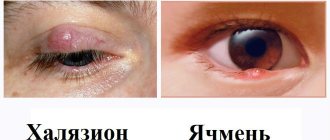
DETAILS: Papilloma on the cheek inside - Medical advice
It should be noted the dangerous manifestations that occur during the course of this disease. If a piece of the retina is partially detached, deterioration in vision and health may occur. If the retina detaches completely, this can lead to complete loss of vision. In this case, it is recommended to visit a doctor, the sooner the better. After all, a person’s vision will depend on this.
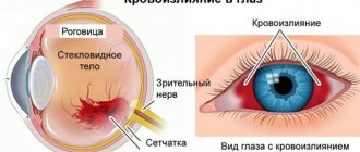
The vitreous body is the fluid medium between the lens and the retina. With a negative impact, dead cells accumulate in it. Large formations cast a shadow on the retina, which is manifested by loss of visual fields.
Black spots that move with your gaze are also known as floaters or blind spots. The pathology is especially clear when you look at a plain surface, such as white paper or a clear sky.
With sudden head movements or tilts, the visual defect disappears, but then appears again.
Prolonged strain on the visual analyzer, for example, when reading a book or driving a car, also makes this symptom more noticeable.
Under normal circumstances, the eyeball has a white or pinkish tint. If a yellow spot is visualized on the eye, a disorder in the process of bilirubin metabolism is suspected.
Similar formations also appear in older people due to physiological aging of the conjunctiva. They are called pingueculae.
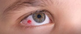
However, most often patients come to see an ophthalmologist with a red spot on the sclera. The cause of this phenomenon may be physical fatigue, as a result of which a vessel in a person’s eye bursts and hemorrhage occurs. In addition, the stain can appear as a result of microtrauma, when using blood thinners, or a lack of vitamin K.
A gray spot on the organ of vision may be a congenital formation, ocular melanocytosis.
The causes of yellow spots in the eyes vary. They depend on the type of yellow spot that appears on the eye, its size, and location. Some common types are presented below.
Pinguecula
In some older people, you may notice a yellow spot that is clearly visible on the white of the eye. As a rule, it is located near the inner corner of the eye. This is a pinguecula.
This is a sign of aging of the choroid. The pinguecula does not reduce visual acuity and does not cause any symptoms if it is not damaged. The factors leading to its occurrence are the following:
- unfavorable professional conditions (smoke, strong wind, sand, dust);
- UV radiation from the sun's rays;
- prolonged work in front of electronic monitors.
The above will not necessarily cause the growth of the pinguecula, but trauma to the optic membrane increases the chances of its growth. Those who wear contact lenses should be especially careful.
Diagnosis is carried out by an ophthalmologist using a slit lamp. Such a yellow spot can rarely be removed. For example, if it is damaged frequently, increases in size, or for cosmetic purposes. For this purpose, microsurgical laser surgery is performed.
Pterygium
When the growth of the mucous membrane of the eyeball is disrupted, a fold forms on the sclera - a pterygium. Most often it is located closer to the inner edge and has the shape of a triangle. This formation grows towards the pupil and, growing on it, can negatively affect vision.
The cause of pterygium is frequent inflammation of the mucous membrane after its irritation due to:
- prolonged exposure to sunlight;
- working in a dusty room;
- dry eye syndrome;
- influence of computer radiation.
For diagnosis, the ophthalmologist uses a slit lamp to determine the size and depth of its growth (biomicroscopy method). Surgical removal is performed as necessary if the patient complains of visual discomfort or decreased vision.
Conjunctival cysts
Conjunctival cysts appear as a raised spot in the eye, sometimes yellow in color. Cysts can be congenital or acquired. A dermoid cyst is a congenital cyst. It appears in childhood and is benign. It looks like a capsule, most often containing fat cells, therefore yellow in color.
Acquired cystic formations develop after inflammatory diseases, injuries, eye surgeries (inflammation of the conjunctiva, sclera, lacrimal duct). They look like fluid-filled nodules and turn yellow over time.
Diagnosing a cyst requires not only an examination by an ophthalmologist, but also a histological examination (studying the composition of the cyst and its tissues under a microscope). As a treatment, doctors suggest surgery to completely remove it or introduce sclerosing (helping to reduce the cyst) solutions into the cavity of the vesicle.
Blind spots in the eye: causes
The vitreous body is the fluid medium between the lens and the retina. With a negative impact, dead cells accumulate in it. Large formations cast a shadow on the retina, which is manifested by loss of visual fields.
Black spots that move with the gaze are also known as floaters or blind spots . The pathology is especially clear when you look at a plain surface, such as white paper or a clear sky.
With sudden head movements or tilts, the visual defect disappears, but then appears again.
Prolonged strain on the visual analyzer, for example, when reading a book or driving a car, also makes this symptom more noticeable.
Causes and types
Conjunctival cysts
A birthmark consists of a cluster of pigment cells - melanocytes, and does not pose a danger to the eye. Based on the amount of pigment, the color of the nevus varies from yellow to dark brown.
A particular type of mole for which surgery is worth considering is a progressive nevus. It grows quickly and may have the ability to undergo malignant transformation.
Leukoma
Leukoma is called a corneal thorn. A fresh formation has a milky tint, but an old leukoma becomes yellow. Its size and shape are quite variable. One of the mechanisms of formation is scarring of part of the eye tissue after injury, including inflammatory nature.
Leukoma can be located:
- on the periphery of the cornea, then the defect does not interfere with vision;
- in the center of the eye, vision decreases;
- completely covers the cornea, total - a person sees only silhouettes.
When examined by a doctor, leukoma is visible to the naked eye. Surgical treatment is required if there is a threat or decrease in vision.
There may be small yellow dots along the edge of the cornea, at the border with the white of the eye. They represent a change in the ocular epithelium. Typically, such small spots appear in allergic conditions, such as allergic conjunctivitis. Once the allergy is cured, they disappear.
Aging and degradation of the macula (age-related macular degeneration, AMD, macular degeneration)
The leading mechanism is dystrophic processes in the retina of the eye, leading to a decrease in the diameter of the feeding vessels.
The following aspects are significant for pathogenesis:
- primary aging of the retinal pigment epithelium and membrane;
- pathological effects of lipid peroxidation products;
- hemodynamic disturbances due to atherosclerotic plaque deposition on the walls of blood vessels;
- genetic predisposition.
Important
The macula has another name: the macula. Its function is to provide central vision and color perception.
In 85%, a dry (atrophic) form of macular degeneration . Atrophy is manifested by yellowish spots - drusen. They can be hard and soft, single and multiple. Loss of visual acuity occurs gradually.
The wet (neovascular) form occurs in 10-15% of cases. More often it is the progression of atrophic AMD. Excessive neoangiogenesis is compensatory in nature and is aimed at increasing trophism and oxygen supply to the retina.
As the pathology progresses, blood cells and fluid leak through the vessels, leading to edema and microhemorrhages, and retinal cells die. One manifestation of this process is the presence of blind spots in central vision.
If the vessels grow into the pigment layer, retinal detachment with scar formation is possible. Loss of vision in this case is irreversible.
Flashing spots before the eyes is only one of the symptoms (more typical for dry AMD). In addition, there may be the following:
- gradual/rapid loss of visual acuity;
- change in contrast (dim colors);
- fog before the eyes;
- fast fatiguability;
- distortion of contours;
- loss of visual fields;
- headache.
Many patients are asymptomatic at the initial stage. With the scar form, not only central but also peripheral vision is lost.
Symptoms
Black spots in the eyes can appear in the form of both single and multiple (filamentous) formations. In some cases, flies are not noticeable at all and do not cause discomfort, while others seriously interfere with a person’s life, turning into full-fledged lines. Dots in the eyes can be a symptom of more serious diseases.
Black dots before the eyes are divided into two types:
- Granular destruction. Hyalocytes (dead eye cells) enter the vitreous body, which die over time, join together and form black dots.
- Filamentous destruction. The development of pathologies and metabolic disorders lead to the death of some collagen fibers. A person begins to see strings or entire webs in front of him, which significantly interfere with object recognition.
DETAILS: After cleansing my face, acne appeared - Face Masks
The main feature of the disease is that when the head is sharply moved to the side, black lines or dots move in the same direction, creating a kind of trail. Pathology occurs after suffering from serious diseases affecting the eyes, when using medications with aggressive substances, or after overcoming 50 years of age. Those who are diagnosed with myopia are also at risk; over the years, changes in vision only contribute to the occurrence of defects.
Rarely, a patient may complain of increased fatigue, nausea, lack of appetite, and increased body temperature.
Factors that contribute to the appearance of floating dark spots
Predisposing factors vary and include:
- age over 55 years;
- previous ophthalmological operations, aggravated medical history;
- diabetes;
- smoking, alcoholism and other intoxications;
- thyroid diseases;
- atherosclerosis;
- hypertonic disease;
- hypotension;
- bacterial and viral infections;
- overstrain of the visual organs;
- avitaminosis;
- prolonged hypoxia (oxygen starvation);
- head injury;
- genetic predisposition;
- osteochondrosis of the cervical spine, causing insufficient blood supply to the head.
Diagnostics
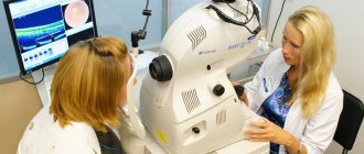
In traditional cases, in order to diagnose pathology, the specialist uses a slit lamp and checks the patient’s visual acuity. A systematic plan for diagnosing the disease under such circumstances is as follows:
- carrying out systematization of data received from the patient;
- carrying out a visual examination of the organ of vision;
- making a preliminary diagnosis;
- implementation of biomicroscopy (for detailed research);
- referral for ultrasound, CT;
- appointment of laboratory analyzes of biological materials.
A competent ophthalmologist will help you correctly recognize yellow spots when they appear in your eyes. To do this, he uses the following methods:
- fundus examination;
- biomicroscopy;
- eye pressure measurement;
- histological examination.
Treatment
When treating this diagnosis, hormonal therapy is used and vitamin C is taken. Sometimes this disease can be caused by some kind of inflammatory process. Anti-inflammatory medications will help in this situation.
You should also remember to adhere to a special diet. The diet should not contain refined cereals, white bread, coffee, over-salted dishes, or foods containing starch. The diet should include seafood, any leafy vegetables, fish, citrus fruits, nuts, etc.
To treat melanosis, which was caused by an inflammatory process, you can use folk remedies.
You need to put 2 tablespoons of cornflower inflorescences in a mug, then pour boiling water over them and leave to steep for 3 hours. The cooled and infused decoction is filtered through cheesecloth and used as an eye lotion. This procedure should be performed daily for 5-7 days.
Oak bark is a good eye wash. It has healing properties. Consider one of the oak bark recipes. To prepare it you will need 500 ml of boiling water. The same amount of oak bark should be added as water. This whole broth is put on fire and boiled for an hour. Afterwards it is cooled and filtered through fine gauze. This product is used as an eye lotion.
Therapeutic measures depend on the underlying cause.
Eye injury
Primary treatment, instillation of antibacterial, analgesic, hemostatic and absorbable drugs.
Atropine or pilocarpine is instilled under the control of intraocular pressure.
In the case of a penetrating wound, emergency surgery is performed; in the future, reconstructive ophthalmological operations are possible.
During the rehabilitation period, vitamin complexes, absorbent agents, and physical therapy are prescribed.
Treatment is predominantly surgical and includes cryopexy (freezing) at the site of injury, laser photocoagulation, removal of the vitreous (vitrectomy), sclerotherapy and pneumatic retinopexy in combination with cryopexy, photocoagulation or laser treatment.
Additionally prescribed medications:
- Emoxipin;
- Taurine;
- Taufon;
- Ophthalm-Katachrome;
- Quinax;
- Emoxy-Optic;
- Papaverine;
- B vitamins;
- Acetylsalicylic acid;
- Pentoxifylline, etc.
The basis of therapy is normalization of blood glucose levels and lifestyle correction.
Drugs for diabetic retinopathy:
- angioprotectors;
- vitamins;
- corticosteroids;
- biological peptides.
Intravitreal injections (Bevacizumab, Ranibizumab) reduce diabetic macular edema and neovascularization of the disc or retina.
Corticosteroids for diabetic retinopathy slow down processes associated with inflammation:
- edema;
- fibrin deposition;
- collagen deposition;
- expansion of capillaries;
- migration of leukocytes and fibroblasts.
Representative – Triamcinolone (synthetic glucocorticosteroid). In combination with laser therapy, the treatment effect is higher.
Surgical interventions:
- Photocagulation;
- Vitrectomy;
- Cryotherapy.
Proper nutrition and physical activity help maintain optimal weight, which further helps control diabetes and its complications.
At the initial stage, specific therapy is not required, but it is important to eliminate/minimize risk factors that contribute to the progression of the pathology.
Some experts consider the use of physiotherapy justified: ultrasound, phono- and electrophoresis, hyperbaric oxygenation.
Drug treatment as the only treatment for wet AMD is ineffective.
Along with drug therapy, laser coagulation of the retina is performed, which is the standard in the treatment of age-related macular degeneration.
Elimination of the main provoking factor – high blood pressure.
Behavior correction: proper nutrition, physical activity, taking antihypertensive drugs.
Medicines:
- vasodilators;
- anticoagulants;
- angioprotectors;
- neoangiogenesis inhibitors;
- anti-sclerotic drugs;
- multivitamins, etc.
In the advanced stage, when hypoxia impairs vision, laser coagulation is performed.
Depending on the provoking agent (virus, bacteria, fungi), appropriate drugs are selected: antiviral, broad-spectrum antibiotics or antimycotics. The drugs can be used both as local and systemic therapy.
Additionally, multivitamins, immunomodulators, etc. are prescribed.
Mishina Victoria, doctor, medical columnist
The course of the therapeutic course is determined after diagnosis and varies significantly.
Pinguecula is a benign formation that does not need to be treated. To eliminate unpleasant symptoms (such as dry eyes, redness and general discomfort), the ophthalmologist prescribes moisturizing drops or mild steroid medications.
A congenital spot is treated only when it comes to a progressive nevus. To remove such formations, laser and radio waves are used.
Red spots on the eyeball caused by infectious processes are treated with medication. In such cases, the doctor prescribes antibiotics.
DETAILS: Spots on the skin of the legs and fingers, what do red and white formations mean
For leukoma, laser removal or keratoplasty will come to the rescue. Implantation of donor cornea is also common today.
A conjunctival cyst can be treated with conservative methods (moisturizing, antibacterial, anti-inflammatory drops) or surgically. In the latter case, they resort to traditional surgery or use a laser.
A yellowish spot on the eyeball is not always a reason to remove it. Surgery is required when:
- There is discomfort when blinking or moving the eyeball.
- Vision decreases.
- The outflow of tears decreases.
- Pronounced cosmetic defect.
- There is a threat of malignant growth.
How to get rid of blackheads in eyes
Vision is a key resource of the body, and you should not treat it with disdain. Treatment of black spots and dots with folk remedies is impossible; the problem itself also does not go away. You are allowed to drink herbs approved by your doctor; they will help you recover faster and prevent the recurrence of vision problems. Do not self-medicate - it is dangerous.
If you suspect the presence of black spots and dots, contact an ophthalmologist who will conduct an examination and confirm or deny the symptom. Contact a public clinic or a trusted private clinic. It happens that black spots arise due to another disease (tumor, infection, etc.), and in a private clinic you can be treated only for symptoms, without paying attention to the pathology itself that caused this condition.
During treatment in the hospital, existing defects are eliminated and the source of the pathology is sought. In most cases, it is possible to get rid of the disease that led to the appearance of black spots and dots, but it is not possible to clear the eye of defects. Dead cells are not removed from the vitreous body on their own.
In rare cases of floaters appearing before the eyes, for minor problems, vitamin drops are prescribed: Taufon, Quinax, ethylmorphine hydrochloride solution. Effective removal of dark spots is possible with the help of potassium iodide drops. If it is necessary to accelerate the regenerative component of the vitreous body, Wobenzym and Emoxipin are used.
If the disease is detected in the initial stages, it is recommended to take multivitamin complexes. The doctor prescribes examinations and procedures that eliminate the causes of vision problems. After getting rid of them, the black spots on the eyes completely or partially disappear on their own. If the disease is advanced, serious measures may be needed.
- Vitrectomy is a surgical procedure in which the vitreous humor is partially or completely removed. It is being replaced by an artificial environment. This extremely dangerous operation can lead to cataracts, retinal detachment, and hypotension. It is used only in cases where other methods have not produced any results.
- Vitreolysis - using a laser, the threads are broken, that is, clusters of dots are destroyed. The eye is completely reanimated. The operation is complex and is performed only by experienced doctors.