Colorblindness - what is it?
Colorblindness
– or color blindness is a condition in which the ability to distinguish colors is partially or completely lost. The degree of anomaly varies - for some, the restrictions concern only one color, while others also see the world in black and white.
Contrary to popular belief, the perception of colorblind people does not replace one color with another: they do not see yellow flowers as blue, or a red umbrella as blue. In their picture of the world, some colors seem to merge, approach each other, while others remain intact. Of course, color perception will depend on the types of defects, which we will discuss below.
The phenomenon of color blindness is named after the English meteorologist, chemist, and physicist John Dalton, who lived in the 18th century. Only at the age of 26, the scientist suddenly realized that he perceived colors differently than others: and this discovery was given to him by his own jacket. Dalton was firmly convinced that the wardrobe item he was using was gray, but in fact the clothes were dark red. The scientist developed the topic and, based on his feelings, described how those suffering from the same feature perceive the world.
It is more correct to call color blindness not a disease, but a feature of the body or a vision defect: it does not pose a threat to the patient’s health. Their difference from ordinary people lies only in the fact that they see some colors - and, accordingly, the general picture of the world around them - differently.
Why does it happen?
Trichromatic vision is inherited in an autosomal recessive manner. This is a genetic disease that does not depend on the influence of external and internal factors on the embryo or environmental influences on an already born child.
Normal trichromasia is present in humans without genetic pathologies of retinal nerve cells. The disease appears as a result of defects in the pigment saturation of rods and cones located in the retina. Based on differences in the loss of one or another receptor, pathology is divided into 3 groups:
- Protanomaly. With it, the receptors for the perception of red color are affected. A person is not able to distinguish even its spectral shades.
- Deuteranomaly. In this case, there is a deficiency of cells that capture green color and its spectrum.
- Tritanomaly. This is a pathological condition in which the entire blue spectral range falls out. This type of color vision disorder is considered the most favorable, since there are few blue shades in nature.
Mechanism of anomaly
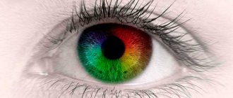
Cones, special nerve cells located in the center of the retina, are responsible for distinguishing colors in the eye apparatus. In healthy people, cones contain three pigments “responsible” for recognizing red, green and blue colors. The combination of these basic colors in the cerebral cortex gives us the wide palette of shades that we enjoy every day. In some cases, the pigment may either be absent altogether or not work properly. It is then that the peculiarities of color perception arise. The disruption of which photoreceptors led to the development of color blindness can be determined by the doctor.
Forms and types of color blindness
In some cases, all three pigments may be present in cones, but the activity of one of them is reduced. In this case we are talking about anomalous trichromasia.
There are three types of abnormal trichromasia:
- protanomaly (with improper development of the “red” receptor);
- deuteranomaly (for problems with the “green” receptor);
- tritanomaly (anomaly concerning the “blue” receptor).
Another option is the absence of one of the three pigments (usually red, much less often blue). This condition is called dichromasia.
Depending on which of the pigments is missing, the following are distinguished:
- protanopia (when there is no red pigment);
- deuteranopia (no green pigment);
- tritanopia (lack of blue pigment).
The rarest manifestation of the disease is the complete absence of all three pigments in the cones - monochromasia or achromasia.
Normal trichromatic vision
Human color perception is provided by special photoreceptors (cones) located on the retina of the eye. At the same time, receptors are divided into several groups, according to the principle of recognizing different lengths of light waves.
Thus, cones capable of recognizing red color react to light whose wavelength exceeds 550 nm, or long-wave radiation, “green” cones react to medium-wave radiation (520 -540 nm), “blue” cones - to short-wave radiation 430 to 470 nm.
Thus, rays entering the retina excite three groups of cones at once, albeit to varying degrees.
Causes of color blindness
Color blindness is a fairly common condition: according to statistics, every eighth man experiences color vision impairment. The likelihood of color blindness in women is much lower: it happens in only 0.4 cases out of 100.
The causes of color blindness can be genetic or acquired.
The color blindness gene is linked to the X chromosome: the anomaly is transmitted through the female line. If the child’s mother is color blind, her son will inherit the same feature, and the daughter will become a carrier and pass it on to her children. In those rare cases where both parents are color blind, the child will suffer from color vision defects. The phenomenon that explains how the trait of color blindness is inherited is called linked inheritance.
Complete color blindness can only be hereditary. The frequency of its manifestation is 1 case per million.
In addition, genetic color blindness may be associated with pathologies such as cone dystrophy and Leber amaurosis.
The acquired form of color blindness is associated with injuries, aging, diseases of the optic nerve, and diabetes. In this case, the abnormality will affect only the affected eye and will tend to get worse.
Vision for a driver's license: features you need to know about
To obtain a driver's license of the established form, a medical certificate is among the required documents.
According to this document, the driver undergoes a medical examination and a medical commission determines whether he is fit to drive or not. According to the order of the Ministry of Health and Social Development of the Russian Federation No. 302n, there is a list of diseases, the presence of which is prohibited from driving. In particular, this also includes some types of visual impairment that could affect the handling of the car.
You can find more complete information on the topic in ConsultantPlus. Free trial access to the system for 2 days.
In short, we can highlight several visual features that affect the possibility of obtaining a license:
- Decreased visual acuity. Usually, sharpness is checked using a well-known table with letters. Naturally, there are craftsmen who try to memorize the letters and name them in the correct sequence. For such citizens, other test tables are provided that can also determine visual acuity. Each category of rights requires its own indicator. Moreover, in the above order, two categories of vision are distinguished: “with correction,” that is, with glasses or contact lenses, and “without correction,” that is, without them. For example, for category B, vision in the “best” eye without correction should be 0.8, in the other eye visual acuity may be absent; if you have contact lenses or glasses for the same category, the “best” eye should see at least 0.5, the worst – 0.2.
- As mentioned above, a future driver can come to a medical examination with an ophthalmologist wearing glasses or contact lenses. But even in this case, not any will be suitable, but only those with diopters not lower than -8 and not higher than +8. The difference between the eyesight should not be more than 3 diopters. But sometimes there may be cases when a healthy eye under glasses can produce distortion and “fall short” of the required number of lines. This is the so-called “lazy” eye. It can be easily corrected in infancy and adolescence, but adults can also “improve” their vision.
- One of the important characteristics of the eye, which is examined during a medical examination to allow a citizen to drive a vehicle, is the color perception of the eye. This characteristic is important so that the driver can at least recognize the colors of the traffic light.
- And finally, there are diseases for which permission to drive a car is not permissible under any circumstances. For example, these may include glaucoma and retinal detachment.
Diagnosis of color blindness
If color vision disorders are suspected, ophthalmologists use special tables, Nagel and Rabkin anomaloscopes, flickering lights and other methods.
Polychromatic tables (Ishihara, Yustovoy, Rabkina) look like an organized cluster of different-sized circles of primary and secondary colors of the same brightness. Some of the circles form a wriggling line, figure or number against the background of the rest. Some tables allow not only to identify the defect, but also to determine the type of color blindness and its degree.
Holmgren's method. The patient is asked to choose from 133 skeins of multi-colored wool, those that belong to the same color, but in different shades.
Correction of color blindness
Unfortunately, a method that could permanently correct the anomaly has not yet been invented. There is no effective treatment for color blindness, as well as methods of prevention. Patients with cataracts and diabetes mellitus – diseases that can cause color blindness – may be advised to undergo examinations with an ophthalmologist every six months.
If the pathology is the result of brain damage, surgical intervention may be prescribed for treatment.
For correction, you can use special optics - glasses or lenses, which enhance the perception of primary colors, bringing the colorblind person’s vision closer to normal. There are also special high-tech cybernetic devices and software that help colorblind people at work. However, it is not possible to completely convert color blindness into normal vision.
Anomalous trichromasia
Anomalous trichromasia is characterized by a decrease in the patient's ability to perceive any of the three primary colors. The ability only decreases without completely disappearing, which is due to a congenital pathology associated with damage to one group of cones. Thus, anomalous trichromasia is divided into:
- Protanomaly, with an anomaly in the development of the first receptor - red;
- Deuteranomaly, with an anomaly in the development of the second receptor - green;
- Tritanomaly, characterized by an anomaly in the development of the third receptor - blue.
- Protanomaly and deuteranomaly are usually classified according to the degree of manifestation: A - a sharp degree of pathology, close to dichromasia (“two-color vision”); B - average degree of pathology; C - weak degree of pathology, close to trichromasia (normal “three-color vision”).