What is color blindness? Causes of pathology
Most often, people with color blindness are unable to recognize one color - red, blue-violet or green.
In this article
- What is color blindness? Causes of pathology
- Types of color blindness
- How to determine color blindness in children?
- How to determine color blindness in an adult?
- Treatment of color blindness
- Correction of color blindness
However, in ophthalmological practice there are cases when patients are not able to distinguish several shades at the same time. This phenomenon is called paired color blindness. In addition, some people cannot distinguish all the colors of the spectrum. But colorblind people with this type of pathology can perceive “invisible” colors, for example, gray. The main cause of color blindness is a disruption of the color-sensitive receptors, which are located in the central region of the retina. These receptors (nerve cells) look like cones. They come in several types, each containing a specific protein pigment. The first is responsible for capturing the green spectrum with a wavelength of 530 nm, the second for perceiving the red spectrum at 552–557 nm, and the third for capturing the blue spectrum at 426 nm. Under normal conditions of development of the visual system, people have all three types of receptors described above, that is, they have the correct perception of the color spectrum. There are two types of color blindness: hereditary and acquired. The first type is associated with a mutation on the X chromosome, which is inherited by the child from the mother. As a rule, this eye disease can occur in men, since their set of genes lacks an additional X chromosome that could compensate for the mutation. Much less often, the mutating gene is passed on to a newborn girl. Statistics show that color blindness occurs in 5-8% of men and only 0.5% of women.
An acquired type of pathology can occur as a concomitant disorder with complications of other eye diseases or with external damage to the visual organs. There are several areas of damage that can directly influence the development of color blindness in a patient - the optic nerve and the retina. In addition, the key causes of the acquired disease include: age-related changes in the visual organs, as well as the use of certain medications. Often, already in old age, a person may show signs of color blindness.
Color blindness test. Rabkin table (full description)
Here is a diagnostic test based on Rabkin’s polychromatic tables, used to identify color blindness, as well as its manifestations. This test is familiar to every male Russian - all conscripts undergo it at the medical examination at the military registration and enlistment office.
We will tell you what each of the 27 pictures above means and what kind of deviation it reveals. The test also contains “check” cards for calculating malingerers.
Rules for taking the test:
- Relax, look at the pictures from a decent distance, preferably about a meter, it is important not to look at them with your nose at the screen.
- Take your time, spend about 5 seconds on each picture.
- Then read the text under the picture and compare with your results.
- If you see deviations in yourself, do not panic. When passing the test from a monitor screen, everything greatly depends on the settings of the image itself, the color of the monitor, etc. However, this is a recommendation to contact a specialist.
Explanation of some terms in signatures:
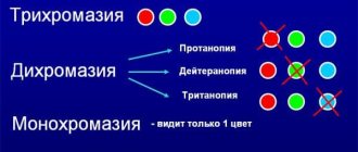
- A person with normal color perception is a normal trichromat;
- A complete lack of perception of one of the three colors makes a person a dichromat and is designated respectively as prot-, deuter- or tritanopia.
- Protanopia is the inability to distinguish certain colors and shades in the areas of yellow-green, purple and blue colors. Occurs in approximately 8% of men and 0.5% of women.
- Deuteranopia is a decreased sensitivity to certain colors, mainly green. Occurs in approximately 1% of people.
- Tritanopia is characterized by the inability to distinguish certain colors and shades in the areas of blue-yellow, violet-red colors. It is extremely rare.
- Monochromites , which perceive only one of the three primary colors, are also rare Even more rarely, with severe pathology of the cone apparatus, achromasia - a black and white perception of the world.
1.
All normal trichromats, abnormal trichromats and dichromats distinguish the numbers 9 and 6 equally correctly in this table (96). The table is intended primarily to demonstrate the method and to identify malingerers.
2.
All normal trichromats, abnormal trichromats and dichromats equally correctly distinguish two figures in the table: a circle and a triangle. Like the first one, the table is for demonstrating the method and for control purposes.
3.
Normal trichromats distinguish the number 9 in the table. Protanopes and deuteranopes distinguish the number 5.
4.
Normal trichromats are distinguished by a triangle in the table. Protanopes and deuteranopes see a circle.
5.
Normal trichromats are distinguished in the table by numbers 1 and 3 (13). Protanopes and deuteranopes read this number as 6.
6.
Normal trichromats distinguish two figures in the table: a circle and a triangle. Protanopes and deuteranopes do not distinguish between these figures.
7.
Normal trichromats and protanopes distinguish two numbers in the table - 9 and 6. Deuteranopes distinguish only the number 6.
8.
Normal trichromats distinguish the number 5 in the table. Protanopes and deuteranopes distinguish this number with difficulty, or do not distinguish it at all.
9.
Normal trichromats and deuteranopes distinguish the number 9 in the table. Protanopes read it as 6 or 8.
10.
Normal trichromats are distinguished in the table by numbers 1, 3 and 6 (136). Protanopes and deuteranopes read two numbers instead: 66, 68 or 69.
11.
Normal trichromats distinguish between a circle and a triangle in the table. Protanopes distinguish a triangle in the table, and deuteranopes - a circle, or a circle and a triangle.
12.
Normal trichromats and deuteranopes are distinguished in the table by numbers 1 and 2 (12). Protanopes do not distinguish these numbers.
13.
Normal trichromats read the circle and triangle in the table. Protanopes distinguish only a circle, and deuteranopes - a triangle.
14.
Normal trichromats distinguish the numbers 3 and 0 (30) at the top of the table, but do not distinguish anything at the bottom. Protanopes read the numbers 1 and 0 (10) at the top of the table, and the hidden number 6 at the bottom.
15.
Normal trichromats distinguish two figures at the top of the table: a circle on the left and a triangle on the right. Protanopes distinguish two triangles at the top of the table and a square at the bottom, and deuteranopes distinguish a triangle at the top left and a square at the bottom.
16.
Normal trichromats are distinguished in the table by numbers 9 and 6 (96). Protanopes distinguish only one number 9 in it, deuteranopes - only the number 6.
17.
Normal trichromats distinguish two shapes: a triangle and a circle. Protanopes distinguish a triangle in the table, and deuteranopes - a circle.
18.
Normal trichromats perceive the horizontal rows of eight squares each in the table (color rows 9th, 10th, 11th, 12th, 13th, 14th, 15th and 16th) as monochromatic ; vertical rows are perceived by them as multi-colored.
19.
Normal trichromats are distinguished in the table by numbers 9 and 5 (95). Protanopes and deuteranopes distinguish only the number 5.
20.
Normal trichromats distinguish between a circle and a triangle in the table. Protanopes and deuteranopes do not distinguish between these figures.
21.
Normal trichromats distinguish the vertical rows of six squares each in the table as being of one color; horizontal rows are perceived as multi-colored.
22.
Normal trichromats distinguish two numbers in the table - 66. Protanopes and deuteranopes correctly distinguish only one of these numbers.
23.
Normal trichromats, protanopes and deuteranopes distinguish the number 36 in the table. Persons with severe acquired pathology of color vision do not distinguish these numbers.
24.
Normal trichromats, protanopes and deuteranopes distinguish the number 14 in the table. Persons with severe acquired pathology of color vision do not distinguish these numbers.
25.
Normal trichromats, protanopes and deuteranopes distinguish the number 9 in the table. Persons with severe acquired pathology of color vision do not distinguish this number.
26.
Normal trichromats, protanopes and deuteranopes distinguish the number 4 in the table. Persons with severe acquired pathology of color vision do not distinguish this number.
27.
Normal trichromats distinguish the number 13 in the table. Protanopes and deuteranopes do not distinguish this number.
Types of color blindness
A visual disorder such as color blindness can manifest itself in different ways. Experts define, as mentioned above, several of its types:
- Dichromia is the inability of the patient's visual system to detect blue and green shades.
- Deuteranopia is the inability to distinguish green colors.
- Disruption of red receptors (the most common problem) - the patient does not recognize red and green colors.
- Complete absence of color vision (the rarest disorder) - the patient can only recognize gray, black and white tones.
Most often, it is possible to determine the presence of color blindness only in an adult. The central region of the retina is equipped with color-sensitive receptors (rods and cones). These nerve cells contain several types of pigments: iodopsin and erythrolab. The cones contain iodopsin, which consists of two pigments: chlorolalab and erythrolab, which ensure the perception of all visible zones of the spectrum. Chlorolab is highly sensitive to the yellow-green region, and erythrolab is highly sensitive to the yellow-red region.
At the same time, the rods contain a pigment such as rhodopsin. It is capable of absorbing certain regions of the spectrum and provides a chemical bond between the opsin and the chromophore that is part of it. There are two maximum points in this spectrum: one is in the blue region and reaches a limit at 278 nm, to which opsin is sensitive, the other reaches a limit at 500 nm, to which the chromophore is sensitive under low light conditions.
How to determine color blindness in children?
As a rule, color blindness in children is a hereditary disease; in extremely rare cases, it is acquired. Ophthalmologists warn that if a pathology is present, the child will not receive enough information necessary for his full development. For example, if he has a form of color blindness such as deuteranopia (failure to recognize the color green by the visual organs), the child may confuse traffic lights.
Diagnosing visual impairment at an early age is quite difficult, since children can only meaningfully learn to identify colors by the age of 3-4 years. Therefore, you can check whether a baby has color blindness only by closely observing his behavior. For example, pay attention to his drawings. If a child paints the sky with green crayons and the grass with red, this may be a reason to be wary. Among other things, you can try simple testing. How can you identify colorblindness in a child yourself at home? Ophthalmologists recommend placing two candies in front of him: one in a black or gray wrapper, the other in a colorful one. If a child chooses the second option without hesitation, he is healthy. In case of an ambiguous choice, at random, it is worth contacting a specialist to find out the reasons and identify the degree of possible color blindness.
It is important to understand that if a child is diagnosed with a degree of color blindness such as dichromia (the ability of the visual organs to recognize two out of three colors), then he will still be able to obtain a driver’s license in the future and will not experience difficulties when choosing a profession and place of work. To date, there is no effective treatment for color blindness in children. Thus, a certain compensatory reaction in a child may occur during logical thinking, inference and memory. For example, he can remember the order of the colors of a traffic light: red, yellow, green.
Scientists are developing experimental treatment methods for various degrees of color blindness. One of them is the introduction of missing genes into the retina using genetic engineering. This method is still at the stage of laboratory testing.
Rabkin tables
The color perception test is carried out by showing the test taker pictures consisting of circles of different colors that form numbers or certain shapes. If there are problems with color perception (color weakness or blindness to certain colors), then the person does not see the figure, does not see everything, or perceives completely different signs.
Conditions for the color perception test:
- the study should be carried out when the person being tested is in normal health,
- in a lighted room,
- at a distance convenient for the person.
- The time for recognition should not exceed 10 seconds.
| Polychromatic tables E.B. Rabkin to test color blindness (color vision). | |
| Table 1 All normal trichromats, abnormal trichromats and dichromats distinguish the numbers 9 and 6 equally correctly in the table (96). The table is intended primarily for demonstration of the method and for reference purposes. | |
| Table 2 All normal trichromats, abnormal trichromats and dichromats distinguish two figures equally correctly in the table: a square and a triangle. Like the first table, it is intended primarily for demonstration of the method and for reference purposes. | |
| Table 3 Normal trichromats distinguish the number 9 in the table. Protanopes and deuteranopes distinguish the number 5. | |
| Table 4 Normal trichromats are distinguished in the table by a triangle. Protanopes and deuteranopes see a circle. | |
| Table 5 Normal trichromats are distinguished in the table by numbers 1 and 3 (13). Protanopes and deuteranopes read this number as 6. | |
| Table 6 Normal trichromats distinguish two figures in the table: a circle and a triangle. Protanopes and deuteranopes do not distinguish between these figures. | |
| Table 7 Normal trichromats and protanopes distinguish two numbers in the table - 9 and 6. Deuteranopes distinguish only the number 6. | |
| Table 8 Normal trichromats distinguish the number 5 in the table. Protanopes and deuteranopes distinguish this number with difficulty, or do not distinguish it at all. | |
| Table 9 Normal trichromats and deuteranopes distinguish the number 9 in the table. Protanopes read it as 6 or 8. | |
| Table 10 Normal trichromats are distinguished in the table by numbers 1, 3 and 6 (136). Protanopes and deuteranopes read two numbers instead: 66, 68 or 69. | |
| Table 11 Normal trichromats are distinguished in the table between a circle and a triangle. Protanopes distinguish a triangle in the table, and deuteranopes - a circle, or a circle and a triangle. | |
| Table 12 Normal trichromats and deuteranopes are distinguished in the table by numbers 1 and 2 (12). Protanopes do not distinguish these numbers. | |
| Table 13 Normal trichromats read the circle and triangle in the table. Protanopes distinguish only a circle, and deuteranopes - a triangle. | |
| Table 14 Normal trichromats distinguish the numbers 3 and 0 (30) in the upper part of the table, and do not distinguish anything in the lower part. Protanopes read the numbers 1 and 0 (10) at the top of the table, and the hidden number 6 at the bottom. Deuteranopes read the number 1 at the top of the table, and the hidden number 6 at the bottom. | |
| Table 15 Normal trichromats distinguish two figures at the top of the table: a circle on the left and a triangle on the right. Protanopes distinguish two triangles at the top of the table and a square at the bottom, and deuteranopes distinguish a triangle at the top left and a square at the bottom. | |
| Table 16 Normal trichromats are distinguished in the table by numbers 9 and 6 (96). Protanopes distinguish only one number 9 in it, deuteranopes - only the number 6. | |
| Table 17 Normal trichromats distinguish two figures: a triangle and a circle. Protanopes distinguish a triangle in the table, and deuteranopes - a circle. | |
| Table 18 Normal trichromats perceive the horizontal rows of eight squares each in the table (color rows 9th, 10th, 11th, 12th, 13th, 14th, 15th and 16th) as monochrome; vertical rows are perceived by them as multi-colored. Dichromats perceive vertical rows as monochromatic, and protanopes perceive vertical color rows - 3rd, 5th and 7th - as monochromatic, and deuteranopes perceive vertical color rows - 1st, 2nd, 4th, 6- th and 8th. Colored squares located horizontally are perceived by protanopes and deuteranopes as multi-colored. | |
| Table 19 Normal trichromats are distinguished in the table by numbers 9 and 5 (95). Protanopes and deuteranopes distinguish only the number 5. | |
| Table 20 Normal trichromats are distinguished in the table between a circle and a triangle. Protanopes and deuteranopes do not distinguish between these figures. | |
| Table 21 Normal trichromats distinguish the vertical rows of six squares each in the table (color rows No. 1, 2, 3, 4, 5, 6) as single-color; horizontal rows (No. 7, 8, 9, 10, 11, 12) are perceived as multi-colored. Dichromats perceive vertical rows as multi-colored, and horizontal rows as single-colored. | |
| Table 22 Normal trichromats distinguish two numbers in the table - 66. Protanopes and deuteranopes correctly distinguish only one of these numbers. | |
| Table 23 Normal trichromats, protanopes and deuteranopes distinguish the number 36 in the table. Persons with severe acquired pathology of color vision do not distinguish these numbers. | |
| Table 24 Normal trichromats, protanopes and deuteranopes distinguish the number 14 in the table. Persons with severe acquired pathology of color vision do not distinguish these numbers. | |
| Table 25 Normal trichromats, protanopes and deuteranopes distinguish the number 9 in the table. Persons with severe acquired pathology of color vision do not distinguish this number. | |
| Table 26 Normal trichromats, protanopes and deuteranopes distinguish the number 4 in the table. Persons with severe acquired pathology of color vision do not distinguish this number. | |
| Table 27 Normal trichromats distinguish the number 13 in the table. Protanopes and deuteranopes do not distinguish this number. | |
| Thus, normal trichromats read all twenty-six tables correctly, protanopes read seven to eight tables (1, 2, 7, 23, 24, 25 and 26), and deuteranopes read nine tables (1, 2, 8, 9, 12, 23 , 24, 25 and 26). Please note that color calibration on your monitor can play an important role, so the classic result will only be obtained by an ophthalmologist, with paper calibrated tables. | |
How to determine color blindness in an adult?
It is important to understand that color blindness can impose restrictions on a person when choosing a profession. For example, drivers, pilots, sailors, military personnel and doctors of some specializations are required to undergo tests for the presence of this disease. Colorblindness can be determined even at home. For example, there are various sites where you can find reliable tests for identifying the disease. In order for the result to be as accurate as possible, you need to place the monitor at eye level and sit at a distance of 80 cm from it. To determine the degree of color blindness during a self-test, look at each picture for at least 5 seconds. Regardless of whether you were able to recognize the picture, click on it with the mouse; in many tests, various explanations and information appear about what people with different diagnoses see in this image. In addition, tests may contain multiple-choice questions. Before determining the degree of color blindness yourself, you need to clearly understand that each monitor has its own settings and color rendering, so any of them can distort the image. That is why it is undesirable to think on your own how to determine color blindness; you need to contact a qualified specialist who will make the correct diagnosis.
Checking color perception using Rabkin's tables online with answers
Rabkin tables for testing color perception are used to test color perception and identify the form and degree of its impairment. The set consists of 48 tables. Tables 1 to 27 are basic, tables 28 to 48 are control tables, for detailing the diagnosis and identifying cases of malingering and aggravation.
A vision test should be carried out according to the following rules: 1. The brightness of the computer screen should be average (a very dim or bright screen can interfere) 2. Rabkin tables should be at eye level and located perpendicular to the gaze (the inclination of the tables may affect the accuracy of diagnosis) 3. Time looking at the table for about 5 seconds (you should not look at the tables for a long time - this can give false results) 4. It is better to write down the answers on a piece of paper to compare them with the correct answers at the end of the article.
Types of color vision impairment and interpretation of the results at the end of the article. To test your vision for color blindness, the first 27 tables are enough; if you are interested in going through all of Rabkin’s tables, then the remaining 20 tables will be presented at the end.
Attention!!! You can check the answer for each table at once. To do this, hover your mouse over the table and you will see a tooltip with the answers.
N - normal trichromats, Pr - protanopes, De - deuteranopes, Pa - protanomalies, Yes - deuteranomalies, Pp - acquired pathology, + correct answer, - incorrect answer, II vertical rows are different, = - horizontal rows are different, A, B, C - strong, medium, weak degree of anomalies.
Normal vision in which there are three primary colors (green, red, blue) and their shades is called trichromasia. A person with normal vision is called a normal trichromat.
A condition in which three primary colors are distinguished, but shades are not distinguished, is called anomalous trichromasia . There are three types of anomalous trichromasia: protanomaly - impaired perception of shades of red, deuteranomaly - impaired perception of shades of green, tritanomaly - impaired perception of shades of blue.
According to the degree of violation, anomalous trichromasia is divided into A, B, C. Degree A is the most severe, grade C is the mildest. A person with anomalous trichromasia is called an anomalous trichromat or color anomalist . According to the colors: protanomal , deuteroanomal , tritanomal .
A visual impairment in which one primary color cannot be distinguished is called dichromasia . There are three types of dichromasia: protanopia - impaired perception of red, deuteranopia - impaired perception of green, tritanopia - impaired perception of blue. A person with dichromasia is called a dichromat . According to the colors: protanope, deuteranope , tritanope .
The complete inability to distinguish colors is called monochromasia . At the same time, a person sees everything in black and white colors and their shades.
Tritanomaly and tritanopia are extremely rare and are usually an acquired pathology. Other types of color vision disorders are congenital pathologies. Answers are given for normal trichromates (N), deuteronaps (D), protonaps (P)
How can you determine color blindness: known tests
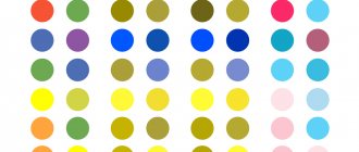
In ophthalmological practice, to diagnose color blindness, specialists most often resort to the Rabkin and Ishihara tests. Rabkin's tables are pictures that consist of small multi-colored circles with identical brightness. If a person has healthy vision, then it will be easy for him to distinguish the numbers and geometric shapes that form these circles. If there are signs of color blindness, images cannot be seen. There is also a type of polychromatic table containing pictures that only people with color blindness can recognize. When taking the Ishihara test, a person needs to see numbers on a colorful background or trace the trajectory of a winding line. Here the numbers and background consist of green and red dots. If a person cannot distinguish red from dark or, on the contrary, light tones, then he has color blindness. Such tests can be carried out at home, however, to determine an accurate diagnosis, it is recommended to seek the help of an ophthalmologist.
Tests to determine color blindness
The test for color perception consists of the ophthalmologist showing the patient a picture and asking him to name the sign depicted on it. Time for this is given 5 seconds. The main pictures are used to identify the presence and type of color blindness, the additional ones – the severity of the pathology. The test also allows you to determine the cause of the disease.
Another test used to determine color blindness is named after the Japanese ophthalmologist Ishihara. Its essence is the same as Rabkin’s tables, only numbers are used exclusively as identifiable images. You can take the Ishihara test yourself on some websites.
When taking the color blindness test, you must follow these rules.
- The person being tested should feel normal. Feeling unwell may interfere with the test result.
- Lighting should be natural.
- Direct rays of the sun should not fall on the drawing, causing reflection.
- The card should be at eye level.
- The distance between it and the patient's eyes should be about 1 m.
- The picture display time is 5 seconds.
Vision is considered normal if at least 90% of the pictures are correctly identified. If color blindness is present, the number of correct answers usually does not exceed 25%.
The picture shows one example of a test for color blindness.
Treatment of color blindness
If the reason for a patient’s lack of color vision is a mutating gene, that is, the disease is hereditary, it is impossible to get rid of it. During the treatment of an acquired form of pathology, with the complete elimination of its root cause, as a rule, good results are observed. That is, the ability to distinguish colors returns to a person. It is important to understand that the treatment of an acquired form of visual system dysfunction depends on the specific factor of its manifestation. For example, if it arose as a result of clouding of the lens due to age-related changes in the visual system, then it is impossible to eliminate the problem. And if the disease is discovered while taking medications, stopping them can help restore healthy vision.
For pathologies such as retinopathy, glaucoma and cataracts, only their correct treatment will make it possible to restore color perception.
What professions are prohibited for colorblind people?
There are quite a few professions that require normal color perception. Laboratory assistants, confectioners, artists, designers, employees involved in color printing, etc.
As for drivers, according to a recently adopted law, only monochromatic people - people with complete color blindness - are not allowed to drive cars. Previously, there were unreasonably harsh requirements for drivers. Even a color-blind driver can see which traffic light is on - top, middle or bottom.
Color blindness does not threaten life; in the worst case, its quality decreases. Usually colorblind people navigate the world around them quite satisfactorily, without experiencing great inconvenience. But everything is determined by the type and severity of color blindness in a particular person. In some cases, there may be a significant deterioration in quality of life.
Video test for color blindness:
Correction of color blindness
To correct impaired color vision, ophthalmologists prescribe specialized contact lenses or glasses to the patient, their surfaces are painted in special colors. Such optical products help to identify differences between specific colors, but may distort some objects. It is known that the cones in the visual system function better in low light conditions. That is why experts have created special glasses for color correction that block light and are equipped with shields or tinted lenses.
For people with mild color blindness, glasses with multi-layer lenses are recommended; they allow you to distinguish between green and red shades.