Strabismus
(
strabismus, heterotropia
) - deviation of the visual axis of one of the eyes from the joint point of fixation in case of impaired binocular vision. The incidence of strabismus is 1.5-2%.
The causes of strabismus can be diseases and damage to the c. n. pp., congenital differences in the anatomical and optical structure of both eyes, ametropia, a sharp decrease in vision or blindness in one eye. Hereditary predisposition to strabismus also matters. As a result of these causes, various parts and sensory-motor connections of the visual analyzer (see), on which normal binocular vision (see) depends, are affected, which leads to its disorder or prevents its formation. With decreased vision or blindness in one eye, K. occurs due to the lack of stimulus for fusion (fusion). If the fusion ability is insufficient, the eye may deviate due to increased (with farsightedness) or weakened (with myopia) accommodation and associated convergence.
There are paralytic Strabismus caused by damage to the nerves (nn. oculomotorius, trochlearis, abducens) innervating the external muscles of the eye, and concomitant Strabismus, when the mechanism of binocular fixation of the object and fusion is upset (see), and the extraocular muscles are almost not affected.
Paralytic strabismus
In the case of an isolated lesion of one of the muscles, the diseased eye deviates in the opposite direction (primary deviation angle). The amount of eye deviation (squint angle) increases as the gaze moves towards the action of the affected muscle. When fixing an object with a paralyzed eye, the healthy eye deviates (secondary deviation angle), and by a much larger angle than the one to which the diseased eye was deviated (the secondary deviation angle is always greater than the primary deviation angle). Eye movements towards the affected muscle are absent or severely limited. There is double vision (usually with fresh lesions) and dizziness, which disappear when one eye is closed. The ability to correctly assess the location of an object in a diseased eye is often impaired (false monocular projection, or localization). There may be a forced position of the head - turning or tilting it in one direction or another.
With paresis (rather than paralysis) of the nerves that control the extraocular muscles, the deviation of the eye and the limitation of its mobility are much less pronounced. Sometimes there is no noticeable deviation at all, but there are complaints of diplopia (see).
A diverse and complex wedge, the picture occurs in cases of simultaneous damage to several muscles in one or both eyes.
With oculomotor nerve palsy, the upper eyelid is drooping, the eye is deviated outward and slightly downward and can only move in these directions, the pupil is dilated, does not respond to light, accommodation is paralyzed (partial ophthalmoplegia).
If all three nerves are affected - oculomotor, trochlear and abducens, then complete ophthalmoplegia is observed (see) - the eye is completely motionless. There are also incomplete ophthalmoplegia - external, in which the external muscles of the eye are paralyzed, but the sphincter of the pupil and the ciliary muscle are preserved, and internal, when only the ciliary muscle and the sphincter of the pupil are affected.
Diagnosis
based on characteristic symptoms. It is important to establish which muscle or muscle group is affected, for which Ch. arr. to the study of double images. In order to determine the localization of the lesion, a thorough neurological examination and electromyography are performed (see).
Treatment
consists primarily of treating the underlying disease. Electrical stimulation of the affected muscle and exercises to develop eye mobility are also carried out. For mild paresis, orthoptic exercises are useful (see Orthoptics). To eliminate double vision, glasses with prisms are used, occlusion of the diseased eye or incomplete occlusion using partially frosted spectacle glass in the part of the visual field where double vision is noted. For persistent paralysis and paresis, surgery is indicated. It is produced no earlier than 6-12 months. after active conservative treatment and stabilization of the underlying process. In case of congenital paralytic K., it is advisable to operate at the age of 3-4 years. For abducens nerve palsy, in addition to resection of the external rectus and recession of the internal rectus muscles, tendon-muscular flaps formed from the superior and inferior rectus muscles are usually transplanted. This operation often restores a certain degree of eye mobility towards the paralyzed muscle.
Method using perimeter
During the diagnosis, the patient fixes his gaze on a candle fixed in the horizontal part of the frame perimeter. In this case, the doctor determines the level of the mark where the second candle should be placed so that it is symmetrically reflected in the other pupil. The study should be carried out in a darkened room. It is advisable to fix the subject's chin on the stand.
During the examination, the patient looks into a special device, while combining two images by moving the handles. Pictures can look different (for example, a dog with ears and a tail, or a circle with a square), they are placed in cassettes, the optical heads of which move until the patient's visual axis coincides with the light beam.
To eliminate adjustment eye movements, you should alternately turn the pictures on and off. The squint angle will be displayed on a special scale.
During the diagnosis, the patient fixes his gaze on a candle fixed in the horizontal part of the frame perimeter. Along with this, the doctor determines the level of the mark where to place the second candle so that it is symmetrically reflected in the other pupil. The study should be carried out in a darkened room. The subject's chin must be fixed on the stand.
During the study, the patient observes through a special device, combining at the same time two pictures by moving the handles. The pictures can be viewed in different ways (for example, a dog with ears and a tail, or a circle with a square); they are placed in cassettes, the optical heads of which move until the patient’s visual axis coincides with the light beam.
To eliminate the installation eye movements, turn the pictures on and off alternately. The squint angle will be displayed on a special scale.
Concomitant strabismus
Rice.
1. Eye position in some types of strabismus: 1—convergent (the visual axis of the left eye is deviated towards the nose); 2—divergent (the visual axis of the left eye is deviated toward the temple); 3—superactive (the visual axis of the left eye is deviated upward). Concomitant strabismus can be congenital or acquired; primary (without visible pathology of the eye) and secondary (developing when the vision of one eye is reduced due to cataracts, corneal cataracts, pathologies of the retina, optic nerve and other diseases of the eyeball); constant and periodic, non-accommodative (not disappearing after correction of ametropia), partially accommodative (decreasing under the influence of correction of ametropia) and accommodative (eliminating by correction of ametropia); monolateral (squints one specific eye); alternating (alternating deviation of the eyes); converging (converging) - the visual axis of one of the eyes is deviated towards the nose (Fig. 1.7); diverging (diverging) - the visual axis is deviated towards the temple (Fig. 1,2); super operative - upward deviation of one of the axes (Fig. 1.3); infraverting - deviation of one of the axes downwards.
Friendly convergent K. usually develops in early childhood and is often periodic at first. Gradually, a restructuring of the child’s entire visual system occurs, adapting to the asymmetrical position of the eyes. Active inhibition of the reaction to adequate stimulation of the central part of the retina of the deviated eye leads to the fact that its images are excluded from visual perception. A functional scotoma occurs (see), eliminating double vision. This scotoma disappears when the fixing eye is turned off from vision.
Violation of normal binocular connections becomes increasingly stronger over time. Under artificial conditions of separation of the visual fields of both eyes, the so-called. abnormal correspondence of the retinas, in which there is a merging of images falling on the central fovea of the retina of one eye and on the paracentral portion of the retina of the other.
Divergent K. occurs much less frequently than convergent K. and is characterized by a later onset and a lower frequency of sensory disturbances.
With monolateral strabismus, the function of the constantly squinting eye is in a state of persistent inhibition, which leads to a sharp decrease in visual acuity of this eye - amblyopia (see). With an intense inhibitory process, the fovea loses its functional superiority over other parts of the retina and incorrect visual fixation occurs.
With friendly K., double vision, as a rule, does not occur. Both eyes (fixing and squinting) make movements in approximately the same volume. The movements of each eye in different directions are usually unrestricted or slightly limited.
Rice. 2. Determination of the strabismus angle in degrees by the position of the light reflex on the cornea (Hirshberg method - explanation in the text)
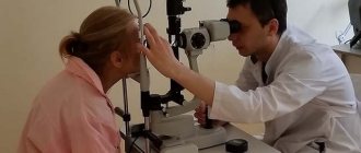
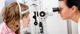
Diagnosis
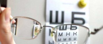
The examination of the patient begins with a medical history (time of occurrence of strabismus, its possible causes, treatment performed, its effect on eye position and vision). The visual acuity of each eye and both eyes together, without correction and with correction, the nature of vision (monolateral or alternating) by means of a test with covering the eyes, the type of vision in the direction of deviation of the eye (convergent, divergent, vertical) and the magnitude of the deviation are determined. For practical purposes, measuring the K angle using the Hirschberg method, which is carried out with a mirror ophthalmoscope, is quite sufficient. K.'s value is estimated in degrees according to the position of the light reflex on the cornea. If the reflex from the ophthalmoscope is located at the edge of the pupil, then the angle of strabismus is 15°, if in the middle of the iris - 25 - 30°, on the limbus - 45°, behind the limbus - 60° or more (Fig. 2). Using a light device, the state of binocular vision is examined. To judge the state of binocular vision in young children, a prism test is used.
Eye mobility is determined by moving a fixation object in front of the patient’s eyes in eight directions. If muscle paresis is suspected, coordimetry and “provoked” diplopia methods are used to judge the state of the oculomotor system.
Using a synoptophore, the ability of the visual analyzer to merge monocular images of objects is studied. In the case of bifoveal fusion, the width of the fusional reserves is determined.
Rice. 3. Diagram of the positions of the fixation object on the fundus during ophthalmoscopic determination of the state of visual fixation; 1—with correct fixation (object in the fovea); 2—in case of incorrect fixation (object on a non-central area of the retina).
Visual fixation is examined (with the second eye turned off) using ophthalmoscopes, into the system of which a fixation object is introduced. When fixation is correct, the object is projected onto the fovea, and when fixation is incorrect, it is projected onto the paracentral areas of the retina (Fig. 3).
The optical media (see Ophthalmoscopy) and the fundus of the eye (see) are carefully examined, and additional methods are used - campimetry (see), a test with a sequential image, a test using the Heidinger phenomenon (see Entoptic phenomena), electrophysiol, studies ( see Electrophysiology of the organ of vision).
Treatment
Treatment is aimed at restoring binocular vision, which returns the fullness of visual functions and at the same time ensures permanent elimination of asymmetry in the position of the eyes.
Complex treatment of friendly eye is generally accepted, which consists of optical correction of ametropia (see), measures to increase the visual acuity of a squinting eye (pleoptics), operations on the eye muscles, pre- and postoperative exercises to restore the combined activity of both eyes (orthoptics) and their ability to correctly estimate the depth of space (stereo-optics).
To correctly prescribe glasses to patient K., an accurate determination of refraction is necessary under conditions of drug-induced relaxation of accommodation. Constant optical correction in combination with orthoptic exercises is the main method of treating accommodative vision, which converges in hypermetropia and diverges in myopia. If, when wearing glasses, the symmetrical position of the eyes is stably maintained or binocular vision is restored, then for small degrees of hypermetropia, optical correction is performed every 6-12 months. decrease by 0.5-1.0 diopters and then cancel.
Treatment of amblyopia in children 3-6 years old, regardless of the state of fixation, should begin with constant switching off (occlusion) of the dominant eye, which should be carried out for at least 4 months. The purpose of such switching off is to achieve equal visual acuity in both eyes and the transition of monolateral strabismus to alternating. It is advisable to combine occlusion with the Avetisov method (1968) - local “blinding” irritation with light of the central fovea of the retina and a variety of visual exercises, the choice of which is determined by the age, level of development of the child, his interests and inclinations. In the absence of an effect from the use of occlusion, as well as in cases of incorrect visual fixation in children over 6 years of age, complex treatment of amblyopia is used, which is based on the Küppers method (1956), which uses a negative sequential image that occurs when the retina of the posterior pole of the eye is illuminated with simultaneous covering foveolar zone using a round mark, and the method of local “blinding” irritation with light of the central fovea of the retina. In the absence of amblyopia or a persistent increase in visual acuity above 0.4, orthoptic exercises are indicated, the purpose of which is to make the foveal retinocortical elements of both eyes dominant and restore their combined activity.
To restore the ability for bifoveal fusion, exercises are performed on the synoptophore, the essence of which is to rapidly alternately stimulate the central fovea of the retina of both eyes, or a method is used based on the use of two monocular sequential images. In the presence of bifoveal fusion, fusion reserves are developed on a synoptophore or (in the case of symmetrical eye position) on a stereoscope with a mirror. Exercises to develop eye mobility can be carried out using a synoptophore, a muscle trainer, or a simplified method by moving an object in front of the child’s eyes in the right directions. In a similar way or using the Convergence Trainer device, convergence is trained at divergent K.
If constant wearing of glasses for 1.5-2 years does not eliminate K. (with the so-called non-accommodative K.), then they resort to surgery (at the age of 5-6 years) with pre- and postoperative orthoptic exercises (development of eye mobility, their ability to merge foveal images of objects, fusion ability, stereoscopic and depth vision).
The purpose of operations on the eye muscles for concomitant K. is to, by changing the muscle balance, i.e., the relative tension of the muscles, to obtain a symmetrical or close to it position of the eyes and thereby contribute to the restoration of normal binocular functions.
Modern tactics of eye surgery are characterized by the use of such types of surgery in which the muscle maintains a reliable connection with the eyeball. If after the first stage of the operation the residual K angle remains, then the second stage of the operation on another muscle of the same eye or on the other eye is carried out after 6-8 months.
When a pronounced horizontal deviation of the eye is combined with a vertical one, it is advisable to first perform surgery on the horizontal muscles, taking into account that vertical deviation can be not only a consequence of muscle paresis, but also an imbalance of the muscles vertically, which often disappears in the primary position of the eye. If the vertical deviation is significant and examination of the oculomotor system indicates a predominant lesion of the muscles of vertical action, then one should first operate on these muscles.
To eliminate strabismus, two types of operations are used - strengthening and weakening the action of muscles.
Rice. 4. Scheme of the muscle resection operation for convergent strabismus: 1— resected section of the external rectus muscle with a stay suture applied to its cut edge (2); 3— passing the suture through the muscle at the site of its anatomical attachment
The operations of the first type include: resection - shortening of the muscle by excision of its section at the site of attachment to the sclera and suturing to the same place (Fig. 4); tenorrhaphy - shortening of a muscle by forming a fold from its tendon; proraphy - movement of the muscle tendon anteriorly (with interventions on the rectus muscles) or posteriorly (with interventions on the oblique muscles) with or without the formation of a fold.
Rice. 5. Scheme of the muscle recession operation for convergent strabismus: 1—place of attachment of the internal rectus muscle; 2— fixation of the muscle posteriorly with two loop-shaped sutures.
The following operations weaken the action of the muscle: free (or complete) tenotomy - intersection of the muscle tendon at the point of attachment without suturing it to the sclera; tenotomy with a restrictive (safety) suture - fixation of the tenotomized muscle at a certain distance from the site of anatomical attachment using a suture passing through this place and the edge of the crossed tendon; partial tenotomy - making two or three incomplete, slightly spaced incisions on the muscle tendon from opposite edges; recession (Fig. 5) - movement of the muscle cut off at the attachment site posteriorly (for interventions on the rectus muscles) or anteriorly (for interventions on the oblique muscles) with its suturing to the sclera; prolongation - lengthening a muscle by completely cutting its tendon in different directions and suturing the cut areas. The free tenotomy operation is performed only on the oblique muscles.
Analysis of indicators
The choice of treatment method depends on the data obtained from studying the Hirschberg strabismus angle. The doctor compares two angles of strabismus - primary and secondary. The primary level determines the angle of deviation of the diseased organ of vision, and the secondary level determines the indicators of the healthy eye.
The strabismus angle is determined based on the following indicators:
- If the glare is located within the pupil, then the angle of strabismus is 10 degrees.
- If the reflection of the glare is fixed at the edge of the pupil, then the angle of strabismus corresponds to 15 degrees.
- If the glare is in the iris area, then the level of pathology corresponds to 25-30 degrees.
Prevention
Antenatal prevention of strabismus consists of preventing intrauterine infection and intoxication, toxicosis of pregnancy, birth trauma, since these factors can cause disruption of the innervation of the oculomotor system in the child.
Prevention and active treatment in children of all possible causes of damage to the nerves that control eye movements are also of great importance for postnatal prevention of K.
The most accessible way to prevent primary concomitant K. is optical correction of ametropia at an early age (1.5 - 2 years). In this way, at least accommodative K can be prevented. It is advisable to prescribe glasses for constant wear for myopia, astigmatism and hypermetropia of 2.5 diopters or more. It is also necessary to strictly adhere to the requirements of visual hygiene, to avoid visual work at too close a distance from the eyes, reading in poor lighting, or lying down.
Methods for identifying pathology and measuring the angle of strabismus at home
With congenital strabismus, you can notice the presence of a problem from the very first days. If the disease is acquired, it is unlikely to immediately notice a slight deviation, given the fact that people rarely go to the clinic for a routine medical examination. After this, the doctor can prescribe treatment at home. To determine pathology, it is not always necessary to visit a clinic - the test can be done at home. To do this, you need to lean back on a chair, fixing your head in a motionless position. Next, the person looks at a stationary object in the window - for example, a sign or a satellite antenna. Focusing of vision on the selected object occurs for 1-2 seconds.
Next, covering his eye with his palm, the person examines the same object for 1-2 minutes. If the fixed object does not jump in different directions when each eye opens, you don’t have to worry about the presence of strabismus - it is simply absent. This is a very simple test, but to obtain more detailed information regarding the condition of the eyes, you should still seek help from an ophthalmologist. He will independently carry out the inspection using appropriate diagnostic equipment and modern techniques. And if, after opening the eye, the object begins to jump from side to side, you definitely cannot do without the help of a specialist - you will have to carry out hardware or surgical vision correction. Maybe,
What to look for when making a diagnosis
When determining the diagnosis, it is necessary to pay attention to the medical history, namely:
- The time when strabismus appeared indicates the etiology. The earlier it occurs, the greater the likelihood of surgical intervention. With late onset, the chances of an accommodative component increase.
- Angle variability is an essential criterion, since the periodic appearance of strabismus makes it possible to understand that binocular vision has been preserved.
- The general condition or abnormal development is of great importance. For example, attention is drawn to the frequency of strabismus in a child suffering from cerebral palsy.
- The specialist needs to check the birth history, become familiar with the indicators during pregnancy, the baby’s weight at birth, pathologies during intrauterine development, or during childbirth.
- Hereditary history also has a strong influence, since in most cases this disease is a congenital pathology.
- As part of testing sensory functions, the level of stability of binocular vision is determined, its acuity, and the presence or absence of bifoveal fusion are revealed. Attention is drawn to the functional scotoma of suppression, the nature of diplopia, and fusion reserves.
When checking motor functions, the doctor analyzes the degree of mobility of each eyeball, characterizes the deviation, and determines the complexity of disturbances in the work of the extraocular muscle of each eye separately.