Classification of hemeralopia
Hemeralopia is classified into 3 types: congenital, symptomatic and essential. The most common form is the essential one. It often occurs due to irrational and insufficient nutrition or excessive consumption of alcoholic beverages.
Symptomatic hemeralopia occurs against the background of certain eye diseases: glaucoma, cataracts, myopia. The congenital form of the disease begins to progress from about 16 years of age.
In addition to the listed species, false night blindness is also distinguished. A person’s vision deteriorates and eye fatigue appears after prolonged work with a TV or computer monitor. This condition is not a disease. It indicates functional eye strain. After sufficient rest, vision is usually restored. But if a person constantly overstrains his eyes and does not give them sufficient rest, this leads to the development of serious ophthalmological diseases and a persistent decrease in visual acuity.
Types of hemeralopia (night blindness)
In medicine, there are three types of hemeralopia: 1) essential hemeralopia 2) congenital hemeralopia 3) symptomatic hemeralopia. With congenital night blindness, the disease is often inherited. Hemeralopia in this case manifests itself early: in children and adolescents. Congenital night blindness can be caused by some hereditary diseases. This may be Usher syndrome or hereditary retinitis pigmentosa. Symptomatic night blindness develops as a result of various eye diseases affecting the retina. Complicated high myopia, glaucoma, tapetoretinal dystrophies, chorioretinitis, optic nerve atrophy, siderosis can cause symptomatic hemeralopia. This type of night blindness indicates organic damage to the retina. In turn, essential night blindness is a functional disorder of the retina, which manifests itself with a lack of or impaired metabolism of retinol (vitamin A), PP, B2. This is caused by various diseases of the liver, digestive system, poor and insufficient nutrition, chronic alcoholism, rubella, toxic damage to the body, and prolonged exposure to bright lighting.
Causes of hemeralopia
The retina of the eye normally contains about 115 million rods and 7 million cones. The immediate cause of the development of hemeralopia is a deviation in the structure of the rods or a decrease in their number.
The causes of congenital hemeralopia include genetic factors.
Night blindness is transmitted by Usher syndrome. It is combined with retinitis pigmentosa, deafness and mental retardation. Causes of symptomatic hemeralopia include:
- periodic or constant increase in pressure inside the eye;
- high myopia;
- retinal damage - retinopathy;
- retinal detachment;
- optical neuropathy, or death of optic nerve fibers;
- inflammatory diseases of the choroid or retina of the eye;
- cataract.
The essential type of hemeralopia is caused by a lack of vitamins – A, B2, PP.
It occurs with anemia, diabetes, diseases of the digestive tract, and liver. A deficiency of these vitamins can occur due to poor nutrition. Essential hemeralopia also develops due to:
- fasting;
- anemia;
- previous chickenpox or rubella;
- poisoning;
- use of drugs that disrupt the metabolism of vitamin A or destroy it;
- prolonged exposure to strong lighting on the organ of vision.
Predisposing factors for the development of visual impairment are:
- infectious diseases;
- menopause;
- vegetarianism;
- strict diets.
Vitamin A reserves in the body last for about a year. It can take up to two years from the development of vitamin deficiency to the first manifestations of the disease.
Pathogenesis and causes of hemeralopia (night blindness)
This disease can develop due to a decrease in the number of retinal rods or their structural changes, as well as when the formation or exchange of the visual pigment rhodopsin, which is contained in the rods and takes part in the perception of light by the eye in poor lighting, is disrupted. The rods in the retina are responsible for the perception of light, including twilight vision, and the cones are responsible for color vision in well-lit conditions. The number of rods significantly exceeds the number of cones, their ratio is approximately 115 million to 7 million. When the relationship between these retinal elements is disrupted, night blindness can occur.
Symptoms of hemeralopia
Symptoms of congenital hemeralopia are noted already in early childhood. Persistent visual impairment cannot be treated.
The disease manifests itself as decreased vision at dusk and at night. In twilight, visual discomfort is noted. A person notices that he does not see objects at dusk and cannot navigate in space. This is especially noticeable when moving from a bright room to a dark one. During the day, as well as in good light, vision remains good.
When there is insufficient lighting, a person’s visual field is significantly narrowed. This means that he sees the world around him as if through a pipe. The patient also cannot distinguish the shape of objects.
With night blindness, there is a noticeable lack of sensitivity of visual receptors to light. It manifests itself in the fact that reading or performing other work requires bright lighting. Turning on a bright light for reading with normal refraction is the first sign of developing night blindness.
Another important symptom is an erroneous assessment of the distance to objects.
This leads to stumbling, hitting doors and other objects. The patient may experience the following symptoms:
- severe visual impairment;
- blue color perception disorder;
- pain and pain in the eyes;
- formation of plaques in the eyes;
- hair fragility;
- baldness;
- dandruff;
- the appearance of a skin rash;
- loss of appetite;
- fatigue and drowsiness.
It is noteworthy that decreased visual acuity in one eye is not a sign of night blindness. The disease affects both eyes.
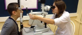
Diagnosis of hemeralopia
An eye doctor diagnoses hemeralopia. First, he collects the patient's complaints, which may indicate a significant deterioration in vision.
If hemeralopia is suspected, a general blood test and an analysis for the amount of retinol in it are prescribed. It is also necessary to diagnose visual acuity using standard tables.
An important study is perimetry. In this case, the doctor determines the patient’s field of vision. With hemeralopia it decreases. This is especially noticeable when studying sensitivity to blue and yellow colors.
The patient is prescribed adaptometry. The doctor examines the adaptation of the visual system of the eye to sudden changes in illumination. Normally, the eye partially adapts to darkness in 5 minutes, and completely in only 40.
Electroretinography is a diagnostics of the structure of the retina of the eye. Changes in the fundus are determined by the development of the underlying pathology. When the cause of hemeralopia is a deficiency of vitamins, then signs of disorders will not be visible on the retina.
Finally, electrooculography is used to diagnose hemeralopia. It reveals the state of the extraocular muscles and the surface state of the retina during eye movement.
These diagnostic methods allow not only to determine the degree of visual acuity impairment, but also to determine the true cause of hemeralopia.
5.1.5.2 Stationary night blindness
Stationary night blindness (hypofunction of the rod apparatus, or hemeralopia). Early psychophysical data showed that there are two forms of congenital stationary night blindness: one form with normal cone function and another with some decrease in it, which has recently been confirmed by electrophysiological studies. Therefore, the classification of congenital forms of stationary night blindness is based on differences in ERG. One type is characterized by a reduced amplitude of the photopic response, which does not increase in the dark (Nougaret type I); in the other type, dysfunction is expressed in the absence of dark adaptation, b-waves in the general ERC
), due to which the a-wave is more pronounced than normal, while the photopic response is reduced (Schubert-Bornschein type II). In type I, with normal visual pigment kinetics, changes in the ERG indicated disturbances in transmission in the internal segment of the photoreceptors. In type II with normal visual pigment and normal ERG a-wave, a disturbance of neuronal transmission in the area of bipolar cells was noted. Electrophysiological and psychophysical data suggest the presence of a primary defect in the interplexiform cells of the inner nuclear layer, which is confirmed, on the one hand, by the absence of structural abnormalities in the photoreceptors, and on the other, by the fact that the normal rod signal does not reach the bipolar cells. Work in recent years has shown that in this type of disease, synaptic transmission at the inverting glutamate synapse between rods and depolarizing bipolar neurons is disrupted [Carr RE, 1992]. In Ogushi's disease (congenital stationary night blindness with changes in the fundus), the ERG b-wave is absent, although some studies have shown the preservation of scotopic function. When using a low-intensity red background, a normal cone response was found, but a reduced amplitude and prolonged rod response. P. Guras (1970) recorded a normal scotopic response in the ERG in this disease to the first stimulus and the disappearance of this scotopic response upon subsequent stimulation. In connection with the found normal regeneration of rhodopsin and its normal photosensitivity, it was suggested that this disease is associated with disturbances in adaptive networks [CarrR.E., Ripps H., 1966], and with fundus albipunctata, changes in ERG and EOG are associated with a slowdown in the regeneration of the rod and cone visual pigments. Congenital stationary night blindness of the Schubert-Bornshein type (Schubert-Boraschein) is divided into two subtypes: complete (with myopia) and incomplete (according to Miyake), according to the degree of preservation of the rod and cone functions in the ERG (either the absence of the ERG, or its partial preservation;).
Tapetoretinal dystrophy (retinitis pigmentosa). The classic pathognomonic sign of this severe hereditary disease, the varied clinical picture of which is caused by mutations of the rhodopsin gene, is the absence of ERG already in the initial stage (
) . In the very early stages of dystrophy with a dominant form of inheritance, the ERG may be subnormal, which makes it possible to separate the cone and rod components of the ERG and study the evolution and localization of the disease. At the same time, an increase in the b-wave culmination time was noted, which serves as a differential sign between stationary night blindness and the sectoral form of pigmentary resinitis, in which the peak time is within normal limits. Electrophysiological changes in retinitis pigmentosa precede changes in the fundus and other functional findings. Attempts are being made, according to ERG data, to separate the types of inheritance in retinitis pigmentosa [BersonE.L., 1981-1991; Apfelstedt-Sylla E., 1996; Cideciyan AV, et al., 1996]. It was found that in the dominant form with full penetrance of the gene, the L-wave of the cone ERG has a normal or slightly reduced amplitude with a normal culmination time, while the rod ERG is significantly reduced and its culmination time is increased. In the dominant type with reduced penetrance, the rod and cone ERG are reduced with a prolonged culmination time. The mitochondrial form of retinitis pigmentosa is similar in functional symptoms to the dominant form. Already in the initial stage, retinitis pigmentosa with an autosomal recessive type of inheritance and sex-linked is characterized by a very weak cone response and a significant increase in the latent period of the b-wave. If the rod response is detected, the time of climax is also significantly increased. It is very important to examine relatives of patients with retinitis pigmentosa along the ascending and descending lines. A decrease in the scotopic b-wave of the ERG, a prolonged time of its culmination, the absence or reduction of oscillatory potentials are typical not only for patients with clinically established forms of retinitis pigmentosa, but also for siblings, heterozygotes, carriers of the pathological gene for retinitis pigmentosa (
). ERG changes detected in early childhood (up to 6 years) can help both in clarifying the diagnosis and genetic form of the disease, as well as in identifying carriers of the pathological gene and genetic consultations. ERG is absent in non-pigmented and small-pigmented unilateral forms of retinitis pigmentosa. . In the case of an atypical unilateral form of the disease, the examination should be repeated after a certain period of time to establish true unilaterality. Electroretinographic responses depend on the extent of the pathological process. Thus, ERG is subnormal in nature and is always present in sectoral forms of retinitis pigmentosa (quadrant, half, central and paracentral).
ERG is absent in retinitis pigmentosa, which is part of the symptom complex (also including a mutation of the rhodopsin gene) of some hereditary ataxic lesions of the cerebellum, hearing loss, Usher syndrome, Moon-Bardet-Biedl syndrome, Hurber-Hunter mucopolysaccharidosis - Neck (Hurber - Hunter - Sheie), some myotonic dystrophies. In Retinitis punctata albescens (severe form), the ERG is significantly reduced, while in retinitis albipunctatus with hemeralopia, the ERG is partially preserved.
Since the cone visual signal in the retina passes through on- and off-biopolar cells, R.A. Sieving (1993) used longer stimuli (150–300 ms) to record photopic ERG in patients with retinitis pigmentosa than other authors, which made it possible, even using total ERG, to identify disturbances in the on- and off-pathways of visual information transmission in the retina . The results of his studies showed that one of the reasons for weakened vision in these patients is pathological signal transmission in the proximal retina, and the functional defect is localized postsynaptically in relation to cone photoreceptors.
Recent genetic findings indicating a rhodopsin mutation in retinitis pigmentosa of an autosomal dominant type of inheritance [DryjaT.P. et al., 1990], suggest that in many retinal dystrophies the primary Defect is at the photoreceptor level. However, it is unclear why in some cases with rod-cone dystrophy (traditionally called abroad “retinitis pigmentosa”), the decrease in the ERG b-wave does not correspond to a sufficiently well-preserved visual field and at the same time the dark adaptation threshold only slightly increases.
- < Back
- Forward >
Treatment of hemeralopia
Treatment for hemeralopia depends on its type. In the symptomatic form of the disease, treatment is carried out for the underlying pathology that led to visual impairment at dusk. For this purpose, surgical methods of treating cataracts or glaucoma can be used. For myopia or retinopathy, laser therapy or careful monitoring of the underlying pathology that led to eye damage is required. After a period of recovery or if compensation for chronic pathology is achieved, vision improves.
Essential night blindness responds well to treatment by taking synthetic analogues of vitamins - A, PP, B2.
To get rid of night blindness, adults need up to 100 thousand IU of the vitamin per day. For children, 1–5 thousand IU of the drug per day is sufficient. The daily amount of riboflavin is 20 mg for children and adults. It is recommended to include in your diet more foods that contain the components necessary for good vision:
- greenery;
- beef and pork liver;
- Cod liver;
- dairy products;
- eggs;
- carrots;
- green onions;
- tomatoes;
- peas;
- different types of cabbage;
- sweet corn;
- all fruits and berries are orange in color;
- cherry;
- gooseberry;
- blueberries;
- black currant;
- dishes made from millet cereals.
You need to follow a diet and take vitamins for several months.
If you have hemeralopia, you should avoid bright light.
In the evening, if you have even a slight degree of myopia, you need to wear glasses. Traditional medicines are also effective:
- To obtain a decoction, mix 2 parts each of blueberry leaves, linden flowers, dandelion, one part each of sea buckthorn and buckwheat leaves. Pour a tablespoon of crushed plants into 200 ml of hot water, boil in a water bath for a quarter of an hour, then leave for half an hour. Drink a glass three times a day after meals.
- Pour a teaspoon of cornflower flowers into 200 ml of boiling water and leave for 60 minutes. Strain the resulting infusion and consume 50 ml three times a day.
- Pour 200 milliliters of boiling water over a tablespoon of blueberries. Take half a glass of this tea 3 times a day.
- Eat sea buckthorn berries in any form in the amount of 2 glasses throughout the day.
- Pour 2 tablespoons of stinging nettle leaves into 200 ml of boiling water, leave for 60 minutes, take a third of a glass three times a day before meals.
- Take 200 ml of carrot juice 2 – 3 times a day.
- It is useful to drink juice from blueberries or grapes.
- Sprout the wheat grains and grind them. Pour 200 ml of boiling water over a tablespoon of the resulting mixture, leave for a quarter of an hour, strain. Drink a third of a glass 3 times a day.
- It is useful to drink fish oil.
{banner_horizontalnyy3}
Diagnostic methods
If you have the above symptoms, you should definitely see an ophthalmologist. What examination methods can the doctor perform?
Adaptometry is based on the Purkinje phenomenon (in twilight lighting, the maximum brightness in the spectrum shifts from red-orange to blue-violet). What is an adaptometer? This is a dark chamber where there is a table with squares of different colors (red, blue, yellow and green). These squares are illuminated with light, the brightness of which gradually increases. At the beginning of the study, the patient cannot recognize the color of the squares, but as dark adaptation occurs, the colors become distinguishable. A stopwatch is used to record the time that passes from the moment the tables begin to be illuminated until the patient sees a lighter square in place of the green one. Normally, this time does not exceed one minute.
In addition to this technique, others are carried out:
- determination of visual acuity (visometry);
- determination of the boundaries of the field of view (perimetry);
- study of color perception (using Rabkin tables);
- ophthalmoscopy - examination of the fundus of the eye.
Complications of hemeralopia
Hemeralopia can cause the following complications:
- injuries due to impaired orientation in space;
- psychological disorders;
- deterioration of vision (even before its loss).
The most disappointing prognosis for genetically determined hemeralopia is that it cannot be cured. In the symptomatic form of the pathology, the outcome of treatment depends on how early therapeutic measures were started. Essential hemeralopia responds well to therapy. The longer the patient neglects treatment, the worse the prognosis.
Nyctalopia, also known as night blindness or night blindness, reduces the ability to see clearly at night or in low-light conditions. Daytime vision does not deteriorate even with nyctalopia.
Nyctalopia is not a disease in itself, but only a symptom of an underlying disease - mainly retinal problems like glaucoma. Also, in some cases, myopia can make it very difficult to see at night.
In dark environments, the pupils dilate to take in more light. This light is received by the retina, which contains cells that help people see colors (cones) and in the dark (rods). When there is a problem with rod cells due to illness or injury, a person has poor or no vision in the dark. Then we are talking about night blindness.
Symptoms of nyctalopia
Night blindness can be noticed under certain circumstances:
- It is difficult to move around the house at night, even with small lights on;
- Avoid going out at night for fear of tripping;
- It’s hard to recognize people’s faces in darkened places like movie theaters;
- It takes a long time for the eyes to adjust to the light when you move from darkness to light;
- It takes a long time to get used to seeing in a dark room.
Causes of night blindness
Nyctalopia can be a symptom of several diseases, including:
- Retinitis pigmentosa
This is a group of rare genetic diseases affecting the retina that can result from changes in any of 100 genes. The retinal band cells are more severely affected in the early stages of these diseases, and one of the first symptoms is night blindness.
- Cataract
Cataracts occur when the lens of the eye becomes cloudy and can cause night blindness. Problems with vision at night are usually one of the first symptoms.
- Glaucoma
Glaucoma occurs when fluid accumulates in the front of the eye and increases pressure on the eye, damaging the optic nerve. Glaucoma first affects peripheral vision and then central vision. Both day and night vision are impaired when parts of the retina stop working.
- Myopia
When eyeballs are too long normally, people develop myopia. This condition impairs the ability to see objects that are far away during the day and at night. Some people may experience blurred distance vision only at night.
In night myopia, weak light makes it difficult for the eyes to focus correctly, or an increased pupil size at night allows more peripheral, unfocused light rays to enter the eye.
- Vitamin A deficiency
To see the full spectrum of light, the eye must produce certain pigments for the retina to function properly. Vitamin A deficiency stops the production of these pigments, leading to night blindness.
- Diabetes
High blood sugar can damage the blood vessels in the retina, causing vision problems such as diabetic retinopathy. Nyctalopia is often one of the first symptoms of diabetic retinopathy.
- Some medications for glaucoma
Some miotics used to treat glaucoma can shrink the pupil and cause nyctalopia.
- Keratoconus
This condition occurs when the cornea becomes thin and bulges out like a cone. Changing the shape of the cornea causes light rays to become out of focus. Night blindness is a symptom of keratoconus.
Treatment of nyctalopia
Treatment will also depend on the underlying condition causing night blindness:
- Retinitis pigmentosa
There is no cure for the disease, but a number of treatments are being studied, such as vitamin A palmitate and an artificial vision device called Argus II. Talk to your eye doctor to find out if these options are right for you.
- Cataract
The only way to remove cataracts is surgery. When the condition does not affect daily activities, glasses can be managed.
- Glaucoma
To reduce the amount of fluid produced by the eye, and therefore to reduce eye pressure, eye drops are prescribed. Surgery is also performed to remove excess fluid from the affected eye.
- Myopia
The most common treatments for nearsightedness are glasses, contact lenses, or refractive surgery such as LASIK. Other options include multiple rigid lenses to realign the cornea (orthokeratology) or low-dose atropine (0.01%) to slow the progression of myopia in children and adolescents.
- Vitamin A deficiency
Oral vitamin A supplements may help resolve this problem. Eating foods rich in vitamin A, such as liver, beef, chicken, eggs, fortified milk, carrots, mangoes, sweet potatoes, and leafy green vegetables, can also help increase your vitamin A intake.
- Diabetes
Treatment will focus on controlling sugar levels and will depend on the type of diabetes. Therapy may include lifestyle changes, regular blood sugar monitoring, insulin and medications.
- Keratoconus
Mild symptoms can be managed with glasses and then with special hard contact lenses. Other treatment options include intaki (small devices that can smooth out the curvature of the cornea), collagen crosslinking (uses special ultraviolet light and eye drops to strengthen the cornea), and corneal transplantation in severe cases.
Forecast
Night blindness is treatable when it is caused by certain conditions such as nearsightedness, vitamin A deficiency and cataracts. But other causes of night blindness, such as retinitis pigmentosa, have no cure, so your eye doctor can offer options that will only reduce symptoms and improve your quality of life.
Prevention of hemeralopia
There are no preventative measures for congenital hemeralopia.
To prevent the progression of symptomatic and essential types of night blindness, it is recommended to follow these rules:
- eat quality and varied food;
- Avoid exposure to bright sunlight;
- observe hygiene when working with a computer;
- avoid prolonged visual strain;
- visit an ophthalmologist 2 times a year;
- promptly diagnose and treat pathologies that can lead to the development of night blindness.
If you suspect deterioration of evening and night vision, you should immediately contact an ophthalmologist.
Hemeralopia is a visual disorder that results in difficulty seeing clearly in low light conditions or at night. With normal vision during the day, a person practically goes blind in the dark. Acquired night blindness is highly treatable if you consult a doctor promptly.
Treatment
A technique that treats night blindness due to the congenital pathology of dark adaptation does not currently exist.
Acquired disorders can be treated if their causes are identified in time. Treatment is carried out for diseases that cause hemeralopia, both local (cataracts, myopia, glaucoma) and general (hypovitaminosis of vitamin A, scurvy, diabetes mellitus).
We recommend reading an article about another possible vision pathology, more precisely about the loss of all colors of light.
To learn more about eye diseases and their treatment, use the convenient site search or ask a specialist a question.