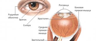
Muscles of the eyeball
Six striated muscles are attached to the eyeball: four rectus muscles - superior, inferior, lateral and medial, and two obliques - superior and inferior.
All rectus muscles and the superior oblique begin deep in the orbit from the common tendon ring, fixed to the sphenoid bone and periosteum around the optic canal, and partly from the edges of the superior orbital fissure. This ring surrounds the optic nerve and ophthalmic artery. The muscle begins from the common tendon ring, subtending the upper eyelid. It is located in the orbit above the superior rectus muscle of the eyeball, and ends in the thickness of the upper eyelid. The rectus muscles are directed along the corresponding walls of the orbit, on the sides of the optic nerve, pierce the vagina of the eyeball and with short tendons are woven into the sclera in front of the equator, retreating 5-8 mm from the edge of the cornea. The rectus muscles rotate the eyeball around two mutually intersecting axes: vertical and horizontal (transverse). The lateral and medial rectus muscles rotate the eyeball outward and inward around the vertical axis, each in its own direction, and the pupil rotates accordingly. The superior and inferior rectus muscles rotate the eyeball around the transverse axis. When the superior rectus muscle acts, the pupil is directed upward and somewhat outward, and when the inferior rectus muscle operates, it is directed downward and inward. The superior oblique muscle lies in the superomedial part of the orbit between the superior and medial rectus muscles. Near the trochlear fossa, it passes into a thin round tendon, enveloped in a synovial sheath, which spreads over a trochlea built in the form of a ring of fibrous cartilage. After passing through the block, the tendon lies under the superior rectus muscle and attaches to the eyeball in its superolateral part, behind the equator. The inferior oblique muscle, unlike the other muscles of the eyeball, starts from the orbital surface of the upper jaw near the opening of the nasolacrimal duct, on the lower wall of the orbit, and is directed between it and the inferior rectus muscle obliquely upward and backward. Its short tendon attaches to the eyeball on its lateral side, behind the equator. Both oblique muscles rotate the eyeball around the anteroposterior axis: the superior oblique muscle rotates the eyeball and pupil downward and laterally, the inferior oblique muscle rotates upward and laterally. The movements of the right and left eyeballs are coordinated due to the cooperative action of the oculomotor muscles.
Protective apparatus of the eye. Accessory apparatus of the eye. Muscular system | Ophthalmology
Description
ORBIT AND ITS CONTENTS
The orbit, or orbit, has the shape of a tetrahedral pyramid with rounded ribs (Fig. 13). The base of the pyramid - its orbital edge is facing anteriorly, the apex - posteriorly, into the cranial cavity. The length of the anteroposterior axis of the orbit is 4-5 cm, the height at the entrance is 3.5 cm, the maximum width is 4 cm. The axes of both orbits converge from front to back and from outside to inside.
The orbit is formed by 7 bones: the frontal, sphenoid, ethmoid, palatine, lacrimal, zygomatic and maxilla. There are 4 walls in the orbit: upper, lower, inner and outer.
The frontal sinus (sinus frontalis) is located in the anterior internal part of the upper wall; its dimensions vary individually. The upper wall of the orbit separates it from the anterior cranial fossa and therefore borders the cranial cavity and the brain.
In the outer corner of the upper wall there is a recess for the lacrimal gland (Jossa glandulae lacrimalis). At the inner edge of the upper wall, at the site of its transition to the inner wall, there is a notch, or bony opening (mcisura, or foramen supraorbitalis), - the exit site of the artery and nerve of the same name.
The lower wall separates the orbit from the maxillary cavity. The outer wall is formed by the lower surface of the zygomatic process of the frontal, the orbital surface of the greater wing of the main wing and the main process of the zygomatic bone and separates the contents of the orbit from the temporal fossa.
The inner wall is formed by the ethmoid bone, its paper plate, in front by the lacrimal bone and the frontal process of the maxilla at the apex of the orbit. On the surface of the lacrimal bone there is a fossa for the lacrimal sac (Jossa sacci lacrimalis). The nasolacrimal bone canal begins from it, which opens in the lower nasal passage at a distance of 3-3.5 cm from the external opening of the nose.
The inner wall separates the orbit from the ethmoid sinus. The paper plate can be very thin and is sometimes represented by two layers of periosteum. It is easily damaged even if you blow your nose carelessly. Damage to this wall causes emphysema of the eyelids and, less commonly, retrobulbar tissue.
Thus, the eyeball is surrounded by the paranasal sinuses. Their pathology is often involved in the development of eye pathology.
The edge of the eye socket (margo superior et inferior) is denser than its bones and. stepping forward. performs a protective function. At the apex of the orbit, in the small wing of the main bone, there is a round optic opening (foramen opticum) with a diameter of 4 mm, through which the ophthalmic artery (a. ophthalmica) enters the orbital cavity and the optic nerve (p. opticus) exits into the cranial cavity (middle cranial hole).
Outward and downward from the optic foramen, between the greater and lesser wings of the main bone, there is a superior orbital fissure (fissura orbitalis superior) covered with connective tissue, connecting the orbit with the middle fossa. Motor nerves pass through the gap to the muscles of the eye: trochlea (n. trochlearis), abducens (n. abducens), oculomotor (n. oculomotorius) and the ophthalmic branch of the trigeminal nerve (ramus ophthalmicus n. trigemini), sympathetic root to the ciliary ganglion, ophthalmic vein (v. ophthalmica).
In case of injury or a tumor of the middle cranial fossa, these formations are compressed or damaged, which leads to the syndrome of superior orbital ptosis (drooping of the upper eyelid), mydriasis (dilation of the pupil), tetraplegia (complete immobility of the eye), anesthesia of the cornea and skin of the eyelid, some exophthalmos, venous stagnation.
In the lower outer corner of the orbit, between the large wing of the main bone and the body of the upper jaw, there is a second fissure - the lower orbital (fissura orbitalis inferior), which connects the orbit with the pterygopalatine fossa. The lower orbital fissure is closed by a connective tissue membrane with smooth muscle fibers (musculus orbitalis), innervated by the sympathetic nerve.
In humans, this muscle is poorly developed, but it still affects the position of the eye in the orbit. An increase in muscle tone can cause exophthalmos (protrusion of the eyeball), while a decrease can cause enophthalmos (retraction of the eyeball). Through the inferior orbital fissure and muscle fibers, an anastomosis of the inferior orbital vein with the venous plexus of the pterygopalatine fossa and the deep facial vein is provided with the possible influence of muscle tone on the venous circulation in the orbit.
In the depths of the orbit, in the main bone, there is a round hole (foramen rotundum), which connects the middle cranial fossa with the pterygopalatine fossa and partly with the orbit. The maxillary nerve (n. maxillaris), the second branch of the trigeminal nerve, passes through the foramen rotunda.
Bone walls limit the entrance to the orbit (aditus orbitae), which is closed in front by the tarsoorbital fascia (fascia tarsoorbitalis), called by some authors the anterior wall of the orbit (septum orbitae).
The tarso-orbital fascia is attached to the edges of the orbit and the cartilage of the eyelids and prevents the spread of infection into the orbit from the eyelids and from the lacrimal sac, which lies in front of it (extraseptally). At the outer edge, the fatty retrobulbar tissue extends beyond the orbit by 3-4 mm.
The edge and walls of the orbit serve to protect the organ of vision. The structure of the orbit determines the characteristics of its pathology. Thus, the anatomical connection of the orbit with the paranasal sinuses often becomes the cause of the transition of the inflammatory process or tumor germination.
The proximity of the optic nerve to the main and ethmoid sinuses during inflammatory processes in them can cause rhinogenic neuritis. In case of injuries to the upper wall of the orbit, damage to the substance of the brain or the spread of the inflammatory process from it to the orbit is possible.
Fractures of the base of the skull can be complicated by damage to the optic nerve canal and complete or partial blindness due to disruption of the integrity or compression of the optic nerve and orbital artery. A characteristic symptom complex is accompanied by a traumatic or inflammatory process in the area of the superior orbital fissure due to impaired venous outflow and nerve function.
Through the venous system of the orbit, the process can spread from the skin of the face or orbit into the cranial cavity. The location of the nerves in the orbit is of great importance in the diagnosis of a number of diseases of the orbit and central nervous system.
The contents of the orbit, in addition to the eyeball, are blood vessels, nerves, and fatty tissue. The latter, like a pillow, plays the role of a shock absorber for the eyeball.
Vascular system of the eye and orbit. Nutrition of the eye and orbit comes from the ophthalmic artery (a. ophthalmica), a branch of the internal carotid artery (a. carotis interna). The ophthalmic artery arises from the carotid in the cranial cavity and, adjacent to the lower surface of the optic nerve, passes with it through the optic foramen into the orbit.
Having penetrated the orbit, the artery is located between the trunk of the optic nerve and the external rectus muscle. Then, bending around the optic nerve from the outside and moving to its upper surface, the ophthalmic artery forms an arch from which most of its branches depart.
The main trunk of the artery divides into terminal branches, which, leaving the orbit, perforate the tarso-orbital fascia and exit onto the skin. The most important branches of the ophthalmic artery: lacrimal (a lacrimalis), medial arteries of the eyelids (aa. palpebrales mediales superior et inferior), frontal artery (a. frontales), artery of the dorsum of the nose (a. dorsalis nasi), supraorbital artery (a. supraorbitales), ethmoidal arteries (aa. etmoidales).
They can be called extraocular, since they do not participate in the vascular supply of the eye. The latter is carried out by intraocular branches: muscular (aa. musculares), posterior long and short ciliary (aa. ciliaresposticae longae et breves), central retinal artery (a. centralis retinae).
Venous circulation is carried out by two ophthalmic veins - the superior (v. ophthalmica superior) and the inferior (v. ophthalmica inferior). These veins collect blood from all tissues of the eye and the contents of the orbit, exit with a common trunk through the superior orbital fissure and flow into the cavernous sinus (sinus cavernosus), located on both sides of the sella turcica.
The superior ophthalmic vein is formed in the upper inner corner of the orbit from the confluence of all the veins accompanying the arteries, the central retinal vein, the episcleral and two superior vorticose veins. Through the angular vein, it anastomoses with the cutaneous veins of the face.
The inferior ophthalmic vein is formed as a venous plexus in the anterior inferior part of the orbit and has two branches. One of them connects with the superior orbital vein in the common trunk, and the other is directed outward downwards and through the inferior orbital fissure opens into the deep vein of the face (v. facialis profunda) and the venous plexus of the pterygopalatine fossa (plexus venosuspterygopalatine).
The inferior ophthalmic vein drains blood from two inferior vorticose veins and some anterior ciliary veins.
Both orbital veins anastomose in the inner half of the orbit. The veins of the orbit do not have valves
. This, as well as the anastomoses between them, the veins of the face, sinuses and pterygopalatine fossa create conditions for the outflow of blood into the cranial cavity, the cavernous sinus; into the pterygopalatine fossa; to the veins of the face, into the deep facial vein.
This creates the possibility of infection spreading from the skin of the face, from the nasal sinuses to the orbit and cavernous sinus through the bloodstream or when vascular walls are involved in the process (endophlebitis, periphlebitis, thrombophlebitis).
Lymph from the orbit flows into the perivascular superficial and deep lymphatic systems of the face, partially into the cranial cavity.
Nerves of the eye and orbit
. The optic nerve (nervus ophthalmicus) is sensory, the first branch of the trigeminal nerve. It departs from the Gasserian ganglion in the middle cranial fossa, enters the orbit through the superior orbital fissure and is divided into three branches: lacrimal (n. lacrimahs). nasociliary (n. nasociliaris) and frontal (n. frontalis) nerves.
The accessory and at the same time protective parts of the eye, in addition to the orbit, also include the eyelids, mucous membrane and lacrimal organs.
EYELIDS
The eyelids (Fig. 16) form the palpebral fissure. They, in the form of movable flaps, cover the front surface of the eyeball, protecting it from harmful external influences. Sliding over the eye with blinking movements, the eyelids evenly distribute tears, maintaining the moisture of the cornea and conjunctiva and washing away small foreign bodies.
The eyelids are formed from the 2nd month of the gestation period and grow towards each other, grow together, and by the end of the 5th month they separate, remaining welded only on the outside and inside at sharp angles.
Inwardly, before connecting, the eyelids make an arched bend, leaving space for the lacrimal caruncle. The eyelids are connected at the corners of the palpebral fissure by the internal and external ligaments (ligamentum palpebrae mediale et laterale). The width and shape of the palpebral fissure vary; normally, the edge of the lower eyelid is on average 0.5-1 mm below the corneal limbus, and the edge of the upper eyelid covers the upper limbus by 2 mm.
These relationships must be taken into account when assessing changes in eyelid position. The palpebral fissure in adults has a length of 30 mm and a vertical dimension of 10-14 mm. In newborns it is approximately 2 times narrower than in adults.
In the inner corner of the palpebral fissure there is a small elevation - the lacrimal caruncle (caruncula lacrimalis), which has the structure of skin with sebaceous and sweat glands and hairs.
The free edges of the eyelids, about 2 mm thick, fit tightly to each other. They distinguish between anterior and posterior ribs, intermarginal, i.e. intercostal space: Eyelashes grow on the anterior rib, into the bulbs of which the excretory ducts of the sebaceous glands of Zeiss open. Moll's modified sweat glands are located between the eyelashes.
The excretory ducts of the cartilage glands (meibomneous) open into the intermarginal space. Their fatty secretion lubricates the edges of the eyelids, promoting their tight fit.
The skin of the eyelids is very thin, delicate and easily folds. The subcutaneous tissue is very loose and almost completely devoid of fat. This explains the ease of occurrence and spread of edema, hemorrhages, air from bruises, local inflammatory processes, diseases of the cardiovascular system, kidneys, etc.
Two horizontal grooves are visible on the skin of the eyelids - the upper orbitopalpebral folds, corresponding to the boundaries of the cartilage of the eyelid. The superior sulcus depends on the tone of the levator palpebrae superioris muscle. The upper eyelid levaggor has 3 legs attached to the eyelid.
The two legs of the muscle are inverted by the oculomotor nerve, and the middle part of the muscle, consisting of piac fibers, is inverted by the sympathetic nerve. Sympathetic nerve palsy causes mild ptosis, while oculomotor nerve palsy results in complete drooping of the eyelid.
Under the skin is the circular muscle of the eyelids, in which the orbital and palpebral parts are distinguished. The fibers of the orbital part make a circle along the edge of the orbit. The palpebral part is located on the eyelids; their contraction leads to the closure of the palpebral fissure during sleep and during blinking.
When you close your eyes, both parts of the muscle contract. Behind the palpebral portion of the orbicularis palpebral muscle is a dense connective plate called cartilage, although it does not contain cartilaginous cells. Cartilage serves as a framework for the eyelids and gives them the appropriate shape.
The thickness of the cartilage contains meibomian glands, which produce a fatty secretion that prevents tears from spilling over the edge of the eyelids. The pinpoint excretory ducts of these glands exit into the intermarginal space. The thinnest film of fatty secretion covers the thinnest layer of tears on the surface of the cornea, delaying its evaporation.
Along the front edge of the eyelid, eyelashes grow in 2-3 rows.
Near the root of each eyelash there are sebaceous and modified sweat glands, the excretory ducts of which open into the hair follicles of the eyelashes. At the medial edge of the eyelids there is an elevation - the lacrimal papilla, at the top of which there is a gaping punctum - the initial part of the lacrimal canaliculi.
The eyelids have an abundant network of widely anastomosing vessels of the orbital (branches of the internal carotid artery) and maxillary (branches of the external carotid artery) arteries.
They form arcades on the eyelids, providing them with good nutrition and regeneration (for injuries, operations). The outflow of venous blood occurs towards the veins of the face and orbit, between which there are anastomoses. There are no valves in the veins, and the blood circulates in different directions.
As a result, the inflammatory process of the eyelids (abscess, phlegmon, barley, etc.) of the upper half of the face into the orbit and cavernous sinus and the development of purulent meningitis are possible. The lymphatic vessels of the upper eyelid flow into the lymph nodes located in front of the auricle, the lower eyelid - into the nodes located at the level of the angle of the lower jaw.
Topographically and anatomically, two layers, or plates, are distinguished in the eyelid: musculocutaneous and conjunctival-cartilaginous. The border between them is the middle of the intercostal space in front of the ducts of the meibomian glands. The inner surface of the eyelids is covered with conjunctiva.
The conjunctiva covers the eyelids, the eyeball, and the cornea with a thin membrane. The conjunctiva of the eyelids is distinguished accordingly. Conjunctiva of the eyeball or sclera (conjunctiva of transitional folds. That part of the conjunctiva of the eyelids, which, forming a vault, passes onto the eyeball, is called the conjunctiva of transitional folds, or vaults.
The conjunctiva of the eyelids is tightly fused with the cartilaginous plate. The epithelium of the conjunctiva of the eyelids is multilayered cylindrical with a large number of goblet cells secreting mucus in the fornix and on the eyeball gradually transforms into multilayered flat, containing few goblet cells.
Upon external examination, the conjunctiva appears as a smooth pale pink shiny transparent membrane. The ducts of the meibomian glands and vessels are visible through it; the conjunctiva performs important physiological functions.
High sensitive innervation ensures its protective role. The secretion of the conjunctival glands constantly lubricates the surface of the eyeball and ensures trophism of the cornea. The barrier function of the conjunctiva is carried out by many lymphoid elements in the submucosa of the adenoid tissue.
ACCESSENTIAL APPARATUS OF THE EYE
LACRIMAL APPARATUS
The lacrimal apparatus consists of a tear-producing part (the lacrimal gland, accessory lacrimal glands of the conjunctiva) and a lacrimal drainage part (lacrimal puncta, lacrimal canaliculi, lacrimal sac, nasolacrimal canal).
The conjunctival sac is a slit-like cavity between the posterior surface of the eyelids and the anterior segment of the eyeball.
The lacrimal gland is located in a depression in the upper outer wall of the orbit behind the tarso-orbital fascia. The secret of the lacrimal gland - the tear contains 98% water, about 0.1% protein, 0.8% mineral salts, a little potassium thiocyanate, epithelium, mucus, fat and lysozyme (an antibiotic of animal origin).
A tear constantly moisturizes the cornea, maintains its optical properties, mechanically washes away dust particles that enter the eye, and plays a trophic role. Lacrimal organs perform an important protective function.
The lacrimal gland provides reflex lacrimation in response to mechanical irritation, a foreign body, lacrimation is provided during emotional crying, when up to 30 ml of tears are released per minute.
Constant, so-called main, secretion is provided exclusively by the accessory lacrimal glands of Krause and Wolfring and amounts to up to 2 ml/day (0.6-1.4 µl/min).
The tear fluid that covers the conjunctiva and cornea in exposed areas is called the tear film. It moisturizes the epithelium of the cornea and conjunctiva, ensuring its metabolism, and protects it from exogenous influences. The tear collects in the recess of the conjunctival cavity at the internal angle - the lacrimal lake. From here it is discharged into the nasal cavity through the lacrimal ducts.
MUSCULAR SYSTEM
There are 8 muscles in the orbit (Fig. 17). Of these, 6 move the eyeball: 4 straight - upper, lower, internal and external (t. recti superior, et inferior, externus, interims), 2 oblique - upper and lower (t. obliquus superior et inferior), the muscle that lifts the upper eyelid (t. levator palpebrae), and orbital muscle (t. orbitalis).
The muscles (except for the orbital and inferior oblique) originate in the depths of the orbit and form a common tendon ring (annulus tendineus communis Zinni) at the apex of the orbit around the optic nerve canal.
The tendon fibers intertwine with the hard sheath of the nerve and pass onto the fibrous plate that covers the superior orbital fissure. The eye muscles form a muscular funnel in the orbit. Inside it are the optic nerve, ophthalmic artery, oculomotor, nasociliary and abducens nerves. The remaining vessels and nerves entering the orbit through the superior orbital fissure pass outside the muscular infundibulum. The functions of the extraocular muscles are determined by their position and place of attachment (Fig. 18).
Thus, the external muscles rotate the eyeball in all directions relative to the vertical and horizontal axes and, according to their physiological action, are divided into 4 groups: abductors - the external rectus and both obliques; adductors - internal, superior and inferior straight lines; levators - superior straight and inferior oblique; lowerers - lower straight and upper oblique.
Fascia of the orbits
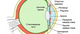
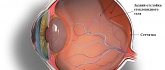
The orbit, in the cavity of which the eyeball is located, is lined with the periosteum of the orbit, which fuses in the area of the optic canal and the superior orbital fissure with the dura mater of the brain. The eyeball is surrounded by its membrane - the vagina, or Tenon's capsule, loosely connected to the sclera. The gap between the eyeball and its vagina is called the episcleral (Tenon's) space. On the posterior surface of the eyeball, the vagina is fused with the external sheath of the optic nerve; in front, the vagina approaches the fornix of the conjunctiva. The vagina of the eyeball is pierced by vessels and nerves, as well as tendons of the extraocular muscles, whose own fascia is fused with this vagina.
Between the vagina of the eyeball and the periosteum of the orbit, around the oculomotor muscles and the optic nerve, lies adipose tissue permeated with connective tissue bridges - the fatty body of the orbit, which acts as an elastic cushion for the eyeball. In front, the orbit with its contents is partially closed by the orbital septum, which originates from the periosteum of the upper and lower edges of the orbit and attaches to the cartilages of the upper and lower eyelids, and in the area of the inner corner of the eye it connects with the medial ligament of the eyelid. The orbital septum has openings for the passage of blood vessels and nerves through it.
Eyelids
The upper eyelid and lower eyelid are formations that lie in front of the eyeball and cover it from above and below, and when the eyelids close, completely covering it. At the level of the edge of the orbit, the skin of the eyelids passes into the skin of adjacent areas of the face. At the border of the upper eyelid and forehead, a transversely oriented skin ridge covered with hair protrudes - this is the eyebrow.
The anterior surface of the eyelid is convex, covered with thin skin with short vellus hair, sebaceous and sweat glands. The back surface of the eyelid is facing the eyeball, concave. This surface of the eyelid is covered by the conjunctiva.
In the thickness of the upper and lower eyelids there is a connective tissue plate, whose density resembles cartilage - this is the upper eyelid cartilage and the lower eyelid cartilage. The age-old part of the orbicularis oculi muscle is also located here. From the upper and lower cartilages of the eyelids, the medial ligament of the eyelid, common to these cartilages, goes to the anterior and posterior lacrimal ridges, covering the lacrimal sac in front and behind. To the lateral wall of the orbit from the cartilages follows the lateral ligament of the eyelid, which corresponds to the lateral suture.
A thin, wide tendon of the levator palpebrae superioris muscle is attached to the upper edge and anterior surface of the cartilage of the upper eyelid. The free edge of the eyelid, limited by its posterior and anterior surfaces, respectively forms the anterior and posterior edges of the eyelids and bears hairs located closer to the anterior edge in 2-3 rows - eyelashes. Closer to the posterior edge, the openings of the modified sebaceous (meibomian) glands of the cartilage of the eyelids open, the initial part of which is located inside the cartilaginous plate of the eyelid. There are more such glands in the thickness of the upper eyelid (30-40) than in the lower eyelid (20-30). The edges of the upper and lower eyelids limit the transverse palpebral fissure, which is closed on the medial and lateral sides by the fusions of the eyelids - the medial and lateral commissures of the eyelids.
The conjunctiva is a connective tissue membrane of a pale pink color. It consists of the conjunctiva of the eyelids, which covers the inside of the eyelids, and the conjunctiva of the eyeball, which is represented by a thin epithelial covering on the cornea. At the junction of the conjunctiva from the upper and lower eyelids to the eyeball, depressions are formed - the upper and lower fornix of the conjunctiva. The entire space lying in front of the eyeball, limited by the conjunctiva, is called the conjunctival sac, which closes when the eyelids close. The lateral angle of the eye is more acute. The medial corner of the eye is rounded and on the medial side it limits a depression - the lacrimal lake. Here, at the medial corner of the eye, there is a small elevation - the lacrimal caruncle, and lateral to it - the semilunar fold of the conjunctiva, the remnant of the nictitating (third) eyelid of lower vertebrates. On the free edge of the upper and lower eyelids, near the medial corner of the eye, outward from the lacrimal lake, an elevation is noticeable - the lacrimal papilla with an opening at the top - the lacrimal punctum, which is the beginning of the lacrimal canaliculus.
The lacrimal apparatus includes the lacrimal gland with its excretory canaliculi, opening into the conjunctival sac, and lacrimal ducts. The lacrimal gland is a complex alveolar-tubular gland with a lobular structure, lying in the fossa of the same name in the lateral corner, at the upper wall of the orbit. The levator palpebrae superioris tendon divides the gland into a large superior orbital portion and a smaller inferior orbital portion lying near the superior conjunctival fornix.
Under the fornix of the conjunctiva, small accessory lacrimal glands are sometimes found. The excretory canaliculi of the lacrimal gland, up to 15 in number, open into the conjunctival sac in the lateral part of the upper fornix of the conjunctiva. The tear (tear fluid) coming out of them washes the front part of the eyeball. Next, the tear fluid flows through the capillary gap near the edges of the eyelids along the lacrimal stream into the area of the medial corner of the eye, into the lacrimal lake. The short (about 1 cm) and narrow (0.5 mm) curved superior and inferior lacrimal canaliculi originate at this point. These canals open into the lacrimal sac separately or connected to each other. The lacrimal sac lies in the fossa of the same name in the inferomedial corner of the orbit. Downwards it passes into a rather wide (up to 4 mm) nasolacrimal duct, ending in the nasal cavity, in the anterior part of the lower nasal passage. The lacrimal part of the orbicularis oculi muscle is fused to the anterior wall of the lacrimal sac, which, when contracted, expands the lacrimal sac, which facilitates the absorption of tear fluid into it through the lacrimal canaliculi.
Development of the orbit and auxiliary apparatus of the eye
Description
Development of the orbit
. The walls of the orbit are part of the human facial skull. Like other structures of the facial skull, they are derived from neural crest cells (ectomesenchyme). As mentioned above, neural crest cells migrate in the early stages of intrauterine development from the area of the neural crests and accumulate under the ectoderm of the head end of the embryo in the form of strips of varying widths. In combination with the ectoderm, they form the following structures: the rudiment of the nose, the oral cavity, the lower and upper jaws, and the walls of the orbit.
Initially, it is necessary to characterize the process of facial formation
(Fig. 5.10.1, 5.10.2).
Rice. 5.10.1.
Formation of facial structures:
a - embryo 5-7 mm; b - embryo 6-7 mm; c — embryo 11.8 mm; d - embryo 14 mm (1 - olfactory fossa; 2 - maxillary process; 3 - mandibular process; 4 - second branchial arch; 5 - third branchial arch; 6 - medial nasal process; 7 - lateral nasal process; 8 - eye rudiment; 9 - left half of the frontonasal process; 10 - rudiment of the external auditory canal
Rice. 5.10.2.
Derivatives of embryonic facial processes (according to Sulic, 1982):
on the left - a scanogram of the head of an embryo at the 6th week of development (1 - eye primordium; 2 - process of His).
The diagonal lines in both figures indicate the derivatives of the maxillary process, and the vertical lines - the nasal process) It occurs as follows. At the beginning of the first month, a frontal process is distinguished at the cephalic end of the embryo, containing the expanded cephalic end of the neural tube (the primary anlage of the brain). In the area of the frontal process, on its lateral sides, the rudiments of the organ of vision are already visible. The first branchial arch at this stage of development is divided into two parts by a groove - the maxillary and mandibular arches. Thus, the entrance to the oral cavity looks like a gap limited by five processes. The upper edge of the oral fissure consists of an unpaired frontal process and, located on the sides of it, the maxillary processes. The lower edge of the primary oral fissure is formed by two mandibular processes.
Soon, depressions called olfactory fossae
. In this case, the frontal process is divided into several sections. The area located in the midline between the olfactory fossae retains the name of the frontal process, and the horseshoe-shaped elevations surrounding the olfactory fossae become the nasal processes - medial and lateral. The lateral nasal process is separated from the maxillary process by the nasolacrimal groove. It connects the orbital sockets with the olfactory fossae and subsequently closes to form the nasolacrimal canal.
At subsequent stages of intrauterine development, the processes come together and “fusion”
. At the same time, the formation of the external outlines of the embryo’s face begins. As a result of the fusion of the medially located nasal process with the maxillary process, the medial, lower and lateral orbital walls are formed. The roof of the orbit is formed by the capsule of the developing brain.
When describing the histogenesis of the bone walls of the orbit, it is necessary to mention the features of the development of the skull as a whole. These data should also help in understanding the development of orbital bones.
The bones of the skull are divided into three different groups based on their origin. The first group includes
bones developing from the primordial skull. They ossify on a cartilaginous basis (chondrogenic). These include the ethmoid bone, part of the main bone and part of the temporal bone.
The second group includes
bones that make up the skull and are phylogenetically younger. They develop from ectomesenchyme, and their ossification occurs on a connective tissue basis (desmogenic). These include part of the ethmoid bone, frontal, parietal, nasal, lacrimal, maxillary and zygomatic bones.
The third group includes
so-called visceral bones (arise from the cartilaginous anlage of the gill arches and form “visceral” bones). They do not take any part in the formation of the bone orbit.
Thus, it becomes clear why the lesser wing of the sphenoid bone, in contrast to the greater wing and other lamellar bones. in the early stages of embryonic development it consists of cartilage.
The process of bone formation itself comes down to the fact that the eye bud, gradually increasing in volume, gradually stretches the surrounding mesenchyme, leading to its compaction
. It is in this compacted mesenchyme that the first areas of ossification appear in the seventh week of embryonic development (Fig. 5.10.3).
Rice. 5.10.3.
Diagram illustrating the number and localization of ossification points of the bony walls of the orbit:
ossification centers appear between the 6th and 8th months of embryonic development.
Ossification gradually spreads to the periphery. The bones come closer together and sutures form between them. Ossification of the orbital walls is completed by the time of birth. The exception is the apex of the orbit. It is also necessary to dwell on the peculiarities of the origin of the soft tissues of the orbit. First of all, this concerns the vascular system. The endothelial lining of all blood vessels of the orbit arises from the mesoderm. Other components of the vascular wall, including smooth muscle, arise from neural crest cells (ectomesenchyme). The external (striated) muscles of the eye also originate from the mesoderm.
There are differences in the origin of other connective derivatives of the orbit. Thus, the structures located in the upper and outer parts of the orbit come from the mesoderm. At the same time, the connective tissue components located on the inner side come from the neural crest cells.
Development of the external eye muscles and soft tissues of the orbit
(Fig. 5.10.4—5.10.6).
Rice. 5.10.4.
Histological section through the eyeball and orbit of a four-month-old fetus:
the partially formed walls of the orbit are clearly visible. external muscles of the eye (1 - eyeball; 2 - eyelids; 3 - retina; 4 - external muscle of the eye: 5 - optic nerve; 6 - optic chiasm; 7 - bony wall of the orbit)
Rice. 5.10.5.
Formation of the external muscles of the eye: condensation of mesenchymal cells is noted. The cells elongate and myofibrils appear in their cytoplasm. Black arrows indicate the external muscles of the eye, and the light arrow indicates the nerve trunk (1 - sensory part of the retina; 2 - pigment epithelium; 3 - condensation of mesenchyme with the formation of the choroid; 4 - condensation of mesenchyme with the formation of the sclera)
Rice. 5.10.6.
Scheme of the structure of the orbit at the time of birth (sagittal section):
a - schematic image;
b - histological section (1 - frontal bone; 2 - levator of the upper eyelid; 3 - frontal nerve; 4 - superior rectus muscle; 5 - periosteum; 6 - Zinn's tendon; 7 - optic nerve; 8 - lower branch of the oculomotor nerve; 9 - inferior rectus muscle; 10—Müller muscle; 11—maxillary nerve; 12—maxillary bone; 13—inferior branch of the oculomotor nerve; 14—inferior oblique muscle; 15—lower eyelid; 16—upper eyelid. Asterisks indicate the distribution of fatty tissue of the orbit) External The muscles of the human eye develop similarly to the muscles of reptiles, birds and mammals. All vertebrates have six extrinsic eye muscles, and some of them have an additional muscle, the ocular retractor, which pulls the eye back for a protective purpose.
Extrinsic muscles of the eye
are an integral part of the connective tissue system of the orbit and have a single histogenesis, namely: they originate from neural crest cells. Neural crest cells migrate towards the paraxial mesoderm. It is here that they are the source of the development of mesoderm, from which the external muscles of the eye are formed.
In mammals, the paraxial mesoderm of the trunk consists of seven tubercles called somatomeres, delimited from each other on the surface by shallow grooves. The eighth somatomer is also the first somite. Both the muscles and the connective tissue of the orbit develop from somatomeres.
How do the external eye muscles develop?
? In the human embryo at the developmental stage of 14 somites (day 25), condensation of the premandibular mesoderm is observed. These are areas of the future external eye muscles innervated by the oculomotor nerve. These include the superior, internal and inferior rectus muscles, as well as the inferior oblique muscle. Two separate clusters of cells in the region of the maxilomandibular mesoderm are the source of the development of the external rectus and superior oblique muscles.
Connective tissue, and not myogenic rudiments, determines the exact location of future muscles. At the same time, the mesoderm from which the muscles emanate is distinguished by strict spatial localization, which is absolutely necessary for the growth of nerve trunks in their direction and clearly localized attachment to the sclera of the developing eye. It is assumed that neural crest cells receive the necessary information for differentiation into muscle tissue from the optic vesicle. From the accumulation of mesenchymal cells of the upper part of the orbit, the superior rectus and superior oblique muscles, the levator of the upper eyelid, as well as the upper half of the internal rectus and external rectus muscles develop.
From a collection of mesenchymal cells located in the lower part of the orbit, arise the inferior rectus and inferior oblique muscles, as well as the lower half of the internal rectus and external rectus muscles.
Primary myofilaments, already containing various types of myosin heavy chain, are formed even before the innervation of future muscles begins. Primary fibers are replaced by secondary myoblasts, which already contain myosin, characteristic of differentiated muscle. Thus, there are two stages of myogenesis
. In the first stage, primary myoblasts containing various types of myosin heavy chain arise from neural crest cells, and their differentiation is determined by interaction with connective tissue. At the second stage, secondary muscle fibers are formed, but only during the interaction of the muscle rudiment with the nervous system.
The developing motor plate of the nerve stimulates the formation of the postjunctional membrane of the motor plate in the muscle with the formation of acetylcholine receptors and the synthesis of acetylcholinesterase in it. The protein agrin is synthesized in the body of motor neurons.
, which is then transported to the nerve endings. In nerve endings, this protein is found both in the active form and in a state associated with the basement membrane of the nerve ending. It is believed that agrin promotes motor neuron-induced synthesis and accumulation of synaptic proteins in the neuromuscular plate.
Differentiation of the extrinsic muscles of the eye occurs in the direction from the apex of the orbit forward, while the sclera differentiates in the opposite direction. It has now been shown that muscles develop simultaneously along their entire length.
Recently, it has been established that the final differentiation of myoblasts depends on the degree of maturity of their motor innervation
. In this case, differentiation of motor neurons of the external eye muscles located in the brain stem (nuclei of the oculomotor nerves) occurs regardless of the degree of muscle differentiation. At the end of the first month of embryonic development, the nerve trunks reach the external muscles of the eye. This happens in a certain sequence. The muscles reach the branches of the oculomotor nerve first, and only then the abducens and trochlear nerves.
It was noted that the initial number of axons of the oculomotor nerve is significantly greater than in the postnatal period. A decrease in the number of axons is associated with the death of some motor neurons during embryonic development. A similar pattern extends to other nerves, in particular to the axons of the retinal ganglion cells that form the optic nerve.
Transverse striation of muscle cells
detected quite early. It appears in the second month, and by the end of the second month, strands of long striated muscle cells have already been formed. Around the 3rd month, muscle cells are surrounded by collagen fibers that form fascia. In parallel, the vasculature and perineural adventitia develop. Connective tissue septa appear somewhat later (by the end of the fourth month).
Mesenchymal derivatives of the orbit
are differentiated last. Beds of capillary vessels and areas of fatty tissue appear between the connective tissue septa (4th month). By the 6th month, the muscles occupy their usual position among the structures of the orbit. Subsequently, there is a predominant increase in muscle mass without the appearance of any new qualitative signs.
As stated above, differentiation of the connective tissue of the orbit begins later than the differentiation of the muscles and walls of the orbit. This is manifested in the fact that condensation of mesenchyme occurs approximately at the 3rd month of development, and the formation of capillaries and fatty tissue - at the 4th. Only by 6 months does the connective tissue of the orbit reach the differentiation characteristic of an adult.
Growth of tendons of the extrinsic eye muscles
occurs parallel to fetal development. Initially, the tendons are attached to the eyeball over a large area (from the edge to the equator). After some time, part of the tendon tissue undergoes reverse development, thereby freeing the surface of the sclera. At the same time, the places of tendon attachment become increasingly clear. The junction of the tendons with the sclera gradually moves back. This process of differentiation of the sites of tendon attachment to the sclera continues for two years after birth. In children with esotropia, there are no abnormalities of the tendons of the internal and external rectus muscles, and there is no correlation between the angle of strabismus and the site of muscle insertion.
Throughout the year after birth, the structure of the external muscles of the eye continues to improve - the ratio of fibers of different diameters changes
, the number of mitochondria increases, differentiation of blood vessels occurs, the ratio of myosin subtypes changes, etc. In parallel, myelination of motor nerves occurs, and nerve endings mature.
Further improvement of the muscular system of the eye occurs throughout the development of human visual functions.
It should be noted that postnatal muscle development may be impaired by inadequate development of visual functions
. So. suturing a cat's eyelids results in significant thinning of the extrinsic eye muscles, decreased activity of redox enzymes, and decreased capillary density in the muscle. Functional changes are also revealed, namely a decrease in the speed of muscle contraction and its endurance. The number of motor neurons in the nuclei of the oculomotor nerve also decreases.
In conclusion, it should be noted that the levator of the upper eyelid is formed from the dorsolateral part of the superior rectus muscle with an embryo size of 22-30 mm and grows in the direction of the upper eyelid. This growth is completed by the fourth month.
Eyelids and conjunctiva
(Fig. 5.10.7—5.10.9).
Rice. 5.10.7.
Development of the eyelids:
a - convergence of the folds of the ectoderm above the cornea; b—fusion of the edges of the eyelids; c—separation of eyelids
Rice. 5.10.8.
Dynamics of eyelid formation (scanning electron microscopy):
a - ventro-lateral surface of the head of a human embryo (8 weeks); b—eyelids of the embryo after fusion; c, d — process of eyelid fusion (higher magnification)
Rice. 5.10.9.
Development of appendages (according to I. Mann, 1966):
a - embryo size 37 mm;
b — embryo size 50 mm; c — embryo size 73 mm; d - embryo size 160 mm (1 - hair follicles; 2 - Moll glands; 3 - tarsal (Meibomian) glands; 4 - Zeiss glands) The soft tissues of the face, including, naturally, the eyelids, develop from ectomesenchyme. Eyelid formation begins at 4-5 weeks (embryo 8-12 mm).
In the morphogenetic sense, the process boils down to the formation of two horizontal folds
, consisting of mesenchyme of neural origin (formed from the secondary arch; Gasser), covered with outer ectoderm. The lower eyelid is formed from the maxillary process, while the upper eyelid is formed from the medial and lateral portions of the frontonasal process. The folds gradually approach each other, covering the eyeball.
At the stage of embryonic development corresponding to an embryonic length of 35-40 mm (ninth week), the upper eyelid and lower eyelid are fused by an epithelial suture above the cornea
. The space that appears behind the fused eyelids is lined with stratified prismatic epithelium. This space is designated by the conjunctival sac. Goblet cells develop, in the cytoplasm of which sialomucin is found. Initially, they appear in the fornix, and then in the conjunctiva of the eyelid and eye. Quite early, muscarinergic and adrenergic receptors of goblet cells appear.
Subsequently, the ectoderm covering the anterior surface of the eyelids turns into skin. Immediately after the eyelids fuse, eyelashes begin to develop
, sebaceous (Meibomian) glands, Zeiss glands (4th month) and modified Moll sweat glands. The connective tissue structures of the eyelid and the striated muscle are formed from the mesenchyme. It is important to note that in the presence of eyelid coloboma, which develops as a result of the fact that the eyeball in the embryonic period is not completely covered with fused eyelids, dermoid transformation of the tissues of this area occurs.
By the fifth month, the eyelids separate
. Apparently, the separation of the eyelids occurs as a result of the onset of secretion of glands that secrete secretions into the area of “sticking together” of the epithelial derivatives of the eyelid.
In the most medial part of the lower eyelid (embryo size 58 mm), between the medial ligament and the lacrimal papilla (papillary projection), a section is separated from the eyelid tissue, which subsequently turns into the lacrimal caruncle
. Histological examination of the lacrimal caruncle can detect almost all the structural elements of the eyelid margin (skin appendages).
One of the main reasons for the separation of the lacrimal caruncle from the lower eyelid is the development of the lower canaliculi. This is confirmed by the discovery of patients in whom the lacrimal caruncle was absent due to underdevelopment of the lacrimal canaliculi.
During the development of the lacrimal caruncle, a semilunar fold
. It protrudes medially in the shape of a crescent and extends towards the conjunctival fornix, attaching to the medial straight ligament and the lacrimal caruncle. The semilunar fold slightly separates the medial part of the eyelid from the eyeball, which creates the condition for the collection of tears in the lacrimal lake.
Disruption of the processes of embryonic development of soft tissues of the face leads to the formation of various anomalies. The main causes of abnormalities are insufficient migration of neural crest cells
or disruption of the processes of fusion of processes both on the lateral side and along the midline. Experimental reproduction of clefting of the hard palate is possible by removing a portion of the neural crest before cell migration begins. Midline facial abnormalities (hypertelorism) result from insufficient fusion of the frontonasal process.
Lacrimal gland
. The lacrimal gland is formed at the end of the second month of embryonic development (embryo size is 25 mm) in the form of outgrowths of the basal cells of the conjunctival epithelium in the upper temporal vault. Neural crest cells accumulate around these cords. Subsequently, these cells form the acini of the gland.
At approximately the 3rd month (embryo size is 60-65 mm), ducts appear. This process is associated with vacuolization of epithelial cells located in the center of the cords.
At the end of the embryonic period, branches of the alveolar-tubular gland are formed from the epithelial buds. Their terminal sections are lined with prismatic epithelium, and the ducts open into the conjunctival sac. Epidermal growth factor stimulates tear secretion, activating the synthesis of prostaglandins, which influence the movement of fluid from the intercellular space into the conjunctival sac.
Tear drainage system
(Fig. 5.10.10—5.10.12).
Rice. 5.10.10.
Scheme of development of the lacrimal apparatus:
1 - lacrimal gland; 2—tarsal (Meibomian) glands; 3 - nasolacrimal duct; 4 - upper eyelid; 5 - lower eyelid; 6 - lacrimal duct; 7 - lacrimal sac; 8 - lacrimal caruncle
Rice. 5.10.11.
Scheme of development of the lacrimal apparatus (according to Duke-Elder, 1963):
a—6th week of development: b—12th week of development: c—3.5 months of embryonic development (1—conjunctival sac; 2—nasal cavity)
Rice. 5.10.12.
Development of the lacrimal system: embryo at the end of the fourth month of development;
a — frontal section of the embryo’s head at the level of the lacrimal drainage system;
b - similar section at higher magnification (1 - lacrimal sac; 2 - lacrimal canal; 3 - lacrimal canaliculi; 4 - conjunctival cavity) Features of the development of the nasolacrimal canal have always been the object of close attention of researchers, since there are still attempts to clarify the main patterns of the occurrence of its congenital obstruction.
As stated above, at the 7 mm stage of embryonic development, a depression appears between the lateral nasal and maxillary processes, directed towards the eye rudiment. It's called the nasolacrimal duct
(naso-orbital groove). The ectoderm in this area thickens and is covered with mesoderm on top. These centrally located ectodermal masses gradually move in two directions - towards the eyeball bud and towards the nose. The edges of the groove soon close, but the lumen has not yet formed, but is filled with epithelial cells. From the proximal end of the formed tube, two arms grow, the future tear ducts. They connect to the edges of the upper and lower eyelids and then open into the enlarged portion of the nasolacrimal duct (the lacrimal sac).
Bone tissue begins to form in the surrounding mesoderm
(maxillary and lacrimal bones), which subsequently forms the bone walls of the nasolacrimal canal.
Canalization of the ectodermal cord
, surrounded by mesoderm, begins when the embryo is 32-36 mm long. The sewerage process occurs in segments. Initially, epithelial cells located in the center of the cord undergo degeneration. As a result, the proximal and distal ends of the resulting tube, fused with the conjunctival and canalicular epithelium, remain closed for quite a long time with a thin membrane. The degeneratively altered epithelium gradually sloughs off, and the resulting detritus accumulates at the lower end of the resulting tube.
Located superiorly, i.e., facing the conjunctival cavity, the membrane usually opens at birth. The lower membrane (Hanser valve) in 35-73% of cases is preserved at the time of birth. The presence of high hydrostatic pressure in the lacrimal sac contributes to the rupture of the lower membrane.
Disruption of the development of the nasolacrimal canal
and the opening of its membranes often leads to the development of anomalies. The most common anomalies in this area are the following: congenital absence of part of the nasolacrimal canal, an excessive number of lacrimal openings, and a fistula.
Knowledge of the characteristics of the origin and development of the bone walls and soft tissues of the orbit is of great practical importance. It is known that the clinical and biological features of pathological processes, primarily tumors, are associated with their histogenesis. Knowing the histogenesis and topography of various formations of the orbit, one can quite clearly predict the likelihood of developing a particular type of tumor.
Violations of the above complex morphogenetic processes that occur during the formation of the orbit lead to the development of quite serious anomalies. Thus, with a defect in the upper bone wall, intense pulsating exophthalmos
. Hypoplasia of the orbital margin has been described, characterized by severe agenesis of the orbital margin, hypoplasia of the eyelid skin and cartilaginous plate of the eyelid, various anomalies of the lacrimal drainage system (ectopia of the superior lacrimal punctum, shortening or absence of the inferior canaliculus, atresia of the nasolacrimal canal). These changes are combined with coloboma of the inner part of the lower eyelid and congenital anomalies of the external muscles of the eye.
The process of proper formation of the orbit is also influenced by the formation of the eyeball. , microphthalmos with a cyst, cephalocellus may develop
(presence of brain tissue and/or meningeal membranes in the orbit).
It should also be noted that enucleation of the eye
in both children and adults it leads to a decrease in the volume of the orbit and improper development of bone and soft tissue formations. This is accompanied by a decrease in the volume of the orbit. Previously, such changes occurred quite often and reached 50% of observed patients. The degree of underdevelopment of the orbit depends on the age of the patient at which enucleation was performed, as well as the length of time that has passed since the operation.
—-
Article from the book: Structure of the human visual system | Vit V.V.
Vessels and nerves of the organ of vision
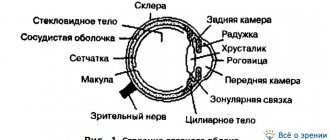
The eyeball and its auxiliary organs receive blood from the branches of the ophthalmic artery, which in turn is a branch of the internal carotid artery. Venous blood from the organ of vision flows through the ophthalmic veins into the cavernous sinus. The retina is supplied with blood by the central retinal artery, which penetrates into the eyeball in the thickness of the optic nerve and gives off superior and inferior branches in the area of the disc. The central retinal vein and its tributaries are adjacent to the arteries of the same name. The short and long posterior and anterior ciliary arteries branch in the choroid. The branches of these arteries in the thickness of the iris anastomose with each other and form two arterial circles: a large one at the ciliary edge of the iris and a small one at the pupillary edge. The sclera is supplied with blood by the posterior short ciliary arteries. From the dense venous network of the choroid proper, 4-6 vorticose veins are formed, which pierce the sclera and flow into the ophthalmic veins. The anterior ciliary veins collect blood from the ciliary body, iris and sclera.
The eyelids and conjunctiva receive blood from the medial and lateral arteries of the eyelids, anastomoses between which form the arch of the upper eyelid and the arch of the lower eyelid, and the anterior conjunctival arteries in the thickness of the eyelids. The veins of the same name drain into the ophthalmic and facial veins. The lacrimal artery goes to the lacrimal gland.
The muscles, fascia, and fatty body of the orbit are also supplied with blood by branches of the ophthalmic artery. Lymphatic vessels from the eyelids and conjunctiva are directed to the mandibular, as well as to the superficial and deep parotid (preauricular) lymph nodes.
The contents of the orbit receive sensory innervation from the first branch of the trigeminal nerve, the ophthalmic nerve. From its branch, the nasociliary nerve, long ciliary nerves extend to the eyeball. The lower eyelid is innervated by the infraorbital nerve, which is a branch of the second branch of the trigeminal nerve. The constrictor pupillary muscle and the ciliary muscle receive parasympathetic fibers of the oculomotor nerve (from the ciliary ganglion as part of the short ciliary nerves), and the pupillary dilator muscle is innervated by the sympathetic fibers of the internal carotid plexus, which reach the eyeball along with blood vessels. The superior, inferior, medial rectus, inferior oblique and levator palpebrae superioris muscles receive motor innervation from the oculomotor nerve, the lateral rectus from the abducens nerve, and the superior oblique from the trochlear nerve.
AUXILIARY ORGANS OF THE EYE AUXILIARY ORGANS OF THE EYE
The auxiliary organs of the eye are the muscles of the eyeball, the lacrimal apparatus, the conjunctiva, and the eyelids.
Orbital cavity,
in which the eyeball and its auxiliary organs are located, it is lined with the periosteum of the orbit, which in the area of the optic canal and the superior orbital fissure fuses with the dura mater of the brain.
The eyeball is enveloped by its connective tissue vagina (vagina bulbi
-
Tenon's capsule),
which is connected to the sclera by loose connective tissue.
On the posterior surface of the eyeball, the vagina is fused with the external sheath of the optic nerve; in front it approaches the fornix of the conjunctiva. The vessels, nerves and tendons of the extraocular muscles pierce the vagina of the eyeball. Between the eyeball and its vagina there is a narrow episcleral (Tenon's) space (spatium episclerale).
Between the periosteum of the orbit and the vagina of the eyeball lies the fatty body of the orbit (corpus adiposum orbitae).
In front, the orbit (and its contents) is partially closed
by the orbital septum (septum orbitale),
starting from the periosteum of the upper and lower edges of the orbit and attached to the cartilages of the upper and lower eyelids. In the area of the inner corner of the eye, the ocular septum connects to the medial ligament of the eyelid.
The eyelids
(palpebrae)
protect the eyeball from the front.
They are folds of skin that limit the palpebral fissure and close it when the eyelids close (Fig. 125). On the sides, the eyelids are connected by lateral and medial commissures, closing the corresponding corners of the eye. The lateral angle of the eye (angulus oculi lateralis)
is sharp, and
the medial angle (angulus oculi medialis)
is rounded.
Due to this, in the area of the medial angle there is a recess - the lacrimal lake (lacus lacrimalis).
From above, the upper eyelid is limited
by the eyebrow (supercilium)
with short, coarse hair.
The lower eyelid drops slightly when the eyes are opened under the influence of gravity. The muscle that lifts the upper eyelid
(m.
levator palpebrae)
approaches which begins together with the rectus muscles from the common tendon ring.
The muscle runs in the upper part of the orbit and is attached to the upper eyelid cartilage (tarsus superior)
- a plate of dense fibrous connective tissue that performs a supporting function. In the thickness of the lower eyelid there is
Rice. 125.
Upper and lower eyelids of the right eye, front view: 1 – eyebrow; 2 – upper eyelid; 3 – iris; 4 – fibrous membrane of the eyeball; 5 – lacrimal caruncle; 6 – medial commissure of the eyelids; 7 – lacrimal punctum; 8 – bottom
eyelid; 9 – eyelashes; 10 – pupil; 11 – lateral commissure of the eyelids
similar cartilage of the lower eyelid (tdrsus infdrior).
cartilaginous glands (gldndulae tarsdles)
that open at the edges of the eyelids .
Closer to the anterior surface, in the thickness of the eyelids, lies the age-old part of the orbicularis oculi muscle. Along the edges of the eyelids, eyelashes (cdilia) are located in 2-3 rows .
of the sebaceous glands (gldandulae sebacdeae)
open into their hair bags The convex anterior surface of the eyelids is covered with thin skin with short vellus hairs. The concave posterior surface of the eyelids is covered with conjunctiva.
Conjunctiva
(tdnica conjunctiva)
is a thin connective tissue pale pink membrane in which the conjunctiva of the eyelids, covering the inside of the eyelids, and the conjunctiva of the eyeball are distinguished (Fig. 126).
At the point of transition of one part of the conjunctiva to another, the upper
and
Rice. 126.
The structure of the century. Frontal section: 1 – conjunctiva; 2 – cartilage of the eyelid; 3 – century-old part of the orbicularis oculi muscle; 4 – ciliary gland; 5 – edge of the eyelid; 6 – eyelash; 7 – leather
lower fornix of the conjunctiva (fornix conjunctivae superior
et
fornix conjunctivae inferior).
The space located in front of the eyeball and limited by the conjunctiva forms
the conjunctival sac (sdccus conjunctivdlis),
which closes when the eyelids close.
The rounded medial corner of the eye on the medial side limits the lacrimal lake. At the medial corner of the eye there is a small elevation - the lacrimal caruncle (cardncula lacrimalis).
Lateral to the lacrimal caruncle is
the semilunar fold of the conjunctiva (plica semilunaris conjunctivae)
- a rudiment of the blinking (third) eyelid found in vertebrates.
The conjunctiva is lined with three-layer non-keratinizing epithelium lying on the basement membrane. Towards the edge of the eyelid, the epithelium becomes multilayered squamous. The epithelium of the conjunctiva contains goblet glandulocytes. The lamina propria of the conjunctiva is formed by loose connective tissue, which contains fibroblasts, macrophages, mast cells, plasma cells, single melanocytes and lymphocytes. The conjunctival sac is moistened by tear fluid secreted by the lacrimal gland.
The lacrimal apparatus
(apparatus lacrimalis)
includes the lacrimal gland and the lacrimal duct system (Fig. 127).
Lacrimal gland (glandula lacrimalis),
consisting of several alveolar-tubular serous glands, located in the fossa of the lacrimal gland of the frontal bone in the superolateral part of the orbit. The levator palpebrae superioris tendon divides the gland into two parts: the larger superior orbital portion and the smaller inferior palpebral portion, which lies near the superior conjunctival fornix. In the fornix of the conjunctiva there are sometimes
Rice. 127.
Lacrimal apparatus of the right eye, front view: 1 – lacrimal gland; 2 – upper eyelid; 3 – lacrimal canaliculus; 4 – tear lake; 5 – lacrimal sac; 6 – nasolacrimal duct
small size accessory lacrimal glands. From 5 to 12 excretory canaliculi of the lacrimal gland open into the superior fornix of the conjunctiva. The tear washes the front of the eyeball and along the lacrimal stream (rivus lacrimalis)
- the capillary fissure, located near the edges of the eyelids, flows into
the lacrimal lake (lacus lacrimalis),
located in the medial corner of the eye.
At the medial corner of the eye, at the edges of the eyelids, where they converge, surrounding the lacrimal lake, the superior and inferior lacrimal papillae (papillae lacrimales) are located.
At the top of these papillae there is a narrow opening -
the lacrimal punctum (punctum lacrimalis).
lacrimal canaliculus (canaliculus lacrimalis),
about 1 cm long and about 0.5 mm in diameter,
originates from the lacrimal punctum The upper and lower tubules flow into the lacrimal sac (saccus lacrimalis),
which faces upward with its blind end.
The lower end of the sac passes into the nasolacrimal duct (ductus nasolacrimalis),
which opens into the lower nasal passage. The lacrimal part of the orbicularis oculi muscle, fused with the wall of the lacrimal sac, contracts and expands it. Thanks to this, tears are absorbed into the lacrimal sac through the lacrimal canaliculi.
Muscles of the eyeball.
The human eyeball can rotate so that the visual axes of both eyeballs converge on the object in question. Movements of the eyeballs are carried out
Rice. 128.
Muscles of the eyeball (oculomotor muscles), front view (A) and top view (B): 1 – superior rectus muscle; 2 – block; 3 – superior oblique muscle; 4 – medial rectus muscle; 5 – inferior oblique muscle; 6 – inferior rectus muscle; 7 – lateral rectus muscle; 8 – optic nerve; 9 – optic chiasm
six striated extraocular muscles: four rectus muscles (superior, inferior, medial, lateral
–
musculi recti superior, inferior, medialis, lateralis)
and two obliques
(upper
and
lower
–
musculi obliqui superior
et
inferior)
(Fig. 128, Fig. 129).
The inferior oblique muscle of the eye begins on the lower wall of the orbit near the opening of the nasolacrimal duct. The rest begin in the depths of the orbit in the circumference of the optic canal and the adjacent part of the superior orbital fissure from the common tendon ring (annulus tendineus communis),
surrounding the optic nerve and ophthalmic artery. The ring is fixed to the sphenoid bone, the periosteum around the optic canal and partially to the edges of the superior orbital fissure. All rectus muscles are directed along the corresponding walls of the orbits, on the sides of the optic nerve, pierce the vagina of the eyeball and are attached to the sclera in front of the equator in various areas according to their names.
Superior oblique muscle of the eye
lies in the superomedial part of the orbit between the superior and medial rectus muscles.
Near the trochlear fossa of the orbit, it passes into a thin round tendon wrapped in a synovial sheath. This tendon passes over the trochlea (trochlea)
in the superomedial corner of the orbit, turns posteriorly and laterally
Rice. 129.
Superior oblique and other muscles of the eyeball, top view. On the right side of the picture, the levator palpebrae superioris muscle has been cut and partially removed. The upper wall of the right and left orbits was removed: 1 – medial rectus muscle; 2 – block (superior oblique muscle); 3 – eyeball; 4 – muscle that lifts the upper eyelid; 5 – lateral rectus muscle; 6 – common tendon ring; 7 – visual chiasm; 8 – lateral rectus muscle; 9 – lacrimal gland
and is attached to the sclera behind the equator of the eye on the superolateral surface of the eyeball. Inferior oblique muscle
attaches to the eyeball on the side also behind the equator.
Rectus muscles
rotate the eyeball in the appropriate direction around two mutually intersecting axes: vertical and horizontal (transverse). The lateral and medial rectus muscles rotate the eyeball outward or inward around the vertical axis, each in its own direction. The pupil rotates accordingly. The superior and inferior rectus muscles rotate the eyeball around the transverse axis up or down. The oblique muscles rotate the eyeball around the sagittal axis: the upper – down and outwards, the lower – up and outwards. Thanks to the friendly action of these muscles, the movements of both eyeballs are coordinated.
Blood supply and innervation of the organ of vision.
The eyeball and its auxiliary organs receive blood from branches of
the ophthalmic artery
(a branch
of the internal carotid artery).
One of the branches of the ophthalmic artery (central retinal artery) supplies blood to the retina and part of the optic nerve, the other - to the sclera and choroid (Fig. 130).
The central retinal artery
penetrates the thickness of the optic nerve into the eyeball, in the area of the disc it gives off upper and lower branches, which spread within the inner granular and ganglion layers of the retina. The capillaries, surrounded by processes of glial and Müller cells, are formed by fenestrated endothelium.
In the choroid of the eyeball, short and long posterior
and
anterior ciliary arteries.
The anterior ciliary arteries in the thickness of the iris anastomose with each other and form two arterial circles: a large one, located at the ciliary edge of the iris, and a small one, adjacent to its pupillary edge.
The sclera is supplied with blood by the posterior short ciliary arteries.
From the dense venous network of the choroid proper, 4-6
vorticose veins are formed,
which pierce the sclera and flow into the ophthalmic veins.
Blood flows from the ciliary body, iris and sclera
into the anterior ciliary veins The eyelids and conjunctiva are supplied with blood from the medial
and
lateral arteries,
which, by anastomosing, form the arches of the anterior and posterior conjunctive arteries in the thickness of the eyelids. The veins of the same name drain into the ophthalmic and facial veins. The lacrimal gland receives blood from the artery of the same name.
The muscles, fascia and fatty body of the orbit are also supplied by branches of the ophthalmic artery.
Lymphatic vessels
from the eyelids and conjunctiva they go to
the submandibular,
as well as to
the superficial
and
deep parotid (preauricular) lymph nodes.
Innervation
The sensitive contents of the orbit are received from
the optic nerve
(the first branch of the trigeminal nerve).
Long ciliary nerves
from its branch, the nasociliary nerve,
to the eyeball The lower eyelid is innervated
by the infraorbital nerve
(a branch of the maxillary nerve).
The sphincter of the pupil and the ciliary muscle are innervated by parasympathetic fibers of the oculomotor nerve
(from the ciliary ganglion as part of the short ciliary nerves). The pupillary dilator receives innervation through the sympathetic fibers of the internal carotid plexus, which approach the eyeball along with blood vessels. Top, bottom and
Rice. 130.
Blood vessels and nerves of the eyeball:
1 – venous sinus of the sclera; 2 – circular groove of the sclera; 3 – anterior ciliary artery; 4 – conjunctival vessels; 5 – whirlpool vein (vein of the eye’s own choroid); 6 – sclera (tunica albuginea); 7 – short posterior ciliary arteries; 8 – central artery and veins of the retina; 9 – arterial circle of the optic nerve; 10 – long ciliary nerves;
11 – long posterior ciliary artery; 12 – large arterial circle of the iris;
13 – small arterial circle of the iris; 14 – cornea
the medial rectus, inferior oblique muscles of the eye and the muscle that lifts the upper eyelid are innervated by branches of the oculomotor nerve,
the lateral rectus is
the abducens nerve,
the superior oblique is
the trochlear nerve.
Conducting path of the visual analyzer.
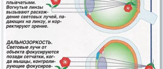
The light beam passes through the cornea, the aqueous humor of the anterior chamber, the pupil, which, depending on the intensity of the light, either expands or contracts, the aqueous humor of the posterior chamber, the lens, the vitreous body and enters the retina. Thanks to light refractive media, a beam of light is directed to the macula of the retina - the zone of best vision. An important role in this belongs to the lens, which, with the help of the ciliary muscle, can increase or decrease the curvature during accommodation. The oculomotor muscles direct the eyeballs towards the object in question, set the axes of both eyes parallel when looking into the distance, or bring them closer when viewing an object at close range.
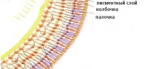
When light hits rods and cones - the processes of the first neurons of the visual pathway - a nerve impulse is generated in them, which is transmitted to bipolar neurocytes, and from them to ganglion neurocytes (Fig. 131). Axons of ganglion cells form the visual
Rice. 131.
Location of neurons in the retina
(diagram): 1 – cones; 2 – sticks;
3 – pigment cells;
4 – bipolar cells;
5 – ganglion cells;
6 – nerve fibers. The arrow shows the direction of the light beam
the nerve that exits the eye socket through the optic nerve canal. On the lower surface of the brain, the optic nerves form a chiasm, but only the fibers coming from the medial part of the retina of each eye cross. Each optic tract contains fibers carrying nerve impulses from the cells of the medial half of the retina of the opposite eye and the lateral half of the eye on its side. Some of the fibers of the optic tract are sent to the lateral geniculate body, where they end in synapses on the neurons located here. Another part of the axons of ganglion neurons, not reaching the lateral geniculate body, is directed through the handles of the superior colliculi to their nuclei. From the superior colliculi, nerve impulses follow to the nuclei of the oculomotor nerve (motor and accessory autonomic), innervating the muscles of the eye, the muscle that constricts the pupil, and the ciliary muscle. Thus, in response to light waves entering the eye, the pupil contracts and the eyeballs rotate in the direction of the light beam.
The axons of the neurons of the lateral geniculate body are directed to the cells of the visual cortex, located in the occipital lobe of the cerebral hemisphere, near the calcarine sulcus (field 17) (Fig. 132).
Conducting path of the visual analyzer
Light entering the retina first passes through the transparent light-refracting media of the eyeball: the cornea, the aqueous humor of the anterior and posterior chambers, the lens, and the vitreous body. The pupil is in the path of the light beam. Under the influence of the muscles of the iris, the pupil either narrows or dilates. Light-refracting media direct a beam of light to a more sensitive place of the retina, the place of best vision - the spot with its central fovea. An important role in this belongs to the lens, which, with the help of the ciliary muscle, can increase or decrease its curvature when seeing at close or far distances. This ability of the lens to change its curvature (accommodation) ensures that the light beam is always directed to the central fovea of the retina, which is in line with the observed object. The direction of the eyeballs towards the object in question is ensured by the extraocular muscles, which set the visual axes of the right and left eyes parallel when seeing at a distance or bring them closer (convergence) when viewing an object at close range.
Light entering the retina penetrates into its deep layers and causes complex photochemical transformations of visual pigments there. As a result, a nerve impulse occurs in light-sensitive cells (rods and cones). Then the nerve impulse is transmitted to the next neurons of the retina - bipolar cells (neurocytes), and from them - to the neurocytes of the ganglion layer, ganglion neurocytes. The processes of ganglion neurocytes are directed towards the disc and form the optic nerve. Enveloped in its own sheath, the optic nerve leaves the orbital cavity through the optic nerve canal into the cranial cavity and forms the optic chiasm on the lower surface of the brain. Not all fibers of the optic nerve are crossed, but only those that follow from the medial part of the retina facing the nose. Thus, the optic tract following the chiasma consists of nerve fibers of ganglion cells of the lateral (temporal) part of the retina of the eyeball on its side and the medial (nasal) part of the retina of the eyeball of the other side. That is why, when the chiasm is damaged, the function of conducting impulses from the medial parts of the retina of both eyes is lost, and when the optic tract is damaged, it occurs in the lateral part of the retina of the same side and the medial part of the other.
Nerve fibers in the optic tract follow to the subcortical visual centers: the lateral geniculate body and the superior colliculus of the midbrain roof. In the lateral geniculate body, the fibers of the third neuron (ganglionic neurocytes) of the optic pathway end and come into contact with the cells of the next neuron. The axons of these neurocytes pass through the sublenticular part of the internal capsule, form the optic radiation and reach the area of the occipital lobe of the cortex near the calcarine sulcus, where the highest analysis of visual perceptions is carried out. Some of the axons of ganglion cells do not end in the lateral geniculate body, but pass through it in transit and, as part of the handle, reach the superior colliculus. From the gray layer of the superior colliculus, impulses enter the nucleus of the oculomotor nerve and the accessory nucleus (Yakubovich's nucleus), from where the oculomotor muscles, as well as the constrictor pupillary muscle and the ciliary muscle are innervated. Along these fibers, in response to light stimulation, the pupil narrows (pupillary, pupillary, reflex) and the eyeballs turn in the desired direction.