Every organ in our body is very important. Without arms, without legs it is very difficult to live, without a heart it is generally impossible. Who is in charge of our entire body? Of course, the head. Do you know the saying: “Bread is the head of everything”? From this saying it is clear that the head is most important.
The brain is located in the head, which is responsible for the functioning of our entire body. If any changes appear in the brain, this immediately affects the activity of the whole organism. Regulation of the body occurs with the help of nerve impulses (nerve endings of the brain) and with the help of special chemicals (pituitary gland) - humoral regulation.
The baby's head consists of two parts: the facial part and the brain part. At an older age, when the head stops growing, the bones of the skull are tightly fused to each other and motionless. In contrast, the bones of a newborn baby’s skull are very mobile and can move not only relative to each other, but also overlap the edge of one bone with another. The place where two bones join is called a suture, the space between the three bones is called the fontanel. In newborn children, 6 fontanelles can be identified. Gradually, throughout life, these fontanelles close. The closure of fontanelles has its own timing, each of them must be closed until a certain point in life.
Many animals are able to move independently and even look for food within a few hours after birth. Our children remain completely helpless for many months. Why is this happening? Everything is very simple: man is a highly organized, social animal. This means that in the course of life the human body learns to perform a large volume of different types of activities: talking, getting food, walking and much more. It is impossible to obtain all this knowledge at once, so a person learns from the example of other people how to survive in this world (social factor). In addition to the social skills that a child acquires during life, there is also innate memory, the experience of previous generations. Such memory protects us from death (instinct of self-preservation). We are instinctively afraid of fire, snakes and bright red insects, although no one has ever bitten us before. In addition to the instinct of self-preservation, the child inherits other reflexes and instincts from his ancestors. Thus, newborn children have a search reflex; they instinctively look for food. If a newborn baby is placed on his mother's stomach, he will crawl to the breast on his own and begin to suck milk.
Why aren't we born already able to walk and talk? It's simple: for this, the child must be in the womb for too long. If a baby develops too long in its mother's womb, its bones will harden and the skull bones will lose their mobility. In this case, the child’s skull loses the ability to change volume, which makes it difficult for the head to pass through the woman’s pelvis, the bones of which are tightly fused and do not move.
After birth, the child begins to develop rapidly. At the same time, doctors distinguish physical and mental development.
What is it and how does it arise
Even if the birth is performed by a highly qualified doctor, no pregnant woman is immune from complications at the time of delivery. According to numerous studies, a hematoma in a newborn in the head area is formed as a result of its compression at the time of movement along the birth canal.
A bruise forms in the area of the head where the child is currently resting. Another source is the difference between intrauterine and external pressures. A rupture of the blood vessel and capillary occurs, and blood exudate spreads under the skin layer.
The tumor often forms not only on the head, but also in other parts of the body, for example, on the face or limbs. According to experts, this is not a dangerous pathology that responds well to treatment and does not have a negative impact on the development of the child. However, during the first 2 weeks the baby must be shown to a neurologist.
Crusts on a child's head
At birth, various substances remain on the baby’s skin that helped the baby develop inside the uterus. This can cause a crust to form on the scalp. This crust can be either in the form of separate small formations or completely cover the entire head. This condition is not a pathology and does not require special treatment. The only thing a child needs is hygienic scalp care. All crusts must be lubricated with Vaseline oil, which softens them, and then carefully removed with a cotton swab. This operation must be carried out daily for 5-7 days.
Difference from generic tumor
On the baby’s head, a birth tumor is similar to cancer, that is, a tumor-like neoplasm. However, there are some differences between these pathologies, which are expressed in the nature and consequences:
- a hematoma occupies any one cranial bone, while a tumor-like neoplasm can develop in several areas of the head;
- the tumor density is higher, internal exudate does not spill during palpation, which is typical for a birth hematoma;
- at the same time, hemorrhage can form not in a single place of the body, but also in other areas that the baby rested on at the moment of leaving the woman’s womb, whereas this is not typical for a tumor-like neoplasm;
Why do hematomas develop during childbirth?
The main reason why a hemorrhage forms on the head of a newborn after childbirth is compression of the cranial bone at the time of exit from the woman’s womb. Due to differences in intrauterine and external pressure, blood vessels and capillaries rupture, and blood exudate enters the space under the skin and accumulates, forming a neoplasm.
Other sources of hematoma formation on the head of a child after childbirth include:
- large fetal head size;
- abnormal presentation of the child - pelvis, buttocks, other parts of the body;
- a woman has a narrow pelvis, which creates difficulties when the baby’s head passes through the birth canal;
- low percentage of amniotic fluid, whereby the fetal head is not sufficiently wetted and makes its way through the birth canal dry, and this increases the risk of injury;
- insufficient qualifications of the obstetrician, when forceps are applied incorrectly, a vacuum is used;
- premature babies born prematurely.
In a hematoma located in the head area, which formed in a baby after birth, fluid does not immediately accumulate in a large volume. In infants, the blood clotting function is not yet fully functional.
For this reason, for several days after delivery, you can observe how the neoplasm increases in size.
The size of the hematoma on the head decreases in approximately 7-10 days. Soon its spontaneous resorption is observed.
What does a newborn look like?
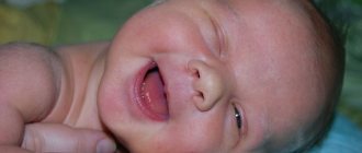
The neonatal period lasts from the moment of birth to 4 weeks of the child’s life. During this time, so many changes occur! A newborn baby is really wrinkled, because he was in the amniotic fluid all the time. But literally an hour or two will pass, and the wrinkling will disappear - a slight swelling of the face, eyelids, labia or scrotum will remain. This swelling will also go away in a day or two. And then the baby will be able to open his eyes - still cloudy. There may be hemorrhages on the sclera (whites) of the eyes, which resolve within a few (3 - 4) days.
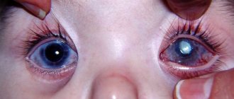
Naturally, you will immediately start figuring out your eye color.
But most newborns, regardless of heredity, have blue eyes at birth. The exception is children of dark-skinned children: they are brown-eyed at birth.
Of course, every mother will carefully examine the visible parts of the baby’s body - his face, his head. Let's take a closer look at this.
Don't be surprised by the purplish- red color of your skin
- do not forget that the baby has made a difficult journey from the uterus through the birth canal, endured extreme stress, enormous tension. When the baby is shown to you for the first time, even before the newborn's first toilet, his skin will be covered with a white creamy substance - a lubricant that helps him pass through the birth canal more easily and protects him from bacteria. 1-2 days after birth, jaundice occurs in 40-45% of newborns. This is due to the fact that in the liver there is an intensive restructuring of fetal (fetal) hemoglobin, which was necessary for the fetus for oxygen exchange through the placenta, into mature hemoglobin, with the help of which gas exchange is carried out in the lungs. Jaundice, as a rule, is not very pronounced - it is a slight icteric staining of the skin, mucous membranes, and sclera of the eyes. It appears on the 2-3rd day of life and usually lasts 7-10 days. At this time, “intrauterine” blood (fetal hemoglobin) is replaced by the adult version.
Many dark-skinned children may have “Mongoloid
spot”
- a bluish spot - on the skin above the buttocks in the sacral area. Don't be alarmed, it's not a bruise. By 5 - 7 months of age, this spot will disappear.
In the place where the baby pushed through the mother's birth canal, there will be a slight swelling - a birth tumor, the so-called cephalohematoma.
It will resolve within 7 days.
As the head passes through the mother's birth canal, the bones of the skull are placed on top of each other. This can be felt on the child's head with a small bony longitudinal protrusion. By the end of the week, the bones will fall into place, and the size of the head will increase by an average of 1 cm. A large fontanel
(the place where the bones of the fetal skull meet, not yet fused, but connected by membranes), and if it is not visible, then it can be palpated. And you will probably notice that the baby’s head is relatively large compared to other parts of the body: so far it occupies 1/4 of the baby’s total “height”.
Some children are born with quite a lot of hair on their heads, and some are “bald as a knee.” Don't worry. Even now, there is hair on the baby’s head—vellus hair; moreover, the baby is covered with hair almost everywhere except the palms, feet, and lips. During the first 3 months of life, vellus hair will be completely replaced by coarser hair. And hair color can also change - even over the course of several years: light hairs can subsequently become quite dark.
Some babies have vellus
can cover the shoulders, back, forehead and cheeks. This is a sign of immaturity or prematurity. This increased hairiness will go away within a few weeks. Another cause of concern for mothers can be the almost complete absence of eyelashes and eyebrows. Unlike hair on the head and body, this is bristly, coarser hair. They grow much more slowly, so a newborn baby may not appear immediately, but after several weeks. Their color may also change.
The newborn's face is somewhat swollen, this goes away on the 2-3rd day. After swelling on the face has passed, some children may develop so-called vascular
in the area of the upper eyelids, bridge of the nose or on the forehead between the eyebrows, above the ears and at the border of hair growth at the back. Typically, such spots do not fade for a long time and gradually disappear at 3-4 years of age.
milia may appear on the baby’s face.
- yellowish pimples on the nose, chin and cheeks of a newborn. Don't be scared! These are dilated sebaceous glands that are visible through the skin. Gradually, as you gain weight and, accordingly, the fat layer, the milia will cease to be noticeable.
Now let's look at those parts of the body that are hidden by the diaper. All babies already have nails at birth. In a full-term mature baby, the nails protrude beyond the nail bed. For immature and premature babies, no. The nails are soft, but they can scratch the baby’s skin very painfully. Therefore, a newborn baby can have a “manicure” - carefully trim his nails.
In the first week of life, the baby’s skin may peel off greatly: exfoliation of the “ intrauterine
and replacing it with a new one.
In the delivery room, a special bracket is placed on the umbilical cord at a distance of 1 cm from the umbilical ring. The remainder of the umbilical cord falls off on the 2-3rd day in the maternity hospital, leaving in its place an umbilical wound - a depression covered with a crust. By the 8th - 10th day the umbilical wound will heal. The little person will just get a navel - a reminder of the intrauterine connection with the mother.
Don't forget to pay attention to body parts such as the mammary glands
and genitals.
On the 3rd - 4th day, both boys and girls may experience engorgement (enlargement) of the mammary glands. There may even be colostrum-like fluid released. Do not squeeze under any circumstances! Apply a dry gauze bandage so that friction with clothing does not injure the delicate skin. By the 10th day this condition usually goes away. This condition may coincide with a phenomenon called a “hormonal crisis” of a newborn in girls, when whitish mucus may be released from the genitals. Sometimes it can be stained with blood. You shouldn’t be afraid of this - it’s the maternal hormones that are “coming out”. This will pass in 2-3 days. Do not be surprised if the labia majora do not cover the labia minora - gradually everything will become the opposite. Some girls may also experience protrusion of the hymen from the vagina. In a few weeks, she will “suck” into place.
As for the boy, you should pay attention to his scrotum. Normally it should be relatively large and hang freely. It should not fit tightly to the crotch. The testicles should already be in the scrotum.
All that remains to be seen is the oilcloth tag on the baby’s hand. It indicates the gender of the child, the mother's last name and initials, the medical history number and the time of birth of your baby. Carefully preserved in the home archive, it will someday remind you of that exciting moment when you first saw your “treasure”.
Why is it dangerous?
The question of whether a hematoma on the head is dangerous can be answered this way: in most cases it does not cause negative consequences and does not affect the general condition and development of children. However, when exposed to certain factors, a purulent process may begin.
In this case, it is necessary to begin therapeutic measures as soon as possible to remove purulent masses when they have a liquid consistency. When the pus hardens, the baby's head becomes deformed.
The consequences of a hematoma on the head of a newborn after childbirth can also be expressed in developmental delays, but this is an exception, which is diagnosed in one case out of 10 thousand infants. Even less often, cerebral palsy occurs against the background of a hematoma.
To avoid such negative consequences, it is necessary to carefully monitor the condition of the newborn and monitor any changes in the condition of the neoplasm. Parents should be alert to the following symptoms:
- increased irritability and restlessness;
- violation of the regime of wakefulness and rest;
- difficulties with breastfeeding, constant regurgitation of eaten food;
- colic that cannot be controlled with medications;
- tilting the head back;
- tilting or turning the head only to one side;
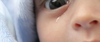
- unilateral lacrimation;
- muscle weakness.
If you notice such symptoms in your baby, you should contact a therapist, who, if necessary, will refer you to another specialized doctor.
Types of formations
Neoplasms in the head area of a child that arise during childbirth are divided into several types. The difference is only in the exact location of the tumor concentration. Based on this, we can distinguish cephalohematoma and other types of hematomas:
- cephalohematoma - hemorrhage in the area between the periosteum and the bones of the head;
- epidural - blood accumulation between the bones of the skull and the dura mater;
- intracerebral - penetration of blood into the brain cavity;
- subdural - hemorrhage in the area between the dura and arachnoid membranes.
Clinical manifestations may vary in each case.
Cephalohematoma
Cephalohematoma in a newborn forms after birth or after 2-3 days. In most cases, the hematoma goes away on its own and does not have a negative impact on the health of the newborn. Its size increases on the first and second days due to a deficiency of blood coagulation function.
The location of a cephalohematoma in an infant is between the periosteum and the skull bone. It does not extend beyond the boundaries of the affected bone and has clear edges. Size ranges from 3 to 7 cm.
When palpating the head, a neoplasm with an elastic and soft consistency and a roller-like compaction along the periphery is discovered. Hemorrhages larger than 8 cm require surgical intervention, because spontaneous resorption in this case is quite rare.
The child often spits up when feeding, becomes restless and irritable.
Epidural hematoma
Epidural hemorrhage due to a violation of the integrity of the vessels of the periosteum does not occur between the periosteum and the skull bone, like a cephalohematoma, but between the skull bone and the dura mater of the brain. In this case, the latter peels off from the cranial bone.
Due to the fact that in infants, children under 2 years of age and in elderly people there is a tight adhesion of the dura mater of the brain to the skull of the head, epidural neoplasms are not often diagnosed during such periods.
Treatment of cerebral edema
How is cerebral edema treated?
In the case of persistent hypertension, regular lumbar drainage (using a thin polyethylene tube), which is inserted according to a technique reminiscent of Seldinger catheterization, can be recommended to treat cerebral edema.
In the case of moderate or normal hypertension (about 140 mm), it is sufficient to treat the brain of a newborn by evacuating the cerebrospinal fluid and administering intravenous glucose 40% (100 ml). With increased cerebrospinal fluid pressure, osmotic diuretics are included in the dehydration complex: maznitol (60-80 g), urea (30-40 g), and also lasix (about 40 g). In complicated situations they are used two to three times a day. Less effective in treatment are hypertonic solutions based on glucose, mercury diuretics, CaC12 (10% - about 200 ml).
Unreasonable extensive use of diuretics in the treatment of severe cerebral edema in a newborn can provoke dehydration and large losses of calcium, but brain injury is accompanied by a fairly high tendency to lose this electrolyte. In case of cerebral edema, daily monitoring of ECG, ionogram, diuresis, water balance in general and water distribution in the main sectors is mandatory.
Is treatment required and what kind?
Before determining treatment for a head tumor in an infant after birth, the doctor prescribes an ultrasound examination, which allows you to determine the volume and identify or refute other pathological conditions. When the hematoma is small in extent, specific therapy is not carried out.
Sometimes doctors prescribe calcium gluconate to tighten the vascular branches and vitamin K, which helps reduce the intensity of bleeding. Help with large hematomas in children (more than 8 cm) involves pumping out the fluid using special equipment.
After pumping, a pressure bandage must be applied. Advice for parents - there is no need to panic when prescribing such therapy. It is considered absolutely safe and does not have a negative effect on the body. However, after the fluid is sucked out, the child must be under the supervision of a specialist for a certain period of time.
How to remove a hematoma on a newborn’s head resulting from a bruise at the time of delivery if it becomes infected? If the integrity of the epidermis in the area with hemorrhage is compromised, this creates excellent conditions for the penetration of pathogenic bacteria. The result is redness of the epidermis, an increase in local temperature, and a purulent process.
In such cases, surgical intervention is immediately performed to drain the purulent contents. Given the severity of the process, the use of antibacterial and disinfectants is prescribed.
Forecast
A hematoma on the head of a newborn after childbirth does not pose a significant danger to his health. Despite this, such babies are observed by doctors in the first months of life. Monitoring is carried out for 2 weeks and if the tumor does not decrease or disappear, appropriate therapy is prescribed.
An unfavorable prognosis is observed in cases where suppuration is not treated in time, which may result in ossification of purulent masses. This is especially dangerous if it is localized in the area of pressure on the nerve endings. As a result, children’s development is delayed, the development of cerebral palsy, and speech disorders.
As you can see, the prognosis is favorable in most cases. However, you should not rely on the fact that the help of doctors is not required in this case. You still need to see a doctor to assess your health.
I write articles in various areas that, to one degree or another, affect such a disease as edema.
Causes of cerebral edema in a newborn
What are the causes of cerebral edema?
Brain swelling can be considered a secondary sign of injury. It can be local or generalized. Brain edema appears in a newborn with a variety of diseases that affect the nervous system: stroke, traumatic brain injury, abscess and brain tumors, meningitis and encephalitis, hypoxia, various forms of occlusive hydrocephalus, various syndromes of deterioration of osmotic balance, general intoxication, infections, body burns, malignant hypertension, etc. It has been proven clinically and experimentally that various etiological factors provoke pathogenetic various forms of brain swelling in a newborn, but the mechanisms of its increase are equivalent.