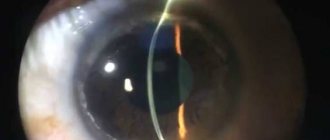
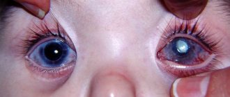
A spot on a dog's eye. Lipoid corneal dystrophy. Labrador. (February 2020).
Corneal dystrophy is a condition that affects some breeds more than others. It is an inherited disorder that gets worse over time and it negatively affects the dog's eyes, with both eyes being affected equally. Unfortunately, corneal dystrophy is one of the eye diseases most commonly seen in dogs, but it is not associated with any other disorders.
Types of corneal dystrophy
Dogs can suffer from any of three types of corneal dystrophy, which are classified by the location they are found in the eye. These are the following:
- Corneal epithelial dystrophy – this form of the condition sees cell formation being negatively affected
- Stromal corneal dystrophy – this form of the condition sees the dog's cornea turn cloudy
- Corneal endothelial dystrophy – this form of the condition affects a cell found on the lining of a dog's cornea
Surgical treatment of corneal dystrophy
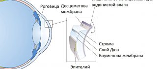
Surgical correction is considered the most effective treatment for corneal dystrophy. If the epithelial layer or Bowman's membrane is damaged, laser surgery can be used to remove the area of dystrophy (PTK - phototherapeutic keratectomy).
For preventive purposes, to “strengthen” the structure of the cornea, a collagen cross-linking procedure is recommended if pathological processes occur in the stroma.
If deeper layers are damaged, it is necessary to perform an operation - keratoplasty with removal of the damaged layer of corneal tissue and transplantation of donor tissue in its place.
There are two types of surgical interventions:
- penetrating keratoplasty – the entire central part of the cornea is removed;
- layered keratoplasty – individual layers of the cornea are removed.
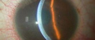
Fig.4 Patient's eye after 6 months. after penetrating keratoplasty surgery
After transplantation of the donor cornea, its cells begin to function instead of their own damaged ones, due to which the transparency of the cornea is restored and the symptoms of the disease are eliminated. In some cases, if a relapse occurs, repeat surgery may be required.
In our ophthalmology center, patients have the opportunity to receive high-tech, world-class ophthalmological care: from doctors from Germany using 100% German technologies!
In addition to penetrating keratoplasty and phototherapeutic keratectomy, we perform unique operations of layer-by-layer anterior and posterior corneal transplantation, including those with femtosecond support:
- DALK – deep anterior lamellar keratoplasty
- DMEK – Descemet's membrane keratoplasty
- DLEK – corneal endothelial transplantation
- DSEK and DSAEK – transplantation of stroma, Descemet's membrane and endothelium
Symptoms to watch out for
If a dog develops epithelial corneal dystrophy, they may well exhibit the following signs that something is wrong:
- Corrosive spasms
- Gray or white irregular or circular shapes, rings or opacities form in the cornea of a dog's eyes
The dog's vision remains normal, and the onset of any symptoms usually begins when dogs are anywhere from six months to six years of age. The condition gradually worsens over time, and the breeds most at risk of developing this condition are as follows:
- Shetland Sheepdogs
- Stromal corneal dystrophy
- When dogs suffer from stromal corneal dystrophy, they usually show the following signs that something is wrong:
- Their corneas develop white, gray or silver round or oval shapes
- Ring opacity (doughnut shape)
- Diffuse Opacity
The condition begins to develop in young mature dogs, and although their vision is not seriously affected, it may be affected to some extent due to opacity in the corneas. The breeds that seem to be most susceptible to this form of both stromal and epithelial corneal dystrophies include the following:
- Afghan Hound
- Airedale
- Alaskan Malamute
- American Cocker Spaniel
- beagle
- Bearded Collie
- Bichon Frize
- Cavalier King Charles Spaniel
- German Shepherd
- Lhasa Apso
- Great Dane
- Miniature Pinscher
- Rough Collie
- Siberian Husky
- Samoyed
- Weimaraner
- beagle
Symptoms of keratitis in cats
The disease can be recognized at an early stage by observing the cat. First, changes appear at the cellular level, then the entire cornea is affected.
We list the clinical signs as the disease progresses:
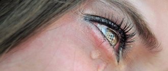
- Corneal syndrome is the “first bell” by which keratitis can be recognized. It is manifested by photophobia, spasm of the corneal muscle (the pupil stops dilating), lacrimation and vascular sprouting along the edges of the cornea.
- Corneal opacification - usually the color of the film is white or light brown. Gradually, due to such changes, vision deteriorates, as a result of which the animal ceases to orient itself in space and restless behavior appears.
- Germination of blood vessels in the cornea - these changes are already irreversible and appear with a prolonged course. If treatment is not started in time, keratitis can be complicated by a breakthrough, the development of secondary glaucoma or cataracts.
Diagnosis of the problem
The veterinarian should have known the dog's complete medical history and, ideally, the dog's lineage, which would aid in diagnosing the problem and determining whether there was a genetic link. The veterinarian will perform an in-depth ophthalmic examination of the dog's eyes and recommend the following tests:
- Blood Chemical Profile
- Complete blood count
- Electrolyte panel
- Analysis of urine
- Tonometer – this will determine if any pressure has built up inside the dog's eye and helps rule out other conditions such as glaucoma
Veterinarians perform a test called slit lamp microscopy, which helps them determine what type of corneal dystrophy a dog may have in the affected eye. To determine if the eye has been damaged in any way, they would use a fluorescent spot that detects abrasions if they have occurred. The spot would also determine whether the dog has any corneal ulcers, which often develop when dogs suffer from endothelial or epithelial corneal dystrophy.
Diagnostics
To make a diagnosis, the veterinarian must examine the cat, check the reaction of the pupils to light, and evaluate the animal’s behavior. You can accurately determine the nature of the lesion and complications using two methods:
- biomicroscopy of the eye;
- bacterioscopic examination.
Based on the examination, the ophthalmologist at our clinic makes a final diagnosis and prescribes therapy. During the treatment, the animal is under constant supervision.
We will help you cure your pet. Call 33-94-45!
Treatment Options
If a dog is found to be suffering from corneal ulcers, the veterinarian will usually prescribe an antibiotic eye treatment to help resolve the problem. If a dog suffers from stromal corneal dystrophy, it usually does not require any form of treatment. Dogs with corneal endothelial dystrophy can be treated with some veterinarians prescribing contact lenses that cover the dog's eyes, or they may recommend performing something called "flap surgery" to correct the problem.
Causes
A hereditary breed predisposition to fatty degeneration of the cornea has been identified: Siberian huskies, beagles, American dogs, spaniels, and German shepherds are at risk. Usually both eyes are affected, and the cause of lipid deposition in the cornea remains unknown.
The disease can also develop against the background of hypothyroidism and concomitant high cholesterol levels in the blood.
Inflammatory eye diseases (dry eye syndrome, uveitis, pannus, etc.) can lead to the development of lipoid corneal dystrophy.
Corneal endothelial dystrophy (Fuchs' dystrophy)
From a clinical point of view, primary and secondary forms of Fuchs' dystrophy are distinguished. The primary lesion of the cornea is congenital. The first manifestations of early dystrophy are detected at birth or in early childhood, but this type of disease is extremely rare. The late version of primary Fuchs endothelial dystrophy most often develops in people over 45 years of age. This type of pathology is characterized by a binocular but asymmetrical course. Secondary dystrophy is an acquired disease in which monocular damage is observed.
Depending on the severity of the pathology, the clinical picture of corneal endothelial dystrophy can be characterized by a latent course or manifestations of bullous keratopathy. Symptoms of the disease progress slowly, so it can take about 20 years from the moment the first signs appear until a detailed clinical picture is formed.
First stage
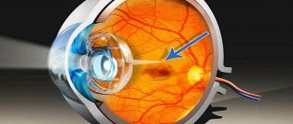
At the first stage of endothelial corneal dystrophy, morphological changes affect only the central parts of the cornea. In this case, specific collagen formations (gutta) of drop-shaped, warty and mushroom-shaped forms appear. No complaints. The only symptom of the disease is a slight decrease in visual acuity in the morning.
Second stage
At the second stage, the number of endothelial cells decreases, corneal edema develops, and single bullae appear. The sensation of a foreign body is replaced by a decrease in the sensitivity of the cornea due to degeneration of nerve endings. Specific symptoms of this stage of endothelial corneal dystrophy are represented by photophobia, eye hyperemia, decreased visual acuity in the morning with subsequent recovery in the evening. This is due to the fact that due to closed eyes during sleep, moisture does not evaporate properly from the cornea, which leads to fluid deposition. During the day, the evaporation of moisture helps reduce swelling and restore visual functions.
The long course of endothelial corneal dystrophy causes a slowly progressive decrease in visual acuity. When the pathological process spreads to the epithelial layer and bullous changes appear, patients complain of a foreign body sensation and increased tearfulness. Pain syndrome, accompanied by severe discomfort in the orbital area, develops when bullae rupture in patients with bullous keratopathy.
Third stage
At the third stage, fibrous tissue is synthesized along the epithelium of the basement membrane, followed by the formation of pannus. The general condition improves somewhat, but the progression of endothelial corneal dystrophy further leads to the appearance of epithelial erosions, ulcers of microbial origin and vascularization of the central part of the cornea.
Signs
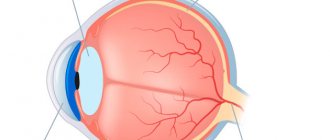
The disease leads to impaired transparency of the cornea. Cloudiness, white or gray spots appear on it, and vascularization (sprouting of blood vessels) occurs. Pigmentation (dark coloring) of the cornea may appear.
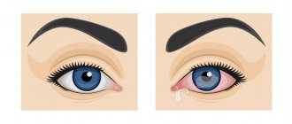
Sometimes the disease develops against the background of dry eye syndrome - in this case, there is dryness and inflammation of the cornea (keratoconjunctivitis sicca). You can also notice signs of other inflammatory eye diseases: redness, discharge, tissue swelling, squinting of the eyes, photophobia, clouding of the anterior chamber of the eye.
Treatment
If a cause is identified that contributes to the development of fatty degeneration of the cornea, then treatment is aimed at eliminating it. Thus, for elevated cholesterol levels, a diet is prescribed; for hypothyroidism, thyroid hormone replacement therapy is used. If eye diseases are detected, they are treated.
Surgical treatment often does not produce results, since the infiltrate usually appears again. There is no effective specific treatment for fatty corneal degeneration in dogs.
Conservative treatment of corneal dystrophy
For corneal erosions, especially when they are repeated frequently, patients are prescribed drug treatment: taking drugs whose action is aimed at restoring the corneal epithelium - keratoprotectors: eye ointments and drops that help moisturize the surface of the cornea. The use of eye drops is advisable during the day, eye ointments - at night, because they have the most prolonged effect.
For therapeutic purposes, patients may also be recommended to wear soft therapeutic contact lenses that relieve pain, eliminate the sensation of a foreign body and create favorable conditions conducive to the restoration of the epithelial layer of the cornea.