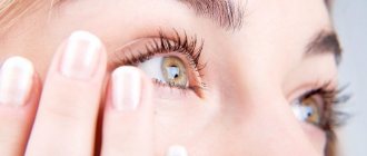
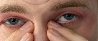
Associated symptoms
People almost never associate a yellow spot on the white of the eye with diseases of the organ of vision or malfunction of internal organs. However, it is a symptom of progressive eye pathology; additional signs are observed:
- Unbearable itching;
- Intolerance to bright light;
- Purulent discharge;
- Split image;
- Painful sensations.
If a yellow spot is a sign of problems with internal organs, then it is accompanied by the following symptoms:
- Loss of appetite;
- Fever;
- Chills;
- Prostration;
- The emergence of a gag reflex.
| As you can see, yellowing of the sclera is not always a harmless deviation; it is often a sign of the development of a serious disease. Therefore, if such an anomaly occurs, immediately contact an ophthalmologist. |
Associated symptoms
Rarely does the appearance of a macular spot appear in isolation. There are usually other symptoms of health problems:
- purulent or watery discharge from the eye;
- pain;
- pain in the eyes, the reasons may be different;
- inflammation of the conjunctival sac;
- red spots under the eyes of a child;
- double vision;
- acute reaction to light.
Signs of general malaise may also be identified:
- lethargy, drowsiness;
- lack of appetite;
- aching joints;
- vomit;
- increased body temperature, chills, fever.
If the child does not yet know how to speak, you need to pay attention to the general state of health.
Proper treatment of inflammatory (non-infectious) eye diseases - read the instructions for use of Diclof eye drops.
Pinguecula – practically never seen in childhood
When vision can be preserved and improved - age-related farsightedness.
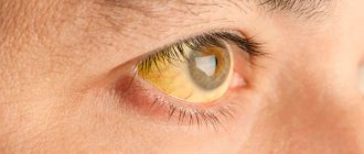
Common Causes of Yellow Spots in the Eye
Yellow spots that appear on the organ of vision are a symptom of the progression of a certain disease:
- Liver dysfunction. A change in the color of the sclera may indicate an increase in bilirubin levels. A similar deviation occurs with jaundice, which is provoked by alcohol abuse, excess weight, gallstones, etc. Coloration of the entire protein in a yellow tint is a sign of the development of hepatitis A;
- Jaundice in a newborn baby also causes discoloration of the sclera. The cause is an increase in the level of red blood cells in the blood. Symptoms should subside within ten days; if signs persist, contact your pediatrician immediately;
- The spot indicates the development of pathologies such as pterygium or pinguecula. In the second case, the corners of the eyes are painted;
- The appearance of a neoplasm. Malignant tumors of conjunctivitis are accompanied by yellowing of the sclera;
- Horner-Trantas spots. The disease is considered a peculiar manifestation of an allergic reaction to external irritants;
- Conjunctivitis cyst. Visually it resembles a small yellow bubble located on the outer epithelium of the organ of vision.
To avoid progression of the disease, go to the clinic as soon as you see changes in the color of the sclera. Return to contents
Causes
Why does macular degeneration occur? Experts have not yet established the exact reasons for the development of this pathology. There are quite a lot of different versions. Some of them are confirmed by research and observations, some remain at the level of theories. Thus, a number of experts argue that with a deficiency of certain mineral compounds and vitamins, a person becomes more susceptible to developing the disease. For example, a number of studies have found that the likelihood of macular degeneration increases several times in the absence of vitamins E and C and antioxidants. Of great importance is the lack of zinc (it is present in the body, but is concentrated in the area of the organs of vision), as well as zeaxanthin and lutein carotenoids. The latter are pigments directly from the macula itself.
Experts name human cytomegalovirus as one of the provoking factors. Some researchers argue that the development of pathology is greatly facilitated by a diet in which the level of saturated fats is very high. In this case, monounsaturated compounds are considered potentially protective. In accordance with some observations, it has been found that it is possible to reduce the likelihood of pathology by taking ω-3 fatty acids. The results of more than ten studies have revealed a statistically significant relationship between macular degeneration and smoking. In this case, the probability of the occurrence of pathology increases by 2-3 times in nicotine abusers (compared to persons who have never smoked). However, five studies found no association.
Possible diseases
Most often, the appearance of a macular spot signals the development of pathologies that do not have a negative impact on the patient’s health and visual acuity.
Pinguecula
Under this name “hidden” is such a deviation as thickening of the mucous membrane. The spots usually appear on the outside of the eye, located symmetrically, in the corners of the eye. Most often, this disease is diagnosed in pensioners, as the conjunctiva wears out and ages.
You can learn more about the pinguecula by watching the video.
Pterygium
The mucous membrane grows and becomes thicker in the inner corner of the eye. The shape of the spot resembles a triangle and may have a yellow or pink tint. In some cases it takes on a rich red color. The cause of the anomaly is the adverse effects of external factors, such as strong wind, ultraviolet radiation, etc.
The spot can grow and if it invades the cornea, this will lead to vision problems. And getting rid of it in an advanced stage is much more difficult than at the initial stage.
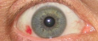
Causes of spots on the white of the eyeball
The spot on the squirrel may be red or yellow-brown.
As a rule, a red spot is formed for the following reasons
:
- High blood pressure or a sharp drop in blood pressure. In this case, one or more microvessels may burst and form a small hematoma. Such spots do not require special treatment, but signal that you need to monitor your blood pressure, as well as check your blood vessels and heart
- Serious physical activity. Due to strong tension, the capillaries on the eye burst, but this is a temporary phenomenon that should not be seriously alarming. Of course, if red spots appear regularly after working out in the gym, for example, you need to reduce the load
- Intraocular pressure. This may be indicated by a stagnant red spot that does not go away for a long time. In this case, a consultation with an ophthalmologist is required to detect existing or stop developing eye pathologies.
Some Patients have congenital spots on the albumen. They resemble birthmarks on our body: they are just a certain pigment that appears in one place or another. Such spots are harmless and do not interfere with vision. If this bothers you aesthetically, consult a specialist to eliminate the darkening.
There are also more complex cases - floating spots
. They are usually detected by moving the eyeball: the spot itself is colorless, but when it gets into the pupil area, the patient feels discomfort and vision becomes blurred. This phenomenon may be a sign of vitreous disease or retinal detachment, which requires quality treatment from an experienced doctor.
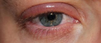
Pinguecula (yellow spot)
- not a disease in itself, but a pathological manifestation of aging of the conjunctiva. As a rule, such spots appear on the inside of the eye, near the bridge of the nose, and are not so easy to notice.
The occurrence of pinguecula is caused by various factors: prolonged deficiency of vitamin A, exposure to aggressive ultraviolet rays, and regular eye strain throughout life.
Yellow spots do not require special treatment, but to make sure they are harmless, consult your doctor.
Diagnosis and treatment
There are many factors that can provoke the development of pathology, so even the best ophthalmologist in the world will not be able to immediately make a diagnosis. You will need to undergo a number of procedures:
- First, the doctor conducts a visual examination and finds out the details of the medical history, this will help make a preliminary diagnosis. If there are problems with the liver, palpation is done to find out whether the organ has increased in size or not;
- Conducting an ophthalmological examination. Biomicroscopy using specialized equipment allows the doctor to study the structure of the eye in detail;
- Ultrasound and CT scan of internal organs located in the abdominal cavity. Diagnostics allows you to determine the location of the accumulation of pathological changes that caused the appearance of a yellow spot. A biopsy is used to confirm the results;
- Examination of the patient's blood, urine and stool. This is necessary to determine the level of hemoglobin and bilirubin.
After establishing the cause that provoked the change in the shade of the sclera, the doctor prescribes treatment.
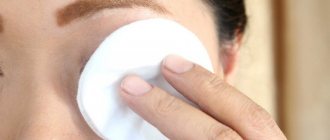
Eye medications
If no abnormalities in the functioning of the internal organs and the visual apparatus are detected, most often doctors diagnose the patient with pinguecula. To eliminate it, eye drops that imitate human tears are prescribed (for example, Oxial). If the pathology is accompanied by an abscess, the doctor prescribes antibacterial and anti-inflammatory medications, such as Tobradex, Diclofenac or Maxitrol.
Contact lenses can damage the tumor and cause inflammation, so avoid wearing them during therapy. Return to contents
Unconventional methods of treatment
Some patients prefer to use “grandmother’s” recipes and be treated with the gifts of nature.
- Blueberries are useful in any form. Eat one hundred grams of berries every day; they contain high concentrations of vitamin C and B. You should eat them only on an empty stomach;
- Prepare fresh juices from carrots, parsley, and celery. To ensure that the body absorbs all the beneficial elements included in their composition to the maximum, add a drop of vegetable oil to the cocktail;
- Eat one hundred grams of raw beets every day, you can make puree;
- In the morning and evening hours, use linden lotions. Leave for twenty minutes, they will soothe the irritated epidermis and relieve inflammation;
- Almond oil lotions help soften the skin;
- Make compresses from beets; they are left on the eyes for a quarter of an hour;
- Blueberry lotions are useful. Squeeze the juice from the berries and soak a cotton swab in it, apply for ten minutes.
| Most often, such methods simply strengthen the visual apparatus, so it is better to use them as preventive measures, and not instead of therapy. |
Laser removal of pinguecula
If the resulting spot causes psychological discomfort to the patient, he can go for surgery. The procedure takes little time and is absolutely painless. Innocaine drops are used as anesthesia. The recovery period after surgery is a maximum of two hours, so there is no need to stay in the hospital.
If no complications arise, the doctor sends the patient home. To protect the eye from adverse external factors, you need to wear a bandage for several days. When outdoors, be sure to wear sunglasses to prevent harmful ultraviolet radiation from affecting your vision.
In some cases, surgery has negative consequences: recurrence of pathology or problems with eye sharpness.
Treatment
Treatment of pinguecula is carried out using conservative therapy. Anti-inflammatory drops are used (Maxitrol, Diclofenac). Eye drops containing boric acid eliminate discomfort and soften the mucous membrane of the eye. If lipid formations still cause discomfort to the patient, then surgical removal of the pathology with a laser beam is possible. The procedure is quite simple and does not take much time. Surgical intervention is also required in the treatment of pterygium.
The treatment method for conjunctival cysts depends on the stage of the disease, the size of the formation and its location. First, the doctor will recommend eliminating the infection that caused it using anti-inflammatory drugs. Surgery will then be performed. During the intervention, the surgeon completely excises the body of the cyst with a laser beam. The operation is performed under local anesthesia.
If the patient has been diagnosed with ocular melanoma, then treatment of the formation should begin as quickly as possible. The most effective method is surgical (complete) removal of the malignant tumor. To prevent the spread of pathology to neighboring, healthy tissues, it is necessary to combine surgery with radiation therapy or systemic chemotherapy.
Failure of metabolic processes is treated with the help of drugs that cleanse the liver and bile ducts of all kinds of toxins and wastes. Conservative therapy helps eliminate blockage of the bile ducts at the initial stage of the disease.
If the yellowness of the whites of the eyes does not go away after this, then surgical intervention cannot be avoided.
Preventive measures
A proper daily routine and a balanced diet will help prevent the development of pathologies or their relapse. Following simple medical recommendations will help you avoid eye health problems:
- Sleep at least eight hours a day;
- Move more, walk in the fresh air;
- When working at a computer for a long time, use safety glasses;
- Take vitamin complexes;
- Use medications to moisturize the mucous membrane;
- Visit your eye doctor regularly.
| On sunny days, wear optical devices marked UV400; they protect your eyes 100% from harmful ultraviolet radiation. |
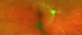
Macular Degeneration: Symptoms
The manifestation of pathology is different in all patients. For example, in some patients, macular degeneration may develop quite slowly. In other patients, on the contrary, the course of the disease is rapid, which leads to significant deterioration of vision. Soreness does not accompany either the wet or dry form of the pathology. Some of the main symptoms of macular degeneration include:
- blurred vision;
- distortion of straight lines (for example, the contours of a doorway may appear curved);
- difficulties in the process of considering details (when reading, for example);
- the presence of a small black dot in the center of the visual field, increasing in size over time.