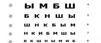
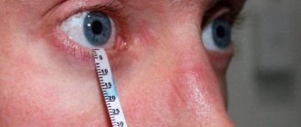
How to test your vision using the Amsler grid
- Sit comfortably on a chair;
- Position the Amsler grid so that the distance between it and your eyes is approximately 25-30 centimeters;
- Put on contacts or glasses if you wear them;
- Cover one eye with a sheet of opaque paper. Do not press your eye with your hand under any circumstances, as this may give a false positive result!
- With your other eye, look at the dark dot in the center of the grid and focus on it for 3-5 seconds;
- Now slowly approach the grid, keeping your eyes on the central black dot, until the distance to the table is approximately 20 cm;
- Analyze the picture in front of your eyes: is there any distortion of the lines and angles? Are all the squares the same shape and the same? Are there places in the table that seem vague or cloudy to you?
- After that, repeat the same steps for the second eye.
A special test is used to check the central field of vision. It is used to determine the quality of the eye's work in the area of the macula. This examination is not available by other methods.
The Amsler grid is a lined table of four hundred sectors. A large dot is indicated in the center of the grid.
The test allows you to detect macular degeneration of the retina. This is a common disease that occurs in people after forty. It reduces visual acuity, makes central objects invisible, and changes the perception of colors. This prevents a person from living a full life. He cannot read, watch TV, drive a car or do other things normally. Often because of this you have to change jobs.
At the clinic, the test is carried out using a grid drawn on a piece of paper. But you can regularly go through it yourself by printing out the grid or drawing it. Or go online on the Internet, displaying a picture with a grid on the full screen.
To perform a vision test using this method, you need to do the following:
- The patient sits on a chair, his back is straight, his gaze is directed straight ahead.
- The Amsler grid is installed in front of the eyes at a distance of approximately 30 cm. Plus or minus 2-3 cm does not matter.
- If the person being tested wears glasses or contacts, they must be worn.
- Cover one eye with your palm or some other handy object. This is done carefully, without pressure, otherwise the disease will not be detected. The person being tested does it himself.
- The patient looks with an open eye at the center of the grid at the point.
- After five seconds, the mesh is moved 10 cm towards the face, the patient continuously looks at the point.
- After another five seconds, the net moves back into place.
The result is recorded and carried out in the same way with the second eye. The entire check takes no more than a minute. Anyone can easily carry it out on their own at home.
Until now, scientists cannot indicate the reasons for the appearance and development of AMD (age-related macular degeneration). One thing is known - people over the age of 45, and especially over 75, are more at risk of developing various eye diseases, including age-related macular degeneration. With age, another problem may arise - pinguecula.
Besides age, there are several other factors that contribute to the occurrence of macular degeneration:
- if there is a family predisposition;
- smoking;
- fatty food;
- female;
- Caucasian nations.
But using this tool does not mean that you should not visit your ophthalmologist, since you can easily miss the first signs of the disease, which only a specialist can detect. Many symptoms are attributed to computer vision syndrome, missing important abnormalities.
The retina is the most important layer of the eye, which is directly responsible for the perception of images, transmitting them to the brain, and providing color vision. Any lesions in the retinal area can lead to deterioration in visual function. The most pronounced vision pathologies are observed when disturbances affect the central zone of the retina - the so-called macula. Here are the light-sensitive structures that provide high-quality central vision.
With age, pathological changes can occur in the macula area - tissue nutrition is disrupted, cell degeneration occurs. This disease is called age-related macular degeneration. According to statistics, AMD is one of the most common causes of complete or partial loss of vision.
Blindness in macular degeneration can be avoided if the disease is detected at an early stage and treatment is started on time.
To test your vision, the Amsler test does not have to be printed out. If you don't have a paper version at hand, you can use the online version of the test grid. The algorithm for checking central vision in both cases will be identical:
- The drawing is placed at eye level at a distance of 30 cm from the face. Some experts recommend first moving the mesh to arm's length and only then gradually bringing it closer to your eyes.
- The test is carried out for each eye in turn, so one eye must be closed.
- The gaze must be fixed on the black dot in the very center of the picture.
- After this, evaluate the evenness of the lines, the size of the squares, and the visibility of the pattern.
- Repeat the diagnosis for the second eye.
After you have performed the Amsler eye vision test, you can make a preliminary conclusion about the presence or absence of defects in the retinal area.
If you saw exceptionally smooth, clear lines in the diagnostic grid with both eyes, all grid squares were the same size, and there were no distortions, discolored or clouded areas, then the risk of macular degeneration is minimal.
If the lines during the test seemed curved, unevenly colored, the squares seemed deformed and unequal in size, as well as other distortions, there is a possibility of degenerative changes in the retina. In this case, you should definitely consult a doctor for a more accurate diagnosis and the appointment of competent therapy.
When testing the condition of the retina and central vision, you need to adhere to several rules:
- It is advisable to carry out the test in the morning, after a full night's rest.
- Those who wear contact lenses or glasses need to undergo the examination wearing optical correction devices.
- The room should be well lit.
It is worth noting that the Amsler grid is a fairly informative test, but it does not replace regular examinations by an ophthalmologist, especially over the age of 45.
In the medical department, everyone can undergo examination using the most modern diagnostic equipment, and based on the results, receive advice from a highly qualified specialist. The clinic is open seven days a week and operates daily from 9 a.m. to 9 p.m. Our specialists will help identify the cause of vision loss and provide competent treatment for identified pathologies.
What is it - Amsler grid
A special test is used to check the central field of vision. It is used to determine the quality of the eye's work in the area of the macula. This examination is not available by other methods.
The Amsler grid is a lined table of four hundred sectors. A large dot is indicated in the center of the grid.
The grid can be drawn with black lines on a white sheet or vice versa, white on black. This does not matter and does not affect the test result. The main thing is that there is contrast and the point in the center stands out well. During the vision test, the patient should look at this point.
The test allows you to detect macular degeneration of the retina. This is a common disease that occurs in people after forty. It reduces visual acuity, makes central objects invisible, and changes the perception of colors. This prevents a person from living a full life. He cannot read, watch TV, drive a car or do other things normally. Often because of this you have to change jobs.
At the clinic, the test is carried out using a grid drawn on a piece of paper. But you can regularly go through it yourself by printing out the grid or drawing it. Or go online on the Internet, displaying a picture with a grid on the full screen.
What is the essence of the method?
The Amsler test is used to test the central visual field. Takes about fifteen seconds. Allows you to identify defects in the central part of the retina. Typically used to monitor age-related macular degeneration. But it may indicate other deviations. For example, for diabetic macular edema (a symptom that accompanies a number of diseases) or damage to the optic nerve.
What is the basis of the test?
A square is drawn on a white sheet of paper, divided into 192 identical cells. In the picture, straight lines, right angles. In the middle of the grid, a black bold dot. The patient needs to focus on this small circle that stands out.
What the table looks like is shown in the photo
The method in question for scanning vision was invented and became famous thanks to the invention of Marc Amsler, a respected specialist from Switzerland. The doctor served as professor of ophthalmology in Zurich. The method has become widespread since 1945. Its advantages, information content and accessibility: for self-control, anyone can take the test without resorting to the help of specialists.
But the study is not able to recognize many visual field disorders, such as scotoma (an area of the retina where visual acuity has decreased or decreased). To identify them, you need scotometry (or in other words, the Amsler-Marinchev test).
Who needs a vision test using the Amsler grid test?
To do this, you need to answer a few simple questions.
- The lines had no breaks, were they straight?
- Were there disappearing/appearing light spots at the intersections of the lines?
- Did you see all 4 corners without looking away from the point?
If the answers to all these questions were positive (that is, the picture was clear, the squares were the same, the lines were parallel, etc.), then everything is fine with your vision and there are no symptoms of macular degeneration.
A healthy person sees the Amsler table like this:
People with age-related macular degeneration may see these Amsler grid images
The preliminary test result can be assessed without special education. Everything is very simple.
If the grid is not distorted during the described actions and appears smooth, then everything is in order. But if the edges suddenly float or become distorted, this means that there are problems. Distortion may be visible in different parts of the grille. This could be the very edge, the middle or a corner. There may be one section or several. The lines may become bent or completely disappear from view. Gray spots or nebulae may appear on the grating.
If you saw a completely clear image in which all the lines are parallel, the squares are the same, and the angles are right, then you have normal vision and there are no signs of macular degeneration.
If the image is distorted, then it is necessary to urgently visit an ophthalmologist’s office.
In this case, the vision test is carried out in the same way as described above. However, here the eye must be brought closer until the red spot disappears from the field of vision. Accordingly, if the right eye is checked, the spot should disappear on the right and vice versa.
Please note that the test on a white background is more informative!
The first version of the Amsler grid (on a white background) is larger in area and is more informative. You can see blurry spots on it, which on a black background may go unnoticed.
The Amsler test should become an integral part of vision testing in people over 45 years of age. It is from this age that macular degeneration most often starts. This is especially true for the wet (exudative) form of the disease, since it can be corrected only at the initial stages of its development.
There are many different ways to test vision and identify various ophthalmic defects. One such type of test is the use of the Amsler test.
This is a simple and informative test that can be used not only by a specialist in the process of diagnosing pathology, but also by the patient himself during self-monitoring and detection of a disease such as age-related macular degeneration.
The Amsler test does not require any physical effort and takes no more than 10 seconds. All the patient’s actions boil down to examining a special grid (a field drawn into squares), in the middle of which there is a thick circle.
For your information! This test checks the central visual field and identifies the possible presence of macular degeneration of the retina, also called macular pathology.
Thus, the purpose of the test is to check the area of the eye that is responsible for central vision.
It is impossible to detect malfunction of the macula area using general diagnostic methods using medical instruments and equipment. A simple method such as the Amsler test is suitable for this.
To carry out testing, a special grid (Amsler table) is used, divided into four hundred sectors.
The Amsler grid (full screen test available at this link) can be applied in black on a white background or vice versa. This is a non-critical difference, which still does not affect the result, the main thing is the contrast between the background and the grid itself.
The patient undergoing the examination will have to concentrate his gaze on the central part of the grid. For convenience, a bold circle or large dot is placed right in its center, which is visible against the background of the sectors.
All lines in the grid are parallel to each other, and the angles are right.
Important! You can draw such a grid manually, or you can, but you can pass the Amsler test on a computer. In the absence of vision problems, a person clearly observes this straightforwardness.
In order for test results to be more or less reliable, certain rules are followed.
- The monitor is placed at a distance of 0.3–5 m, always at eye level, so that the surface does not reflect glare.
- Testing is carried out when a person feels well and does not feel tension in the eyes.
- Turn on general lighting.
- The verification table must be clear and contrasting.
- They sit straight, don’t squint, don’t strain their eyes.
- The test is carried out “with a fresh eye” - before going outside, reading, or other work that creates a load on the organ of vision.
Rules for testing vision using a computer
If you have seen a clear picture where the lines are continuous, smooth, without distortion or blur, then you most likely do not have defects in the central zone of the retina. The image should be clear to both eyes.
If there are curvatures and breaks in the lattice, discoloration of different areas, illusory holes in the squares, black spots, it is necessary to be examined by an ophthalmologist, there is a suspicion of macular degeneration.
Amsler grid for retinal testing
to detect malfunction of the macula area using general diagnostic methods using medical instruments and equipment . A simple method such as the Amsler test is suitable for this.
To carry out testing, a special grid (Amsler table) is used, divided into four hundred sectors.
The Amsler grid () can be applied in black on a white background or vice versa. This is a non-critical difference, which still does not affect the result, the main thing is the contrast between the background and the grid itself.
The patient undergoing the examination will have to concentrate his gaze on the central part of the grid. For convenience, a bold circle or large dot is placed right in its center, which is visible against the background of the sectors.
All lines in the grid are parallel to each other, and the angles are right.
Important! You can draw such a grid manually, or you can print it out, but you can also pass the Amsler test on a computer. In the absence of vision problems, a person clearly observes this straightforwardness.
Conditions for the eye test
One of the characteristic symptoms of macular degeneration is a decrease in central visual acuity. Sometimes a person can see a darkening right in the center of the visual field. If such symptoms are detected, it is recommended to undergo the so-called Amsler test.
It is a table laid out in a special way; it is an Amsler grid. Depending on what the patient sees on this table, we can conclude about the presence or absence of defects in the macula area.
In order for the test results to be accurate, the following conditions must be met:
- Eyes should not be tired from prolonged reading or bright light. It is better to carry out the test at the beginning of the working day, rather than at the end.
- If the patient wears glasses, they must put them on before being tested. This also applies to lenses, they must be inserted.
- You should pay attention to the lighting in the room where the test will take place. The light should be normal, not too bright and not dim.
- The grid should be viewed at a right angle. Tilts of the head down or up are not allowed. The image must be strictly right in front of your eyes.
- The patient should not squint or look closely. He needs to try to perceive the whole picture as a whole, and not its individual details.
- The test is carried out for each eye separately. The second eye is covered with the palm of your hand or a sheet of paper. You cannot put pressure on it, but simply cover it, otherwise the result will be distorted.
If the patient has different quality of vision in each eye, the test begins with the eye that sees better.
If a person takes the test on his own and discovers any deviations from the norm, he should immediately contact an ophthalmologist. This may be a signal of the onset of the disease.
Before taking the test, you must ensure that certain conditions and requirements are met:
- A person should be well rested, so it is better to take the test before work or school, rather than at the end of the day.
- The test is performed in the optics that the patient usually wears.
- When taking the test, the room in which this procedure will be carried out must be well lit.
- You must look at the Amsler grid, on which you are taking the test, at a right angle, without tilting your head.
- No need to squint trying to see all the details. The essence of the test is not this, but the perception of this image as a whole.
- The test is performed with each eye in turn. The eye that is not being tested should be covered with the palm of the hand.
If one eye clearly sees better, you need to take the test starting with it.
The Amsler test is performed as follows:
- it is necessary to position the grid (Amsler grid) so that it is at eye level when the patient sits on a chair with a straight back;
- the distance between the mesh and the patient’s eyes should be 30 centimeters (plus or minus a couple of centimeters in this case is not a critical value);
- if the patient uses medical optics, they must be worn;
- one eye is covered with the palm of the hand, and the other one needs to look at the circle in the center of the grid, while pressing on the closed eye with the palm of the hand is prohibited: due to the physiological characteristics of the organs of vision, this can lead to a good result even in the presence of a disease;
- You need to look at the central circle for five seconds, after which you slowly need to bring your head ten centimeters closer to the grid.
After looking at the central circle from a distance of 20 centimeters for another five seconds, you need to take the starting position and repeat the process with the second eye.
It is easy to interpret the result at the end of the Amsler test. In the absence of eye pathology, all angles and lines of the grid will be straight, without bending or distortion.
If a certain area (or areas) of the mesh is distorted, all the signs of macular degeneration are present, and this requires contacting an ophthalmologist and appropriate treatment.
The test allows you to identify abnormalities in the macula area, which leads to macular degeneration. This simple check is recommended from time to time for people aged 45 years and older.
This is an important test that allows you to immediately notice the initial signs of the disease, and it is at these stages that it is best treated.
The Amsler test is designed to detect problems in the central parts of the retina - testing the central field of vision. It is also quite often called a test for determining macular degeneration.
To quickly assess your vision, as well as identify the first signs of age-related macular degeneration, you need to regularly perform the Amsler test online.
If you often wear contact lenses or glasses, you will need to wear them.
2. It should be located in front of the Amsler grid at a distance of 20-30 cm.
3.Close 1 eye with your hand.
4.Concentrate your gaze on the central black dot for 2-5 seconds, then slowly approach the monitor (stop about 15 cm from the monitor), without moving your gaze away from the center of the reticle. Do the same procedure with the other eye.
Do the same for the other eye.
After performing the Amsler test online with a normal central field of vision, the picture should be completely identical - there should be no curvature, spots, or any distorted lines, all lines should be straight. All this indicates normal vision.
Amsler grid (norm)
Pathology of the retina
If you notice changes, you need to contact your attending ophthalmologist, as they may indicate macular degeneration - a pathology of the central region of the retina.
The Amsler test (also known as the macular degeneration test) makes it possible to identify pathology of the macula, which is responsible for central vision. Typically, this pathology can be a consequence of an independent disease (macular degeneration, which occurs in older people) or a symptom of other ailments (for example, diabetes).
So what is the Amsler table? Essentially, it is a black square on a white background, consisting of 4 hundred small squares. The point in the middle of the square is the place where a person should focus his gaze. All lines in this case are smooth and do not intersect; all angles are 90 degrees. You can use the grid below (to make it more convenient), but you can also take the Amsler test online.
Amsler grid
- The test must be taken with glasses or contact lenses if you wear them;
- The test picture should be at a distance of 35-30 cm from the eyes;
- It is best to pass a vision test using a test printed on a piece of paper, but if this is not possible, then you can use a computer;
- Have your vision tested 2-3 times a year.
Rules for preparing for the Amsler test
Before taking the test, you must ensure that the following conditions are met:
- The best time to carry out the test is in the morning or on a weekend day, when a person feels most rested. It should not be done at the end of the day, after hard work or study.
- If a person wears glasses or contact lenses, they do not need to be removed during the Amsler test.
- Do not take the test in a dimly lit room.
- You need to look at the table at a right angle, without tilting your head too much.
- There is no need to squint too hard trying to see the details.
- You need to look at the table with each eye in turn. The eye that is not involved in testing must be covered with your palm. If the patient knows that one of the eyes sees better, then the test should be started with that one.
What to consider when testing
To obtain accurate results, you must follow a number of rules:
- good health is necessary, stress can cause distorted results,
- before the test, strong medications or substances that alter the perception of the sensory organs are not allowed,
- first check the eye that sees better,
- If you wear glasses or lenses, first wipe the optical devices,
- don't squint, don't move your head,
- keep your eyes strictly on the point in the middle of the table,
- good lighting in the room is necessary (natural is preferable),
- It is advisable to first examine the functioning of the right eye, then the left.
Features of passing the test
To get more accurate results, follow a few simple rules:
- take the test while you are in good health (some factors - such as stress, taking medications or drinking alcohol - can distort the test result);
- check the eye with the best vision first;
- if you use glasses or lenses, before starting the test, be sure to check them for cleanliness, and only then put them on;
- do not squint or move your head when taking the Amsler test, and do not look away from the point on the table;
- It is recommended to take the test in a room with good lighting (preferably natural).
How to pass the Amsler test
1. Approach the image with the test with your eyes at a distance of 25-30 cm.
2. Cover one eye;
3. Focus your attention on the point located in the middle of the grid, while contemplating the lines surrounding the point, paying attention to the following points:
- Are the lines in the square smooth and straight or not?
- Are there any curvatures in the lines?
- Are there any gray spots, fogging, or fuzzy patterns?
4. Having recorded the result, now check the other eye.
1. If you observe clear lines of the square, then the central part of the retina of your eye is in perfect order.
2. If, when examining the lines of a square, you observe any irregularities, distortions, gray spots, blurriness, for example, as in the figure below, then you need to contact an ophthalmologist. Such deviations may indicate pathological changes in the central part of the retina.
Basic rules
In order for the test results to be accurate, the following conditions must be met:
- Eyes should not be tired from prolonged reading or bright light. It is better to carry out the test at the beginning of the working day, rather than at the end.
- If the patient wears glasses, they must put them on before being tested. This also applies to lenses, they must be inserted.
- You should pay attention to the lighting in the room where the test will take place. The light should be normal, not too bright and not dim.
- The grid should be viewed at a right angle. Tilts of the head down or up are not allowed. The image must be strictly right in front of your eyes.
- The patient should not squint or look closely. He needs to try to perceive the whole picture as a whole, and not its individual details.
- The test is carried out for each eye separately. The second eye is covered with the palm of your hand or a sheet of paper. You cannot put pressure on it, but simply cover it, otherwise the result will be distorted.
If the patient has different quality of vision in each eye, the test begins with the eye that sees better.
If a person takes the test on his own and discovers any deviations from the norm, he should immediately contact an ophthalmologist. This may be a signal of the onset of the disease.
Why is it important to get tested regularly?
Step one. First, put on your glasses/lenses (if you use them all the time, of course).
Step two. With one eye, look carefully at the point located in the table.
Check the eye that sees best first
Step three. Smoothly, without taking your eyes off, approach the table until the distance is reduced to approximately 20-30 cm.
Step four. Analyze what you see, see if the corners or lines are distorted. See if the squares are the same and if they have the correct shape. Also look for any cloudy or blurry areas in the reticle.
Step five. Check the other eye in the same way.
There are several types of Amsler test
Amsler grid
- If you often wear contact lenses or glasses, you will need to wear them.
- It should be located in front of the Amsler grid at a distance of 20-30 cm.
- Close 1 eye with your hand.
- Focus your gaze on the central black dot for 2-5 seconds, then slowly approach the monitor (stop about 15 cm from the monitor), without moving your gaze away from the center of the reticle. Do the same procedure with the other eye.
Why is it important to get checked regularly?
The Amsler test is required twice a year (however, at least every day is acceptable) for people over 45 years of age. They have a high risk {amp}amp;#171,of getting{amp}amp;#187,macular degeneration. This is especially true for the exudative form of pathology, which can be corrected only at the initial stage. Therefore, it is important to identify the first symptoms as early as possible.
| After 60 years of age, it is recommended to use the proposed central vision self-test method monthly. |
The disease more often affects older people. In pathology, the outlines of objects are distorted, and hollow, empty areas in the center of the visual field are disturbing. The cause is damage to the macula: the very center of the retina, where beams of light are concentrated. The area, also called the macula, allows us to see clearly when driving a car and doing everyday activities. Makes it possible to distinguish people's faces and the colors of surrounding objects.
Another version of the Amsler table
The grid described above may look slightly different.
Variant of Amsler grid with black background
Despite the fact that the testing procedure is no different from that described earlier, the eyes in this case should move closer to the picture until the red spot disappears completely (the left one if the left eye is being tested, and the right one when testing the right one). Also note that the first version of the table is larger and is considered more informative, because it allows you to see blurriness that goes unnoticed on a black background.
Age-related macular degeneration is a chronic degenerative (dystrophic) process in the choriocapillaris layer, Bruch's membrane and pigment epithelium
Note! This test should be regularly taken by people over 45 years of age (the fact is that it is at this age that in most cases the development of macular degeneration begins). If you are over 60, you should take the Amsler test at least every month.
Take another test
What is the retina
The human eye is a complex organ that is both part of the brain and a delicate optical device with which we see the world around us. The retina is an important structure of the eye with a very complex structure. It can perceive light waves, is responsible for the interaction of the optical system of the eye and with our head, receives and transmits impulses to the brain. Simply put, the retina can be compared to the thinnest matrix of a video camera or a screen that perceives the image and transmits it further. The quality of our vision directly depends on the proper operation of this complex optical system.
Receiving light beams from the surface layers, the retina converts this energy into electrical energy and transmits impulses along nerve fibers directly to the cerebral vision department. This process is ensured thanks to the coordinated work of photoreceptors - rods and cones. Cones are receptors for detailed perception. Due to them, the eye distinguishes shades and halftones, sees small details and elements. Rods belong to the group of hypersensitivity receptors. They help the eye see objects: in low light or out of focus, that is, in the periphery. They support the function of lateral vision, providing a person with a panoramic view.
How to find out what pathologies exist
Most often, we do not see the problem with the retina and do not realize the need for treatment. It is the doctor’s task to guide and convince the patient. But we have the power to pay attention to deviations from the norm - when something wrong happens to the picture of the world, periodically carry out simple self-diagnosis and check the functioning of the retina.
When from time to time there are lightning in the eyes, floaters, floating lines, curtains, distortions, flashes, clouding, difficulties when reading with glasses - this is a possible pathology. This is what a person with suspected retinal disease sees.
If at the initial stage it, like retinal detachment, can be asymptomatic, then over time the patient experiences a veil, sparks, and then the letters in the book, objects become distorted, and individual parts of the whole fall out of sight. Someone notices that their vision has worsened after sleep. The fact is that when the body is in a horizontal position, the retina returns to its place, and when a person gets up, that is, takes a vertical position, it again moves away from the choroid and vision defects resume.