Retinal detachment
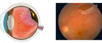
is a severe pathological condition of the eye, in which the neurosensory part of the retina, responsible for vision, peels off from the layer of its cells that is located outside the nerve sheath (from the pigment epithelium). Normally, the retina and choroid of the eye are in close contact with each other.
Detachment is most often caused by tears in the retina. In this case, fluid accumulates in the resulting space, which penetrates from the vitreous body and contributes to retinal detachment.
CAUSES OF RETINAL DETACHMENT
Detachment is the separation of the rods and cones, what we call the neuroepithelium, from the underlying pigment epithelium by the accumulation of fluid between them.
In this case, the nutrition of the outer layers of the retina is disrupted, which leads to rapid loss of vision. The possibility of detachment is due to the structural features of the retina, I wrote about this in previous posts.
Retinal detachment by type can be dystrophic (rhegmatogenous), traumatic and secondary. Secondary is not considered as an independent clinical form, but is only a complication of the underlying eye disease - inflammation, tumor, vascular or congenital diseases.
The cause of rhegmatogenous (regma - break) retinal detachment, or, as they say, primary detachment, as is already clear, is a rupture or ruptures of the retina. As a rule, the rupture occurs somewhere in the periphery, close to the equator of the eye, in the area of thinning and dystrophy.
Types of dystrophies that are dangerous in terms of detachment have already been mentioned in earlier posts:
- “Flying midges” and “glass worms” in the eyes, or where do “broken pixels” in the vitreous body come from?
- How to “sew up” the retina and is it necessary to do it?
The names of the dystrophies are non-standard: “Lattice dystrophy”, “snail track dystrophy”, “traction”, “frost-like”, “white without depression”, “perforated ruptures with and without a cap”, “valvular ruptures” and others.
This is what dystrophic lesions that require laser coagulation look like (before and after the procedure).
Complications and consequences
Complications after surgery are rare, but they do occur. This is mainly due to a violation of the technique of the operation. The most common consequences include:
- Deterioration in the transparency of the cornea due to its drying out.
- Burns of the cornea and iris.
- Perforation or rupture of the membranes of the eye.
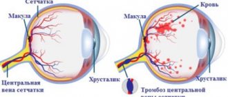
- Hemorrhages in the tissue of the eye. This is observed when the vorticose vein is damaged or when large vessels in the optic organ are damaged.
- Entry of filling material under the retina or under the conjunctiva.
- Strabismus or ptosis is observed when the tissue is stitched incorrectly.
- Cutting seams when applying a filling. Most often this is due to the sutures being placed very close to the place of filling.
- Poor blood circulation in the tissues of the eye. This leads to loss of light perception. Basically, this condition is short-term.
Some time after the operation there may be a relapse. This is due to incorrect surgical technique or incorrect suturing. After surgery, patients are registered with an ophthalmologist and are examined every 2-3 months.
PREVENTION OF RETINAL DETACHMENT
I started with prevention deliberately, because a timely preventive procedure can reduce the risk of detachment by an order of magnitude.
Since previous posts described in detail how this happens, I want to draw your attention to some points.
1. We say that, as a rule, laser coagulation is not a painful procedure, but there is always an adjustment for individual sensitivity. In some cases it can be painful, and in those who are especially impressionable, even very painful. The following factors play a role:
— the volume of coagulation and localization of zones, — the type and model of the laser, — the patient’s position and behavior during it, — the anatomy of the palpebral fissure (“deep-set” eye, large nose, etc.) — the experience of the laser surgeon and the correct choice of contact lens.
Conclusion: if you have a high pain threshold, are afraid or feel uncomfortable during the procedure, be sure to notify the doctor in advance or during it; with the help of medications, we can significantly facilitate the laser coagulation process.
Contraindications to surgery and risk groups
Each type of surgical intervention has a number of contraindications. Vitrectomy cannot be done in the following cases:
- If there is clouding of the cornea.
- If there are pronounced pathological changes in the tissues of the visual organ.
The filling operation can also not be performed in all cases. It is not done if there is clouding of the vitreous body or protrusion of the sclera.
There are also a number of contraindications to laser treatment. Laser operations cannot be performed in the following cases:
- If the retina is detached over a large area.
- If certain tissues of the eye are opaque.
- If pathologies of the iris vessels are diagnosed.
Any types of surgical interventions are contraindicated if a person is intolerant to painkillers or does not tolerate anesthesia well. It is unacceptable to perform operations for infectious and inflammatory diseases of any etiology in the acute stage.
Before choosing a treatment method and surgery, the doctor carefully studies the patient’s medical record and conducts a detailed examination.
DIAGNOSIS OF RETINAL DETACHMENT
The process of optical coherence tomography of the retina (OCT) in our clinic
Diagnosis of retinal detachment includes, first of all, a complete ophthalmological examination with testing of vision, intraocular pressure, examination of the fundus in various ways - contact and non-contact. Inspection in both vertical and horizontal positions may also be necessary.
Special additional diagnostic methods are:
1. Perimetry. A common symptom of detachment is a “veil” or “curtain” in front of the eye, it looks like this:
2. Ultrasound scanning in 2- or 3-dimensional mode. Allows you to determine the detachment through the opaque optical media of the eye or areas inaccessible to inspection... You can determine its height, the contents under it, the relief, the thickness of the membranes - this is important.
3. Electrophysiology - like an eye cardiogram - records electrical potentials from the functioning areas of the retina, indicating the degree of its damage.
4. Optical coherence tomography – we obtain linear sections of the retina to determine the anatomical parameters of the smallest sections in the central and areas close to it. The result can be a two- or three-dimensional image.
5. X-ray CT scan of the retina - provides visualization of the structures of the eye, down to the smallest details.
6. MRI - shows the degree of impairment and makes it possible to create a three-dimensional image of the eye.
SYMPTOMS OF RETINA DETACHMENT
Comparing the eye with a camera containing film, we can say that somewhere on the edge of the frame a scratch has appeared on the emulsion layer. So what of this, you say, because almost the entire frame and most importantly - the center of the “composition” - are still clearly visible. It turns out that this is not entirely true. Fluid begins to penetrate through the gap, flowing under the retina and thereby peeling it off from the underlying choroid. On photographic film, it looks as if the emulsion layer around the scratch begins to swell with bubbles and peel off from the substrate. At this moment, a person sees a rather characteristic picture of a “gray curtain” at the edge of his field of vision. Depending on the location of the gap, the “curtain” can either quickly (over several tens of hours) spread, covering the entire field of view, or creep more gradually (over weeks, and in some cases months) to the central part of the field of view.
Vision during the development of retinal detachment
Quite characteristic of fresh retinal detachment is the symptom of “morning improvement”, when a person in the morning (after a long sedentary lying position) discovers a significant improvement (reduction of the curtain, its paleness and the ability to see through it). By lunchtime it gets worse again, and in the evening it gets even worse.
It is clear that if the rupture is located in the upper parts of the eye, then the liquid quickly falls down and peeling occurs rapidly. If the gap is located below, then the detachment slowly “creeps” upward, and progression will be slower. However, in this case, adhesions between the retinal zones and scars will be more pronounced - there will be more time for their formation.
TREATMENT OF RETINA DETACHMENT
Treatment is necessary, and only surgical, there is no other way out.
No drops, ointments, tablets, injections, or absorbable agents help, but only take time, which allows the detachment to develop further and further. The earlier competent surgical treatment is carried out, the better results it gives and the more vision can be restored. The goal of surgical treatment was formulated more than 100 years ago and is to close (block) the retinal tear. At the initial stage of the disease, there is usually no need to go inside the eye, and surgery consists of local external indentation in the projection of the rupture. For this purpose, special fillings made of soft silicone are used, which press against the area of the rupture, thus blocking it.
Scheme of episcleral filling
As soon as the hole in the retina closes, everything miraculously improves, the “curtain” disappears, and vision begins to be restored. Peripheral vision is restored first; the person discovers that the “vision” is almost normal; later it actually becomes normal. The periphery of the retina is quite stable, and as soon as it returns to its anatomical place, it immediately begins to “work” and recovers well even with long-term retinal detachment. With central vision, things are not so simple. The most favorable cases are when the detachment has not had time to “crawl” to the center. For example, if vision in the center remained 1.0, and half of the field of vision was already covered by a “curtain,” after a successful operation, vision can be restored to 1.0, and the curtain will disappear.
If the detachment has managed to close the central zone, after a successful operation, central vision, unfortunately, cannot be fully restored. What visual acuity will be after surgery in this case depends on a number of factors. The most important of them are the time during which the central zone of the retina was detached, and the state of the blood supply to the retina, which directly depends on age and the degree of myopia (if any).
Recovery of central vision occurs slowly and is usually almost complete by 3 months. In the future, improvement may continue, but at an even slower pace, and we observe that both after a year and after 3 years, visual acuity improves slightly.
Changes in the structure of the retina after detachment, worsening the “picture”
If a person with a retinal detachment is not operated on in time or is operated unsuccessfully, then the detachment persists and continues to develop, in addition, the so-called “proliferative process” begins in the vitreous body.
The eye, as you know, has the shape of a ball, and we already know that it has a lens, a photographic retina, and in addition, the inside of the eye is filled with liquids. These liquids are almost 98-99% water, but with very significant additives. The anterior part of the eye is limited by the cornea on one side and the iris-lens unit on the other. This part of the eye is more responsible for optics and is filled with anterior chamber intraocular fluid. In its properties and appearance, it is almost no different from simple water with the addition of a complex set of minerals and salts.
Another thing is the fluid in the posterior part, limited by the lens, ciliary body and retina. This fluid is called vitreous humor and has the consistency and appearance of a gel or frozen jelly. In addition, the vitreous body is based on a frame in the form of a three-dimensional lattice of collagen fibers.
In case of retinal detachment, the vitreous body never remains indifferent. In the initial period, only slight disturbances in its structure are observed, manifested in the form of various inclusions floating in the field of view. With a long-term detachment, strands develop in the vitreous frame, which, like ropes, are attached to the surface of the retina and, slowly contracting, pull the retina to the center of the eyeball. This process is called vitreoretinal proliferation, which ultimately leads to the formation of the so-called “funnel-shaped” retinal detachment. In such a situation, reconstructive surgery is required, the quality of which is of a much higher level. It is almost impossible to close such a gap with fillings, and it is not enough. The main task is to cleanse the surface of the retina from vitreous cords, straighten it, and then block the tear.
Picture of a severe form of proliferative retinopathy.
For this purpose, special methods are used, the so-called vitreoretinal surgery. Its essence lies in the fact that through pinpoint punctures with long and thin instruments, the surgeon enters the inside of the eye and removes the strands, freeing the retina and straightening it. The process itself is very reminiscent of the painstaking work of a master who, through the neck of a bottle with long tweezers and scissors, assembles a model of an 18th-century sailing ship inside the bottle.
Schematically, this is what a vitrectomy operation looks like
. This operation is very delicate and complex, if you remember that the retina is extremely delicate and fragile nervous tissue, and almost every part of it is responsible for some area of vision. During the operation, the doctor looks inside the eye through its anterior segment, looking “through the pupil.” This requires high transparency of the optical media, that is, the lens-cornea and lens must be as transparent as possible. If the lens is cloudy, that is, the patient has a cataract, then, as a rule, at the initial stage, the lens is replaced with an artificial one, and only then the “repair” of the retina begins.
We discussed how this is done in these posts:
- Cataract: it awaits you personally (if you live, of course)
- We implant an artificial lens (you will need this after 60 years)
In addition, the natural lens, due to its anatomical location, often interferes with working on the peripheral parts of the retina.
In these cases, it is also necessary to change the lens to an artificial one, otherwise the uncleaned areas of the peripheral retina may not allow it to achieve its anatomical fit. The operation is performed in a “dark room”, that is, only a light guide in the surgeon’s hand or an additional light source “chandelier” illuminates the working field like a chandelier, the microscope light is turned off.
Externally, it happens like this (photo from the operating room)
After complete cleansing of the retinal surface from the vitreous strands, it must be straightened and placed on the choroid, that is, its anatomically correct position inside the eye must be obtained.
For these purposes, so-called “heavy water”—a liquid perfluoroorganic compound (PFOS)—is often used. This substance in its properties is almost no different from ordinary water, but due to its higher molecular weight it acts like a press on the surface of the retina, smoothing and pressing it. “Heavy water” copes with detachment very well, in addition, it is absolutely transparent, and the eye filled with this liquid begins to see almost immediately. The main disadvantage is that the eye cannot tolerate it for a long time. A maximum of a couple of weeks, but in practice it is not advisable to leave this liquid in the eye for more than 7-10 days. This means that immediately after straightening the retina, it is necessary to close, “seal” all the breaks in the retina, so as not to get a detachment again after removing the “heavy water”.
Recovery period
The recovery period depends on the chosen operation:
- If laser coagulation was used , then in the postoperative period it passes without visible complications, and the patient is prescribed: Gymnastic exercises for the eyes.
- Avoid physical activity and sports for the first month.
For the first two or three days, wear a blindfold (blindfold).
- Visiting the bathhouse, sauna, solarium.
The minimal recovery period occurs after laser surgery. In other cases, it can last from a week to a month.
After surgical treatment of retinal detachment, the majority of patients (approximately 80% of all operations performed) experience positive dynamics, vision is restored, and the unpleasant symptoms of flashes and sparks before the eyes are eliminated.
Complete restoration of visual function can occur within 2 or 3 months after surgery. At this stage of rehabilitation, the patient is prescribed glasses.
The result of the operation is also affected by the area of retinal rejection; if the macula is involved in this process, then complete restoration of vision is impossible.