The disease is caused by dystrophic and degenerative disorders in the drainage zone of the limbus. These changes are always found to some extent in glaucoma. It is they who cause the low capacity of the drainage system of the eye, which is responsible for the outflow of fluid.
Open-angle glaucoma is considered milder than closed-angle glaucoma. It is less dangerous, more treatable and has a favorable prognosis.
Risk factors for developing open-angle glaucoma are:
- race (representatives of the Negroid race are 2-3 times more susceptible to glaucoma);
- elderly age;
- diabetes;
- arterial hypotension;
- early farsightedness;
- disorders of glucocorticoid metabolism;
- pigment dispersion syndrome.
The earliest stages of open-angle glaucoma are characterized by thickening of the trabecular plates, narrowing of the intrabecular fissures and Schlemm's canal. At first, these changes are minimal, but as the pathology develops, the trabecula degenerates, leading to complete closure of the cracks and overgrowth of Schlemm's canal and most of the collector canals.
Constantly increased intraocular pressure leads over time to secondary degenerative changes - a functional block of the venous sinus of the sclera (displacement of trabeculae towards the outer wall of Schlemm's canal). The degree of this complication largely depends on the anatomical features. Eyes with an anterior location of the venous sinus of the sclera, with weak development of the scleral spur and a posteriorly displaced ciliary muscle are more predisposed to sinus block.
Organic changes in glaucoma are always associated with vascular, endocrine and nervous disorders. Therefore, a significant predisposition to glaucoma is observed in diabetes mellitus, hypertension, and lesions of the subtubercular region.
In addition, the factor of genetic inheritance is important. Because it determines the structure of the eye, open-angle glaucoma often runs in families.
open-angle glaucoma
The danger of glaucoma is that its early stages do not manifest themselves in any way. With increased intraocular pressure and associated functional disorders, a person does not have vision problems. Only when organic changes develop does visual acuity begin to decline. This occurs in advanced and advanced stages of glaucoma, which are difficult to treat.
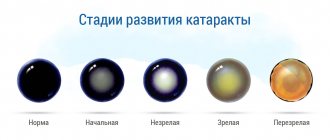
Based on symptoms and subjective sensations, glaucoma can be confused with incipient cataracts. Differential diagnosis should first of all include monitoring of intraocular pressure. If it is normal, glaucoma is less likely. The diagnosis of “cataract” is also confirmed by the fact that when examined in transmitted light, the pink glow of the pupil is weakened, against which zones of opacification (spots, streaks, stripes) are visible.
The following clinical picture is typical for glaucoma:
- increased IOP;
- hemorrhages on the optic nerve head may be detected;
- vision progressively narrows from the side of the nose;
- the pupil glows bright pink in transmitted light;
- the optic disc is colored gray;
- the vessels at the edges of the optic nerve are bent (glaucomatous excavation);
- the presence of pericapillary atrophy;
- pale neuroretinal ring against the background of shallow excavation of the optic disc;
- there are gliosis-like changes in the retina;
- Vascular changes in the conjunctiva are possible.
Glaucoma can occur against the background of normal cerebrospinal fluid pressure in the retrobulbar portion of the optic nerve. In this case, there are chronic disturbances in the blood supply to the optic nerve head (microthrombosis, venous discirculation) and acute conditions associated with hemodynamic disturbances (vascular spasms, drop in blood pressure at night, hemodynamic crises).
Acute-angle glaucoma - what is it?
Acute-angle glaucoma Source: Linza.Guru Acute-angle glaucoma develops as a result of disruption of the drainage system of the eye.
This leads to an increase in pressure in the anterior chamber and the development of a typical glaucomatous attack with a characteristic clinical picture. The prevalence of acute-angle glaucoma is higher among women of post-menopausal age due to changes in hormonal balance and an increase in degenerative changes in the eyes. This is confirmed by clinical studies of ophthalmologists, who revealed damage to the trabecular meshwork and a high frequency of glaucoma attacks in 72% of examined women with signs of this disease.
Overlapping of the trabecular meshwork occurs as a result of displacement of the lateral edge of the iris (pigment) of the eye, its layering on the lens, or as a result of organic diseases of the visual apparatus. The causes of acute-angle glaucoma are:
- uveitis;
- hypervascular iridocyclitis;
- proliferative keratitis;
- granulosa conjunctivitis;
- cataract;
- eye injuries.
Hereditary factors play an important role in the development of glaucoma symptoms; such a predisposition negatively affects the overall prognosis of the disease. A severe, steadily progressive course of open-angle glaucoma with a rapid increase in symptoms negatively affects the patient’s health and requires early surgical treatment to prevent irreversible vision loss.
Acute-angle glaucoma is characterized by a gradual increase in the intensity of attacks, headaches and an increasing change in visual acuity. The pathology is dangerous due to complications, severe disability of patients, and blindness.
Acute-angle glaucoma develops as a result of destructive-dystrophic damage to the drainage system, leading to disruption of the outflow of intraocular secretions. An increase in pressure in the anterior chamber of the eye provokes atrophy of soft tissues, an increase in apoptosis and a deficiency of cellular respiration.
This provokes depression of the optic nerve head, retinal detachment or microhemorrhages. The pathology is accompanied by severe attacks and low effectiveness of drug treatment.
What causes the disease and acute attack
There are common reasons that lead to pathology:
- anatomical features: small diameter of the cornea, eye length less than 25 mm, enlarged lens;
- age-related changes;
- race (more often among Asians);
- hereditary factor (presence of the disease in close relatives);
- various diseases with vascular damage;
- hormonal imbalance (premenopausal women).
There are factors that need to be feared because they are the ones that can provoke the acute attack that we are so afraid of:
- taking certain medications (such as tranquilizers);
- long position in an inclined position;
- sudden stress or emotional overexcitement;
- Even being in the dark for a long time can lead to an attack.
Of course, these conditions may not harm everyone, only those who have already experienced any changes in the structures of the eye.
Classification of the disease
Glaucoma is the concept of several cumulative eye lesions leading to blindness, a group of interrelated diseases. There are about 60 varieties. All of them are combined into several common features:
- Periodically or constantly increased intraocular pressure, above the tolerant level.
- Deterioration of blood supply to eye tissue (ischemia).
- Insufficient supply of oxygen to parts of the eye.
- Development of damage to the optic nerve, characteristic of glaucoma: optical neuropathy. This leads to optic nerve atrophy.
- Visual impairments characteristic of glaucoma.
The disease itself can occur at any age, even be congenital. However, as a rule, the disease manifests itself most often after the age of 60. This disease is the main cause of all leading to incurable blindness. It ranks first in this regard and has a high value.
Glaucoma varies in degree, type, type, stage. There are many classifications of glaucoma. From all this diversity, four forms of the clinical course of primary glaucoma have been identified: open-angle, closed-angle, mixed, and a disease with normal pressure inside the eye.
All of them come in different stages, and the effectiveness of treatment depends on the stage of the identified disease. Let's talk about closed-angle glaucoma, previously known as acute-angle glaucoma or narrow-angle glaucoma. The initial stage of development of angle-closure glaucoma was previously called cornerstone glaucoma.
It received its modern name in 1975 at the All-Union Forum of Ophthalmologists. It was then that the following were classified:
- Acute-angle glaucoma or angle-closure glaucoma, in which the pressure inside the eye increases due to the closure of the anterior chamber angle.
- Open-angle glaucoma, in which the drainage system of the eye is affected.
- Mixed glaucoma, when there are 2 mechanisms of the disease.
All these processes can be stabilized or unstabilized. In the first case, the disease does not progress quickly and is treated with a positive result. In the second case, the behavior of the disease is unstable, sudden improvements and deteriorations in the course of the disease are possible, and it is difficult to treat.
Causes of PACG
Factors predisposing to increased pressure inside the eye and the development of angle-closure form of pathology are:
- heredity;
- nervous overstrain, overwork, stress;
- refractive errors: development of farsightedness, myopia;
- traumatic injuries, bruises, eye burns, postoperative conditions;
- rheumatic diseases;
- diabetes;
- bronchial asthma;
- hypotension or hypertension with crises;
- allergic conditions;
- oncological neoplasms of the eye.
Forms of glaucoma
There are several forms of glaucoma. The most common is primary open-angle glaucoma (POAG). Forms of glaucoma such as angle-closure, normotensive, congenital, pigmentary, secondary and others are less common.
Primary open angle
Chronic glaucoma, also known as primary open-angle glaucoma (POAG), is often called the “silent thief of vision” because it develops asymptomatically. The pressure in the eye slowly increases, and the cornea adapts to this without any protrusion.
Therefore, most often the disease goes unnoticed. If there is no pain, then the patient usually does not even suspect that he is slowly losing vision until the disease reaches a later stage. However, vision gradually deteriorates and the damage becomes irreversible.
In open-angle glaucoma, there is an imbalance between the production and outflow of clear fluid (aqueous humor) filtering through the anterior chamber of the eye. This can occur if the ciliary body produces very large amounts of this moisture or the drainage channels in the anterior chamber are blocked, causing increased intraocular pressure.
As a result of increased IOP, pressure increases on the fibers of the optic nerve, which transmit visual images to the brain. This leads to a deterioration in blood supply, depriving tissues of oxygen and nutrients. Over time, high blood pressure leads to irreversible damage to the optic nerve and loss of vision.
However, more than 2/3 of patients with increased intraocular pressure (more than 21 mm Hg) do not experience loss of visual fields or widening and deepening of the optic disc excavation. This condition is called ocular hypertension.
Cause of increased pressure
It is known that factors in the development of open-angle glaucoma are trauma, uveitis, and treatment with steroid drugs. While steroid therapy of any kind can increase intraocular pressure, topical and parabulbar steroids are more likely to increase intraocular pressure.
POAG is a chronic disease that can be inherited. There is currently no treatment for this pathology, but its progression can be slowed down or stopped. Due to the lack of symptoms, many patients find it difficult to understand the need for lifelong use of expensive drugs, especially when taking these drugs is burdensome and has a lot of side effects.
As with other forms of glaucoma, treatment includes anti-glaucoma eye drops. Laser or other surgical treatments may also be recommended as a way to lower IOP.
Taking prescribed medications regularly is extremely important to prevent vision-threatening damage. Therefore, it is important for the patient to discuss side effects with the doctor in order to choose the most suitable drug for himself.
Closed angle
At the front of the healthy eye, fluid (aqueous humor) builds up and flows out of it, creating enough pressure to maintain the correct shape of the eye without damaging it. That is, the amount of new intraocular fluid that is constantly formed in the eye is balanced by the amount that just as constantly flows out of it through a certain place called the “angle of the anterior chamber.”
It received this name due to the fact that in this place the iris is directly adjacent to the cornea. In fact, the drainage function in the eye is possible at an angle of at least 30 degrees.
If the angle is blocked, fluid will continue to be produced at a normal rate, but will not be able to drain from the eye, causing the pressure to reach dangerous levels. This is the mechanism by which one type of glaucoma develops, known as angle-closure (narrow-angle) glaucoma.
At an angle of 15 degrees, there is a very small anterior chamber between the iris and cornea and a small space between the iris and Schlemm's canal. The reasons for the abnormal location of the iris in narrow-angle glaucoma may be the following:
- pupillary block
- flat iris (Iris plateau syndrome)
- narrow anterior chamber angle
- tumors and other causes
intraocular fluid is produced by the ciliary body, which is located behind the iris. As a rule, it easily flows through the pupil into the anterior chamber of the eye. But if the lens is tightly attached to the back surface of the iris, this outflow is blocked. The fluid remaining behind the iris then pushes it forward until the anterior chamber angle closes.
in this condition, the iris is attached to the ciliary body too close to the trabecular meshwork involved in the outflow of aqueous humor. When the pupil dilates, the resulting folds of the peripheral part of the iris in the angle of the anterior chamber can close this network, causing a rapid increase in IOP. This type of exacerbation of narrow-angle glaucoma can be caused by excessive pupil dilation due to various reasons.
when the length of the eye is less than normal (which is the cause of farsightedness), the anterior chamber is shallow and, accordingly, its angle is also less than normal. This increases the risk of developing angle-closure glaucoma when the pupil dilates or with age-related changes.
A tumor located behind the iris, swelling associated with inflammation of the ciliary body (uveitis), and changes in the shape of the eye after surgery for retinal detachment can also cause angle-closure glaucoma.
Prevention
It is impossible to protect yourself 100 percent from angle-closure glaucoma. If you have a predisposition, then follow the general rules:
- avoid stress;
- eat right;
- do not overload the body with weights and intense training;
- do not be in a completely darkened room;
- avoid bright light;
- do not watch TV in a dark room;
- do not drink a lot of water at one time;
- visit an ophthalmologist regularly for preventive care;
- treat somatic diseases.
The causes, symptoms, treatment and prevention of angle-closure glaucoma are determined by a specialist. PAH cannot be self-medicated, as there is a high risk of developing blindness.
Additionally, read the article about contraindications and restrictions for glaucoma.
Tell us what you know about angle-closure glaucoma? Share the article with your friends. Take care of your eyesight and visit your doctor on time. Be healthy.
Reasons for appearance
As is known, the anterior chamber of the eye is limited in front by the cornea, and in the back by the iris and lens. Acute-angle glaucoma develops in those patients who have an anatomically small anterior chamber, especially in the periphery, since it becomes even smaller as a person ages.
People suffering from farsightedness are more prone to this type of glaucoma, since they tend to have smaller eyes. A person's predisposition to angle closure is determined by examining it with a slit lamp.
The condition of the organ of vision is best determined by gonioscopic examination of the anatomical structure of the corner of the eye; it is recommended to use special corneal contact lenses.
Intraocular pressure increases when the root of the iris of the eyeball moves forward and blocks the trabecular network of fluid outflow pathways. Intraocular fluid is continuously produced in the organ of vision. It flows through channels located in the corner of the anterior chamber.
The upper limit of pressure inside the eyeball is 20 mm. Hg Art. If the outflow tract is obstructed, intraocular pressure rises to fifty or sixty millimeters of mercury within a few hours.
Most often, symptoms of intraocular hypertension initially appear in only one eye, despite the fact that pathological changes may also occur in the second eyeball. If the pressure inside the organ of vision increases too quickly, then there is redness of the eyeball, blurred vision, pain around and inside the eye, severe headache, nausea and vomiting.
Before experiencing blurred vision, most patients see lights and areolas around them. The cause of this phenomenon is corneal edema. During an acute attack of acute-angle glaucoma, the eyeball affected by the pathological process becomes more sensitive and hard on palpation than a healthy one.
As intraocular hypertension continues to increase, nausea and vomiting become so severe that doctors sometimes suggest acute abdominal surgery or food poisoning.
Irreversible vision damage can occur rapidly, over a period of time not exceeding seventy-two hours. This depends on the degree of increase in intraocular pressure. An attack of acute-angle glaucoma is a critical condition that requires urgent consultation with an ophthalmologist.
“Risk groups” for glaucoma include:
- people over 60-70 years of age who do not even have eye complaints;
- people over 40 years of age who:
- intraocular pressure is in the upper limit of normal;
- the difference between the intraocular pressure of the right and left eyes is more than 5 mmHg. Art.;
- the difference between intraocular pressure measured in the morning and evening is more than 5 mmHg. Art.;
- people with a high degree of myopia after 40-50 years, with a high degree of farsightedness (especially women after 50 years);
- people with increased intraocular pressure, regardless of age;
- people with low (relative to age norm) blood pressure;
- people with diabetes, endocrine, nervous and cardiovascular diseases;
- people who have suffered eye injuries, inflammatory diseases (uveitis, iridocyclitis, etc.) of the eyes, eye surgery;
- relatives (including distant ones) of patients with glaucoma with similar structural features of the eye;
- people undergoing a long course of treatment with hormonal drugs.
Glaucoma can occur at any age, but the disease most often develops in older people.
Treatment methods
If you find yourself or someone around you experiencing the onset of an acute attack of the angle-closure form of the disease, then immediately call an ambulance.
Tactics during an acute attack of PAH
First aid for an acute attack of angle-closure glaucoma:
- Avoid situations that dilate the pupil. That is, you cannot cover your eyes with a bandage, you cannot be in a darkened room.
- Instill Pilocarpine drops every 15 minutes.
- Apply a compress to your shins and immerse your feet in hot water. This is required to reduce pressure.
During an acute attack of PAH, the main goal is to reduce IOP. For this, medications are prescribed: Timolol drops, Pilocarpine, Acetazolamide and Mannitol injections. To relieve pain, painkillers or injections "Analgin" may be prescribed, and "Metoclopramide" for nausea and vomiting.
Find even more drops here.
Treatment after the attack has stopped
After the acute attack is over, surgery is performed to create pathways for the outflow of aqueous humor. Artificial pathways normalize the “synthesis-outflow” balance of aqueous humor, leading to stabilization of IOP.
Operations for angle-closure glaucoma:
- Laser peripheral iridotomy. The doctor uses a laser beam to create small holes in the iris.
- Laser cyclocoagulation is the “cauterization” of areas of the ciliary body with a laser. Defects form through which aqueous humor can flow out.
- Surgical iridectomy. The essence of the intervention is to remove part of the trabecular tissue.
During the interictal period of angle-closure glaucoma, treatment consists of following preventive measures and using eye drops: Okumol, Timolol, Dorzopt.
In addition, watch a video tutorial on how laser glaucoma surgery works:
Risk factors
The occurrence of acute-angle glaucoma is due to the peculiarity of the anterior segment of the eye and, in particular, the structure of the angle. An acute, beak-shaped, closed angle occurs when the size of the lens is relatively large, as a result of which the iris turns out to be more moved anteriorly, towards the cornea.
This angle creates a certain obstacle to the path of intraocular fluid into the drainage network. And due to the fact that the main amount of fluid flows through it, the slowdown at this stage of outflow leads to an increase in intraocular pressure.
An anomaly in the structure of the anterior chamber angle in acute-angle glaucoma is directly related to the following factors:
- Senior and elderly age (in addition to degenerative processes of a general nature, with age a thickening of the lens occurs, which increasingly presses the root of the iris to the drainage network, narrowing the angle of the anterior chamber).
- Female gender (acute-angle glaucoma affects women 2 times more often).
- Nationality (in Asians, due to the anatomical structure of the eyelids, acute-angle glaucoma is more common).
- Farsightedness (wearing plus-value glasses since youth).
- Disturbances in the central and peripheral circulation.
- Small anterior chamber.
In the early stages, acute-angle glaucoma, as a rule, has no characteristic symptoms: normal vision remains, pain and other changes in well-being are absent. Sometimes patients may complain of the temporary appearance of rainbow circles before the eyes and asthenopia.
An undetected disease for a long time subsequently leads to deterioration of peripheral vision, when a person clearly sees objects located directly in front of him, but does not notice those located to the side or at an angle.
Initially, the narrowing of the visual fields mainly occurs from the side of the nose, but later, it gradually begins to cover the peripheral parts concentrically, which ends in its complete loss. In addition, patients with acute-angle glaucoma note the appearance of transparent or translucent spots in their field of vision.
Dark adaptation or color perception may be impaired. Sometimes there is an uncorrectable decrease in vision, which indicates an advanced stage of acute-angle glaucoma, with gradual atrophy of the optic nerve. But the most severe manifestation of acute-angle glaucoma is an acute attack of the disease.
An acute attack usually occurs suddenly, without preliminary symptoms. Its immediate causes are not precisely known. The occurrence of an attack may be preceded by emotional overload or stress, prolonged stay in a dark room, the use of drops to dilate the pupil, etc.
Severe form of the disease
In an acute attack of acute-angle glaucoma, sharp pain occurs in the eye and temple area, blurred vision occurs and its sudden decrease. The condition is accompanied by nausea and vomiting, headache, and redness of the eye.
The pupil is dilated. Due to the blockage of the outflow pathways of intraocular fluid by the protruding root of the iris, an uncontrolled, persistent increase in intraocular pressure occurs, leading to irreversible damage to the optic nerve fibers.
Such a situation requires immediate vision saving, and literally hours are counting. Hospitalization to the ophthalmology department and emergency measures are required.
The main causes of glaucoma
As you know, in the eye of an absolutely healthy person a special pressure of approximately 20 mm Hg is regularly maintained. This is possible solely due to the balance on one side of the inflow and, accordingly, outflow of the existing fluid. Subsequently, with the development of glaucoma, this kind of balance inevitably collapses.
Thus, the above-mentioned fluid consistently accumulates, and as a result, intraocular pressure increases. The optic nerve itself and some other nearby tissues begin to experience constant stress, and a disruption occurs directly in the blood supply to the eye itself. As a result of all the processes described above, the optic nerve gradually atrophies, which in turn does not allow signals to enter directly into the human brain. Thus, problems with vision arise, the usual zone of visibility is limited, and as a result, complete blindness may be diagnosed.
Glaucoma, according to qualified specialists, is an irreversible disease . This is why it is so important to start the treatment process in a timely manner. Doctors also pay special attention to the fact that as a result of an acute attack of the disease, instant loss of vision is possible.
Symptoms
But one should not think that this disease is only for older people. There are a number of signs when you need to see a doctor for diagnosis when they appear. Early detection of the disease gives an increasingly greater chance of successful treatment.
A delayed visit to an ophthalmologist can cause complete blindness due to the impossibility of recovery. So, the initial possible symptoms of the onset of glaucoma are:
The disease develops very quickly, so you need to be attentive to your health and, knowing about deviations in the field of vision, you must urgently consult a doctor for treatment.
In the early stages, most cases of open-angle glaucoma are not accompanied by any symptoms or manifestations: normal vision is maintained, there are no pain or other changes in well-being. Sometimes patients may complain about the temporary appearance of rainbow circles before the eyes, the phenomenon of asthenopia.
Since they are not signs specific only to glaucoma, this may lead to underestimation of the condition and, as a result, a delay in diagnosing the disease. However, although there are no symptoms in the early stages of the disease, irreversible damage can occur to the optic nerve.
If glaucoma remains undetected for a long time, the symptoms described below may subsequently appear. The main one is deterioration of peripheral vision. A person sees well straight ahead, but may not notice objects located to the side and at an angle.
Initially, the narrowing of the field of vision occurs mainly from the side of the nose, and later it can concentrically cover the peripheral parts until it is completely lost. It is also possible that a translucent or opaque spot may appear in the field of view.
The patient may notice a decrease in dark adaptation, consisting in deterioration of vision when quickly moving from a brightly lit room to a darkened one, as well as, sometimes, the appearance of color vision disturbances. In some cases, an uncorrectable decrease in visual acuity is observed, which already indicates a severe, advanced stage of the disease, which is accompanied by gradual atrophy of the optic nerve fibers.
The most striking symptoms are observed during an acute attack of angle-closure glaucoma. In this case, the following manifestations of the disease may be detected:
- pain in the eye and headaches radiating along the trigeminal nerve (frontal, zygomatic, temporal regions);
- blurred vision;
- rainbow circles around light sources;
- photophobia;
- redness of the eye;
- nausea and vomiting;
- decrease in heart rate.
It should be noted that often the general symptoms are more pronounced than the ocular ones. Patients are often restless, in some cases they may experience pain radiating to the heart and abdomen, similar to the manifestation of cardiovascular pathology. Slit lamp examination reveals corneal opacity due to edema.
The pupil is greatly dilated, the reaction to light is sharply weakened or absent. On palpation, the eyeball is hard as a stone.
All of the above symptoms of an acute attack of glaucoma require emergency medical attention. If the pressure is not reduced with medication or surgery within the next few hours after the attack develops, the eye faces permanent loss of vision!
Causes of exacerbation
Very often, the causes of acute attacks of the disease can be:
- Extremely high emotional arousal, stress.
- Taking medications that cause pupil dilation.
- Large amounts of fluid consumed.
- Working for long periods of time in an awkward inclined position.
- Long-term use of antidepressants.
- Facial injury.
- Hypothermia of the body.
- Alcohol abuse.
- Frequent overeating.
The general symptoms of acute-angle glaucoma and open-angle glaucoma are almost the same. In either case, the disease affects both eyes, possibly asymmetrically. However, the closed-angle type of glaucoma occurs unevenly, with periods of remissions and exacerbations. It is with acute-angle glaucoma that there are so-called acute attacks.
Acute-angle glaucoma can have several forms: acute, subacute and chronic. The acute form of glaucoma is a serious and dangerous condition, often leading to complete loss of vision. According to statistics, out of 65 million people with angle-closure glaucoma, 7 million patients are completely blind.
A recurrent and complicating cause of acute-angle glaucoma is diabetes mellitus. Persons diagnosed with diabetes should be checked more often by an ophthalmologist, at least once a year. The symptoms of glaucoma are often confused with gastrointestinal diseases, when vomiting and dizziness begin.
Incorrect and untimely diagnosis can deprive a person of his sight. This also needs to be taken into account. Modern diagnostic methods rarely allow this. The main thing is not to waste precious time, during which a complete or partial cure is possible.
We detect the disease at an early stage
Screening and preventive examinations can detect the disease with minimal changes. Especially, such examinations are important for those who have predisposing factors:
- the presence of chronic diseases: diabetes mellitus, cardiovascular pathologies;
- history of eye or head trauma;
- age after 40 years;
- children whose parents suffer from glaucoma.
Diagnostic methods:
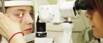
- the easiest way to identify changes is to examine the fundus; an experienced specialist may suspect a diagnosis after this simple manipulation;
- tonometry is a method of measuring intraocular pressure, carried out quickly within 1 minute and painlessly.
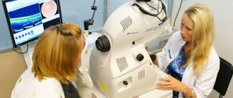
- Computed tomography is one of the most modern and effective methods. Allows you to assess the condition of all eye structures simultaneously;
- perimetry is a diagnostic that reveals whether there is a narrowing of the visual fields or their loss.
Correct and timely diagnosis will help you preserve your vision and avoid serious consequences.
You may be interested in: What is acute-angle glaucoma and how to treat it effectively
Diagnostics
There are several methods for diagnosing acute-angle glaucoma:
- Tonometry. While lying down, the pressure inside the eyes is measured. The higher the range of pressure fluctuations, the greater the likelihood of developing a disease. If the pressure constantly exceeds the norm, then we may be talking about an incipient disease.
- Elastometry. Pressure is determined using several tonometers of different weights. Make a graph and determine the value using the Maklakov method. If everything is in order, the formed abscissa will be straight, otherwise it will be a curve.
- Electronic tonography. It determines the hydrodynamics of the eye. All manipulations are performed electronically, the outflow coefficient and the Becker aqueous humor coefficient are calculated.
- Perimetry. Study of the boundaries of the visual field of the person being diagnosed. These parameters are measured at the slightest suspicion of glaucoma. This diagnosis has 2 types: isopperimetry and campimetry. With the help of the latter, the presence of disturbances in the visual field in the center is detected. This is a very important method for diagnosing glaucoma.
- Gonioscopy. This is an examination of the structural process of the anterior chamber angle, precisely the place where the cornea of the eye smoothly passes into the membrane of the white. This diagnosis is made using a special lens. Using this method, it is possible to determine the condition of the trabecula and the positive outcome of possible surgical intervention, including using laser devices.
- Additional diagnostic measures are: examination of the fundus, programming of the disc funnel, changing the retinal zone and other methods.
To correctly assess the quality of the optic nerve and adjacent areas, various methods of laser ophthalmic tomography and radiography are used.
Diagnostic measures
In order to determine the presence of this disease, measuring intraocular pressure alone is not enough. The specialist must examine the fundus itself, as well as the optic nerve head, in as much detail as possible. Thus, it becomes clear that extensive diagnostic measures are usually required to confirm the diagnosis.
In most modern clinics, specialists conduct a diagnostic examination using a whole complex of special computerized equipment, which means the following:
- determination of field of view;
- measuring pressure inside the eyeball;
- refraction assessment (this is the ability of the optical system itself, thanks to which it can refract all rays of light);
- Ultrasound;
- determination of the size of the lens and the anterior chamber itself;
In addition, the patient must undergo perimetry and determination of visual fields.
Treatment
Source: Glaza.guru Therapy (treatment) begins with a gradual decrease in eye pressure with the help of ophthalmohypotensive drugs that accelerate the outflow of intraocular secretion by the drainage system of the eye. The drugs have a number of contraindications and a high risk of side effects due to their powerful hypotensive effect.
In patients with arterial hypotension, the prescription of antiglaucomatous drugs that lower blood pressure is contraindicated due to the risk of fainting. In this case, drugs are prescribed that reduce the production of intraocular secretion, which leads to the creation of a balance between partial physiological outflow and low fluid production.
For hypertension, drugs that lower blood pressure and loop diuretics are prescribed in combination with ophthalmohypotensive drugs. Patients monitor the level of fluid they drink and excrete during the day, limit the consumption of salt and raw smoked foods.
Limiting stress exposure and increased visual load at work reduces the risk of adverse effects on neuromotor function of the eye. If symptoms persist, laser therapy is prescribed. Surgical treatment is carried out with laser or classical tissue excision.
Laser is a priority treatment method for glaucoma due to the possibility of tissue coagulation without scarring in the postoperative period. After surgery, antibacterial drops and antiglaucomatous drug treatment are used in full.
Controlling attacks of glaucoma allows you to avoid the progression of the disease and prevent the onset of irreversible vision loss.
Prevention and prognosis
Glaucoma is one of those diseases that develops faster in the presence of favorable factors, but can occur even in a healthy person. Risk factors include bad habits, especially smoking, because tars have an extremely negative effect on all structures of the eye. The risk of glaucoma increases in people with hypertension and diabetes. Working with your head down for a long time can contribute to the development of the disease.
It is important to periodically visit an ophthalmologist and measure intraocular pressure once a year. People who are at risk need to undergo tonometry twice a year.
Risk group for acute-angle glaucoma:
- older and elderly people (dystrophy of eye structures, thickening of the lens);
- women;
- Asians (features of eyelid anatomy);
- people with farsightedness;
- people with circulatory disorders.
With timely treatment, glaucoma can be controlled, but in most cases the prognosis remains average. It all depends on the characteristics of the disease in a particular patient and the rate of its progression.
For 65 million people with elevated intraocular pressure, 7 million are permanently blind. The insidiousness of the disease lies in the fact that even with regular examinations it is very difficult to detect it at the initial stage. It is important to pay attention to every symptom, even the most minor one.
Sources used:
- Kvasova M. D. Vision and heredity. - Moscow / St. Petersburg: Dilya, 2002.
- Modern ophthalmology. Management. - M.: Book on Demand, 2009.
- Introduction to clinical ophthalmology / E.E. Somov. - M.: Leningrad Pediatric Medical Institute, 1993.
- IU School of Medicine Department of Ophthalmology
Drug therapy
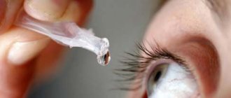
If specialized help is not available at the time of the attack, it is necessary to instill a two percent or four percent solution of pilocarpine four or five times into the conjunctival sac every five minutes. Take 250 mg of diacarb (acetazolamide) and 40 ml of 50% glycerol (at the rate of 1.0 - 1.5 grams per kilogram of body weight) orally.
The doctor's first actions
The patient must be referred to an ophthalmologist and provided with urgent hospitalization. Drug therapy for acute-angle glaucoma in most cases leads to normalization of intraocular pressure.
However, surgical intervention using a surgical laser is considered a radical method of treating the disease. The only effective way to get rid of the disease is laser iridotomy.
The operation consists of making a small hole in the root of the iris with a laser beam. This leads to the fact that the pressure on both sides of the iris is equalized, and the iris does not block the outflow of intraocular fluid.
It is possible to treat the disease using other procedures. The advantage of laser iridotomy is that it can be performed directly in the ophthalmologist's office. The operation lasts several minutes.
If it is performed within twenty-four hours of the onset of an attack of acute-angle glaucoma, that is, before adhesions develop between the iris and the trabecular meshwork, then subsequent attacks can be avoided.
The first signs of acute-angle glaucoma should alert a person. It should be remembered that intraocular hypertension leads to irreversible changes in the organ of vision over a short period of time.
How does glaucoma manifest?
The symptoms of closed-angle and open-angle glaucoma are almost the same, but the former has an uneven course, with periods of improvement and exacerbation. With the angle-closure form, an acute attack is possible.
At an early stage, glaucoma does not manifest itself in any way. Vision can remain normal for a long time, there is no visual discomfort. Sometimes people only notice the appearance of rainbow circles in their field of vision.
The first symptoms of glaucoma:
- blurred vision;
- fogginess;
- the appearance of a glow around illuminated objects;
- pain in the eyes and face;
- headache;
- nausea;
- lacrimation;
- redness and swelling of the eyes.
Since the clinical picture is very blurred, glaucoma is often confused with other diseases, which leads to delayed treatment and greater damage to ocular structures.
The main symptom of glaucoma is impaired peripheral vision. A person sees ahead without problems, but does not notice objects on the sides or at an angle. The narrowing of the field begins from the nose, covering the peripheral parts. Translucent or dark spots may also appear in the field of vision.
Some with glaucoma suffer from reduced dark adaptation, where vision deteriorates when moving from bright light to a dark room. There may also be a disturbance in color perception.
Acute-angle glaucoma can be acute, subacute or chronic. Acute is the most dangerous, as it often leads to irreversible blindness. In the advanced stage, the decrease in visual acuity is not corrected. Atrophy of the optic nerve begins with the loss of certain areas from the field of vision.
Glaucoma surgery
Some patients with glaucoma are treated with surgery to improve the flow of intraocular fluid, thereby reducing eye pressure.
- Laser trabeculoplasty. This surgery is often used for open-angle glaucoma. There are two types of trabeculoplasty: argon laser trabeculoplasty (ALT) and selective laser trabeculoplasty (SLT).
- Laser iridotomy. Laser iridotomy is indicated for the treatment of patients with angle-closure glaucoma or a very narrow anterior chamber angle. The laser makes a small hole about the size of a pinhead in the upper part of the iris and thus improves the outflow of aqueous humor through the angle of the anterior chamber. This hole is hidden by the upper eyelid, making it not noticeable from the outside.
- Peripheral iridectomy.
When laser iridotomy is unable to relieve an acute attack of angle-closure glaucoma or is not possible for other reasons, peripheral iridectomy may be performed. A small section of the iris is removed, allowing intraocular fluid access to the drainage system of the eye. Because most cases of angle-closure glaucoma can be cured with glaucoma medications and laser iridotomy, peripheral iridectomy is rarely used. - Trabeculectomy. During trabeculectomy, a small valve is formed from the sclera (the white tissue that covers the eye). A filter pad, or reservoir, is created under the conjunctiva, a thin tissue covering the sclera.
Once formed, the pad appears as a bulge or bubble on the white part of the eye above the iris, usually hidden by the upper eyelid. As a result, aqueous humor can drain through the valve created in the sclera and collect in the pad, from where it will be absorbed by the blood vessels of the eyeball.Ocular pressure is effectively reduced in 3 out of 4 patients who undergo trabeculectomy. Although regular checkups with an ophthalmologist are necessary, most patients do not need to use eye drops for a long time.
If the newly formed filter channel closes or too much fluid begins to drain from the eye, additional surgery is necessary.
- Surgical interventions using drainage devices (shunt surgery)
During ALT, the laser creates thin, evenly distributed burns in the trabecular meshwork. It does not create new drainage holes, but stimulates the outflow system to operate more efficiently. In SLT, the laser is used at different frequencies to allow operation at low power levels.
In this case, a certain type of cell is affected, and the filtering channels, like a network surrounding the iris, remain intact. SLT may be effective in patients who have not been treated successfully with conventional laser surgery or eye drops.
Even if laser trabeculoplasty is successful, most patients continue to take the medications. For them, this method does not have a long-term effect. Approximately half of those who underwent this intervention again experience an increase in intraocular pressure within 5 years.
Many patients who have had successful laser trabeculoplasty are forced to undergo it again. Laser trabeculoplasty may also be used as a first-line option in those patients who are unwilling or unable to use antihypertensive eye drops.
If trabeculectomy cannot be performed, surgery using drainage devices is usually successful in reducing intraocular pressure. A shunt is a small plastic tube or valve connected at one end to a reservoir (a round or oval plate).
It is an artificial drainage device implanted into the eye through a thin incision. When IOP increases above certain numbers, the shunt redirects aqueous humor into the sub-Tenon's space (under the Tenon's capsule covering the eyeball outside the palpebral fissure), from where it is absorbed into the bloodstream. When everything has healed, the reservoir can only be seen if you lift your eyelid when looking down.
Is it possible to avoid surgery?
Depending on the extent and condition of your eyes, the ophthalmologist will offer 3 treatment options:
- drug therapy;
- laser correction;
- surgical correction, in the form of surgery.
Conservative therapy
If it is possible to identify the disease at the initial stage, drug therapy will have a positive effect. The goal of this treatment is to reduce intraocular pressure using eye drops, oral or even intravenous medications. A decrease in pressure is achieved by increasing the outflow through the drainage system or reducing secretion.
Laser Application
When grade 1 of the disease is missed or not detected, more serious manifestations occur that require laser correction. This intervention is called laser iridotomy.
The procedure is very popular because on average it takes a few minutes. Using a laser, a small puncture is made in the iris, through which fluid can flow freely, helping to reduce pressure.
Other types of laser treatments:
- Peripheral iridectomy - a small area of the iris is cut off and a path is opened for normal drainage of aqueous humor.
- Trabeculectomy is a procedure in which a valve and filter are created. A valve made of scleral tissue allows intraocular secretion to pass into the formed filter, it accumulates it and then sends it into the blood vessels.
The disadvantage of laser treatment is that over time, the holes, filters, and valves become clogged with cells or pigment and drainage is again impaired.
Read on topic: Providing first aid during an attack of glaucoma
Surgical exposure
It involves installing a shunt in the eye. A shunt is a tube that serves to drain fluid when pressure increases. It moves moisture into the blood vessels.
If you are faced with an acute attack of narrow-angle glaucoma, you cannot treat it on your own or with traditional methods. Every minute is precious. You need to urgently call an ambulance or see an ophthalmologist. Vision that is lost during an attack cannot be restored; no methods can cope.
Still, people will always turn to traditional medicine, seek treatment with folk remedies, despite all the recommendations of doctors. Therefore, we will try to list herbs that will help improve the situation, but only on the condition that they will be used as additional therapy:
- Eyebright is rich in microelements and essential oils. Has an antispasmodic effect, relieving pain.
- Blueberry extract has an antioxidant effect that may protect the optic nerve from further damage.
- Ginkgo biloba - due to its composition of flavonoids, improves blood microcirculation and reduces tissue hypoxia.
This also includes the use of various dietary supplements that help the main treatment: vitamin C, fish oil, omega-3 polyunsaturated acids, multivitamins and microelements.
Causes of angle-closure glaucoma
Anatomical and functional changes lead to the appearance of pathology. Anatomical factors - causes of glaucoma - include:
- short anterior-posterior axis of the organ of vision (small size);
- farsighted refraction;
- shallow anterior chamber of the eye and its narrow angle;
- large lens, including due to swelling due to cataracts;
- increase in the volume of the vitreous body.
Angle-closure glaucoma also occurs for functional reasons: due to pupil dilation, increased secretion of aqueous humor, or intraocular vessels overflowing with blood.
Causes of the disease
Risk factors for developing the disease include myopic refraction (myopia), age over 60-65 years, diabetes mellitus, heredity, thyroid disease, hypotension, and diseases of the nervous system.
The reasons for the development of secondary glaucoma may include:
- eye tumors
- surgical interventions on the eyes
- contusions, wounds and burns of the eyes
- dystrophic eye diseases (consequences of hemophthalmos, progressive atrophy of the iris, etc.)
- cataract
- lens dislocation
- inflammatory eye diseases (scleritis, uveitis, keratitis)