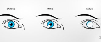
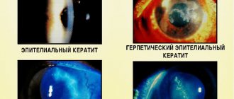
Causes and types of corneal damage
The cornea of the eye is very thin and practically unprotected, except for the movable eyelid. It can be damaged accidentally or intentionally, as a result of an accident or failure to follow safety precautions when wearing contact lenses and glasses.
The main causes of corneal damage are divided into external and internal. External ones occur due to foreign objects, substances, thermal or wave effects entering the eye. Internal - due to congenital or acquired diseases that affect the condition of the cornea and provoke a violation of its integrity.
External causes of corneal damage are considered the most numerous and varied. They are divided into several groups depending on the type of effect on the cornea:
- Mechanical injuries of the cornea
. They are sharp and dull. An acute violation of integrity is characterized by the fact that a foreign object penetrates through the layers of the cornea and reaches the vitreous body and the internal structures of the eyeball. Most often this is a scratch, puncture or cut of the cornea. Blunt eye injuries are expressed by a bruise of the cornea of the eye, which is not accompanied by radical dissection of the tissue. - Thermal damage to the cornea
is a burn of the outer shell by a hot object, excessively heated air, or steam. In rare cases, thermal burns of the eye shell are recorded due to its contact with objects that are too cold. - Chemical damage
. This cause of corneal damage is considered the second most common after mechanical trauma. It occurs when drops of acids or alkalis enter the eye. - Radiation injury to the cornea
, which occurs when unprotected visual contact with excessively bright light (so-called “bunnies”), magnetic, electrical or radiation waves.
Ophthalmologists call primary infections of the eye shell another cause of external corneal damage.
Internal processes leading to the development of pathology do not have a clear classification. These include:
- disruption of metabolic processes in the body or directly in the tissues of the eye, which cause thinning of the membrane, detachment of its outer layer from the membrane;
- dry mucous membrane due to high load on the organs of vision, wearing contact lenses, allergies or the functioning of the lacrimal glands, which provokes premature accelerated wear of the outer layer of the cornea;
- autoimmune thinning of the cornea, during which protective cells attack the tissue of the stratum corneum;
- a genetic anomaly affecting the formation of collagen in the body, as a result of which the cornea loses its firmness and elasticity and becomes brittle;
- instability of intraocular pressure, which causes a loss of strength of the cornea, provokes micro-tears that can become a gateway to infections;
- age-related changes.
They all have the same nature - they cause thinning or a decrease in the protective properties of the eye membrane. Such reasons often provoke erosion, detachment or rupture of the cornea.
Unlike external causes, which appear in one eye, internal ones can affect both eyes simultaneously or sequentially.
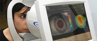
PRK method for the thinnest corneas
For very thin corneas, the thickness of which is 450 microns, the PRK method can be used. This technique is used only for medical reasons, since the postoperative period is painful and longer in comparison with other modern methods.
If laser vision correction is required, and the thin cornea does not allow the use of other methods, PRK surgery is performed. During the correction, the upper flap is not cut off, since the laser beam affects the upper layers of the cornea. Due to the large area of the cornea that is subject to evaporation, the rehabilitation period is painful for the patient. After correction using this method, additional use of a protective lens is required to prevent infection of the eye.
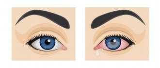
Main symptoms
There are many nerve receptors on the surface of the cornea, which is why the slightest impact on it is accompanied by severe discomfort. This protective reaction helps reduce the duration of traumatic contacts and protects against penetrating damage to the cornea.
Characteristic symptoms of injuries and damage to the outer shell of the eye are:
- intense lacrimation;
- intolerance to bright light;
- blepharospasm (uncontrolled tension of the eyelid muscles);
- pain that feels like sand in the eyes;
- partial loss of vision, inability to focus;
- redness of the mucous membranes of the eyelids, the appearance of a vascular pattern on the sclera.
With deep penetrating wounds of the eyes, when the destructive process affects the optic nerves, the clinical picture is complemented by intense headache, nausea, and dizziness.
Have questions? Ask:
Is it possible to have vision correction during pregnancy or lactation?
It is necessary to determine the timing of pregnancy. But today the approach is such that you can do Smile almost during the entire period of pregnancy and lactation.
But are complications with Smile possible?
Theoretically, yes, as after any manipulation. Therefore, it is important to choose experienced specialists. Our clinic has already performed hundreds of Smile operations, without any complications. There are rare cases when there are contraindications to Smile. This is clarified at the diagnostic stage, which is necessarily carried out before laser vision correction. If there are contraindications to Smile, then we use FemtoLasik or even PRK.
Once again, what is the correction range of the Smile method?
If we talk about myopia, then from -1 to -11. Astigmatism up to -6.0. For more severe deviations, there are other technologies: for example, phakic lenses
. But this is a subject for another discussion. The main thing that the patient should know is that today there are techniques that can solve a wide range of vision problems.
Farsightedness
We talk about myopia all the time. What about farsightedness? Is laser vision correction only applicable to myopic eyes?
Farsightedness is a more difficult problem to solve technologically. Smile, unfortunately, does not work here yet. We can use Femto Lasik or PRK. It allows you to adjust up to 6 diopters.
What is farsightedness? This means that the eye is shorter in length than necessary for a clear focus on the retina. There is another physiological issue with farsightedness: the lens is such a flexible body that in youth it can compensate for up to 3-4 diopters of farsightedness. And after 40 years, when the flexibility of the lens ligaments is lost, farsightedness appears. Sometimes in such cases we change the lens to an artificial one and get excellent results.
Are there many clinics in Moscow now offering Smile technology?
About 6 clinics, including government ones. But I am sure that this technology will soon be everywhere. Moreover, competing lasers are on the way.
By the way, about competitors. How many companies now produce such lasers?
Today, lasers for Smile are made by only one company - Carl Zeiss. This German company is still a monopolist and because of this it sets its own financial conditions.
But now lasers from other companies, also from Germany, are on the way: Zimmer and Schwind. When they enter the market, they will have to choose different names for their methods, since Smile is already a patented technology. But their operating principle will be similar.
What will happen next? Are there any disadvantages to Smile? In what direction will laser vision correction develop?
Over the years, computer software has been improved many times, but the procedure itself remains unchanged. Smile has proven itself to be the best technology, with no particular complaints. When the first Smile laser was invented, the manufacturers did a study on a reference group: they recruited volunteers (60 people throughout Germany), tested them and gave them Smile correction. This was 10 years ago.
This year they published their current status - for all of them, the corneal refraction has not changed in any way since the operation.
Of course, the evolution will be even more accurate, even more convenient, even more predictable, but Smile is great and will last for a long time.
Thank you, Oleg Vladimirovich, for introducing us to the intricacies of the most high-precision field of medicine. We hope that vision correction will become more accessible every year, and more and more clinics around the country will master these progressive methods.
First aid
Correctly provided first aid is the guarantee that the damage will be stopped and eliminated without significant consequences for health. There is a clear algorithm for performing actions after receiving an injury or damage of another nature. First you need to stop contact of the cornea with the traumatic factor:
- If a foreign body gets into the eye, you must carefully remove it with clean hands, a sterile cloth or a stream of lukewarm water. It is not advisable to use tweezers, as they can further damage the membrane of the eye and mucous membrane. Cotton wool is also not suitable, as it leaves microscopic fibers on the surface tissues.
- If the eyes are damaged by chemicals, rinse them with running water for 10-15 minutes, spreading the eyelids with your hands.
- In case of a thermal burn, it is necessary to cool the eye by applying a cloth soaked in ice water to it.
In case of a penetrating wound, it is strictly forbidden to remove the foreign object yourself. As a result, vision will be lost forever.
After eliminating the source of injury, it is necessary to isolate the eye from the external environment. No matter how badly the cornea is damaged, only sterile gauze should be used to apply a bandage. It is applied as an application, without pressing it to the eye socket. You can secure the tissue with a plaster or a loose bandage made from a wide bandage. To get rid of reactive blinking, the remaining eye should also be closed.
After providing first aid, it is important to take the victim to the hospital as quickly as possible. Its treatment will be carried out by an ophthalmologist-therapist or an ophthalmologist-surgeon, depending on the extent of the damage.
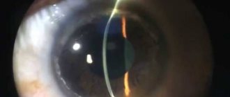
Femtosecond support for correction
If the patient has a thin cornea, vision correction is often performed using the Femto-Lasik method, during which a corneal flap is created using a femtosecond laser without contact. The femtolaser delicately affects the thin tissues of the cornea at a given depth. As a result of laser exposure, microbubbles are formed in the cornea, which combine with each other to form a clearly defined surface of the future corneal flap.
The valve formed in this way has individual parameters that are selected for each patient personally. The technique is suitable for patients with thinned corneas, dry eye syndrome, and other corneal pathologies who previously had to be denied surgery. Femtosecond support during Lasik surgery significantly expands the capabilities of the ophthalmologist to provide the patient with high-quality vision even in difficult cases.
The Femto Lasik technique allows you to create a corneal flap about 90 microns thick. Thus, the minimum corneal thickness for correction using this method can be about 470 microns.
Treatment
The treatment regimen depends on how severe the damage to the cornea is. For superficial injuries of the eye shell, conservative therapy is used. On the first day, to prevent complications, the following is used:
- an antibiotic or antimicrobial drug in the form of an ointment, most often tetracycline;
- antiseptics (chlorhexidine and its analogues) in the form of solutions for instillation (rinsing) of the eye;
- non-steroidal anti-inflammatory drugs of systemic action in the form of tablets.
To speed up regeneration, eye drops with antioxidants (vitamins A and E), B vitamins, collagen, peptides and amino acids, and hyaluronic acid are used.
When the membrane of the eye is deeply destroyed, radical measures are resorted to, most often corneal plastic surgery (keratoplasty). After recovery, a course of antibiotic therapy is prescribed, and drugs with antihypoxic and anti-inflammatory effects are used to prevent neovascularization, scarring and trophic pathologies.
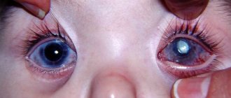
Penetrating lesions on the cornea can only be eliminated surgically. After removing foreign elements that have entered the eye, the displaced elements are repositioned: the iris, lens, etc. If the lens is damaged, it is replaced with a prosthesis. At the end of the operation, nylon purse-string sutures are placed on the eye capsule. They are left for at least 1.5 months. If the cornea is crushed and cannot be collected with sutures, artificial patches or an autograft (a piece of cornea from the surviving eye) is transplanted in its place.
After the operation, victims will undergo long-term rehabilitation, after which the doctor will assess the condition of the eye capsule and, if necessary, prescribe a correction: laser removal of scar defects on the cornea or vision correction.